Respiratory Alkalosis: PaCO2 and Some Rapid Breathing

A respiratory alkalosis occurs when a person breathes too rapidly. The result is a decrease in PaCO2 (ie: the amount of CO2 dissolved in the blood). This causes the blood to become alkalotic (less acidic), which is manifest by an increase in the blood’s pH. The reason the blood becomes less acidic is based on Le Chatelier’s principle. If we take a look at the following equation:

HCO3 + H+ —> H2CO3 –> CO2(g) + H2O

You’ll notice that CO2 (on the right most part of the equation) is what is exhaled via the lungs. When a patient is hyperventilating there is much less CO2 than normal in the blood stream. The body compensates by combining HCO3 and H+ to form more CO2. The resulting decrease in H+ causes the alkalosis (ie: rise in pH).

Causes of Respiratory Alkalosis

So what could cause someone to hyperventilate? The most common things are pain, anxiety, and fever. If the patient is in the ICU and being mechanically ventilated then a respiratory alkalosis may develop if the ventilator is set to give too many breaths per minute.

The most common causes of hyperventilation are:

  • Fever
  • Pain
  • Anxiety
  • Overventilating a mechanically ventilated patient

Acute Versus Chronic and Kidney Compensation

A respiratory alkalosis can be either acute or chronic. The difference depends on how much the kidney compensates for the change in pH. How exactly does the kidney compensate? It dumps HCO3 (aka: bicarb) into the urine. This helps offset the alkalosis and brings the bodies pH back to normal limits.

How do we determine if the kidney is acutely or chronically compensating? We measure the bicarb level. The kidney is acutely compensating if the HCO3 level is decreased 1 to 2 mmol/L per every 10 mmHg drop in the PaCO2 level. The kidney is chronically compensating if the HCO3 level is decreased 4 to 5 mmol/L per every 10 mmHg drop in PaCO2.

For example, if a patient’s PaCO2 on blood gas analysis is found to be 20 mmHg (a normal level is around 40) we would say there is a 20 mmHg drop present. If the HCO3 (determined by a chemistry panel) is at 19 (for argument sake we’ll say a normal bicarbonate level is 23) then that is a 4 mmol drop in the bicarb level for the 20 mmHg drop in CO2, or approximately 2 mmol drop in bicarb per 10 mmHg drop in CO2. This would mean the patient’s kidney is acutely compensating for the respiratory alkalosis.

  Bicarbonate Level (HCO3)
Acute Compensation Decreased by 1-2 mmol/L for every 10 mmHg decrease in the PaCO2
Chronic Compensation Decreased by 4-5 mmol/L for every 10 mmHg decrease in the PaCO2

Why is it important to determine if acute or chronic compensation is occurring? For starters, it gives us a better idea of what may be causing the respiratory alkalosis. If the kidney is acutely compensating we know that the respiratory alkalosis is new. The patient is likely having an acute reaction to something (ie: pain, anxiety, panic attack, etc.). If the compensation is chronic then we know that the patient has been breathing at a faster than normal rate for a prolonged period of time. This may be seen in pregnancy, COPD, and emphysema.

Treatment

Treatment is very straightforward: eliminate the underlying cause! If the patient appears in pain then give pain medication. Fever? Hit them with some acetaminophen. If the patient is mechanically ventilated then decrease the respiratory rate. Once the stimulus for hyperventilating is removed the respiratory alkalosis should improve.

Overview

A respiratory alkalosis occurs when a patient is breathing too rapidly, which cause too much CO2 to be removed from the bloodstream. There are numerous causes including anxiety, pain, and fever. The kidney can acutely or chronically compensate for a respiratory alkalosis depending on how long it has been present. Treatment is to fix the underlying cause.

A Little More Learning…

References and Resources

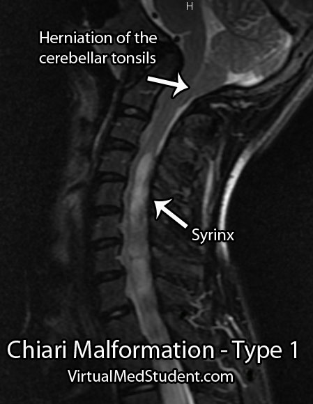
Chiari Malformation: Type1, Tonsils and Syrinx

The Chiari malformations are a group of disorders characterized, at least in part, by herniation of hindbrain structures through the foramen magnum at the base of the skull. They are categorized as type 1, type 2, and type 3 depending on clinical and radiographic findings.

This article will focus on type 1 Chiari malformations. The definition of this malformation has been debated, but most agree that the combination of herniated cerebellar tonsils (usually defined as greater than 5mm below the foramen magnum), with or without a syrinx, in the setting of referable symptoms is sufficient to make the diagnosis.

So why do the cerebellar tonsils herniate? Nobody knows for sure! We do know that type 1 malformations can be congenital or acquired. One theory is that tonsillar herniation is a result of an abnormally small posterior fossa (ie: the bones that compose the base of the skull). A small posterior fossa may be caused by under-development of the occipital somites in-utero (the fetal precursors that form bone and connective tissues), premature fusion of the cranial bones (ie: craniosynostosis), or medical conditions that promote abnormal bony growth.

Other experts advocate that abnormal cerebrospinal fluid pressures between the brain and spine may cause the tonsils to herniate downwards.

Regardless of how you slice it, we can say with certainty that there are multiple potential etiologies for type 1 Chiari malformations.

Type 1 Chiari malformations are commonly associated with a finding known as a “syrinx”. A syrinx is an abnormal fluid filled cavity that is seen in the cervical and/or thoracic spinal cord. It may represent an enlargement and extension of the central canal of the cord, in which case it is termed hydromyelia; it may also represent a complex glial lined cavity, which is referred to as syringomyelia. Regardless, syrinxes are found in 30% to 70% of type 1 Chiari malformations.

For unclear reasons, type 1 Chiari malformations with a syrinx are associated with scoliotic curves of the spine (especially left sided curves). It is believed that the syrinx puts pressure on the motor pathways of the spinal cord. This results in weakness of the paraspinal muscles causing the vertebral column to curve.

Signs and Symptoms

The most common presenting symptom of a type 1 Chiari malformation is pain. The pain is usually located at the back of the head and upper neck. Additionally, a cape-like sensation loss, as well as problems with vision and/or hearing may also be present.

Myelopathic signs or symptoms may be present if there is a syrinx. Myelopathic patients present with a combination of gait dysfunction, hand clumsiness, weakness, abnormally brisk reflexes, spasticity, and Lhermitte’s sign.

Diagnosis

Type 1 Chiari Malformation
Diagnosis of a type 1 Chiari malformation is made when an MRI shows abnormal herniation of the cerebellar tonsils, with or without an associated syrinx, in the context of appropriate signs and/or symptoms.

Treatment

The treatment of Chiari malformation is with surgical decompression. Most commonly this involves "shaving" off part of the occipital bone and removing the C1 lamina. This effectively decompresses the spinal cord and cerebellar tonsils.

If an associated syrinx is present, many neurosurgeons will open the dura (ie: the lining of the spinal cord) and perform a "duraplasty"; during the duraplasty a patch is sewn into place to give the spinal cord and cerebellar tonsils more room. Duraplasty generally improves the size and severity of the syrinx over time, but adds risk and complications to the procedure.

Some neurosurgeons will surgically shrink the cerebellar tonsils after opening the dura. This is done with bipolar electrocautery and serves to further decompress the area.

Overview

Type 1 Chiari malformations are hindbrain abnormalities characterized by herniation of the cerebellar tonsils below the foramen magnum. They are associated with cervicothoracic syrinxes as well as neuromuscular scoliosis. Symptoms can range from pain to neurological deficits. Treatment is with surgical decompression, although the exact type of decompression has been the subject of intense research.

References and Resources

SOCRATES: Thinking About Pain

When I was in medical school one of the most useful mnemonics I came across was "SOCRATES". The mnemonic is designed to figure out the characteristics of someone’s pain. The characteristics of pain help the clinician develop a differential diagnosis from which testing can be ordered, and then hopefully, treatment can be given.

So what does each letter in the mnemonic SOCRATES stand for??? Let’s go letter by letter…

S   O   C   R   A   T   E   S

The first “S” stands for “site”. What body part or parts are involved? Is the pain in the leg? Is it in the abdomen? Is it a general sense of overall discomfort? The site of pain helps you fine tune your subsequent physical exam and diagnostic decision making.

The next letter is “O”, which stands for “onset”. When did the pain start? Asking about the onset of the pain is extremely important! For example, if someone has had chronic low back pain for 10 years that invokes a much lower sense of urgency than someone complaining of the sudden onset of severe belly pain or headache.

S O C R A T E S 


S – Site
O – Onset
C – Characteristics
R – Radiation
A – Associated
T – Timing
E – Exacerbating/
      Alleviating
S – Severity

“C” stands for “characteristics”. What are the characteristics of the pain? You want the patient to describe the pain in their own terms without influencing them too much. The pain may be sharp, dull, heavy, burning, etc, or a combination of descriptors.

The next letter is “R”, which represents “radiation”. I typically ask if the pain stays at the site or if it travels somewhere else in the body. For example, someone with chest pain radiating to the left arm might be experiencing a heart attack. Back pain that is associated with radiation down the leg might indicate a herniated lumbar disc that may require surgery. Back pain radiating to the abdomen could be intraabdominal pathology. Radiation of the pain is an important component to help guide your decision making.

“A” stands for associated symptoms. What other symptoms are present with the pain? For example, if the patient is complaining of belly pain do they also have nausea or vomiting? If they have a headache do they also complain of double vision or photophobia? Associated symptoms can provide a wealth of information to help you hone your differential diagnosis even more.

“T” stands for timing. When does the pain occur? Does it happen at specific times of the day, or is it constant? Does it happen during a certain movement? All of these can give you an idea of the origin for the pain.

The letter “E” represents “exacerbating” factors; grouped within this is also alleviating factors. The patient should be probed as to what makes their pain better or worse. Certain physical positions, medications, etc. may make the pain better or more unbearable. These factors can all provide historical clues about the root cause.

The final “S” stands for “severity”. In most hospitals this is formulated on a 1 to 10 scale with 10 being the most severe pain they’ve ever experienced. This can be a tricky one to gauge because many patients will describe 10 out of 10 pain when they are lying comfortably in bed; therefore, it is often necessary to ask more pointed questions and place pain in a context.

Overall, the answers obtained when using the mnemonic SOCRATES can provide a solid framework from which to order new testing and treatments.

More Good Stuff…