Parkinson’s Disease: Pathology of Movement

Parkinson’s disease is caused by the death of neurons in a part of the brain known as the substantia nigra.

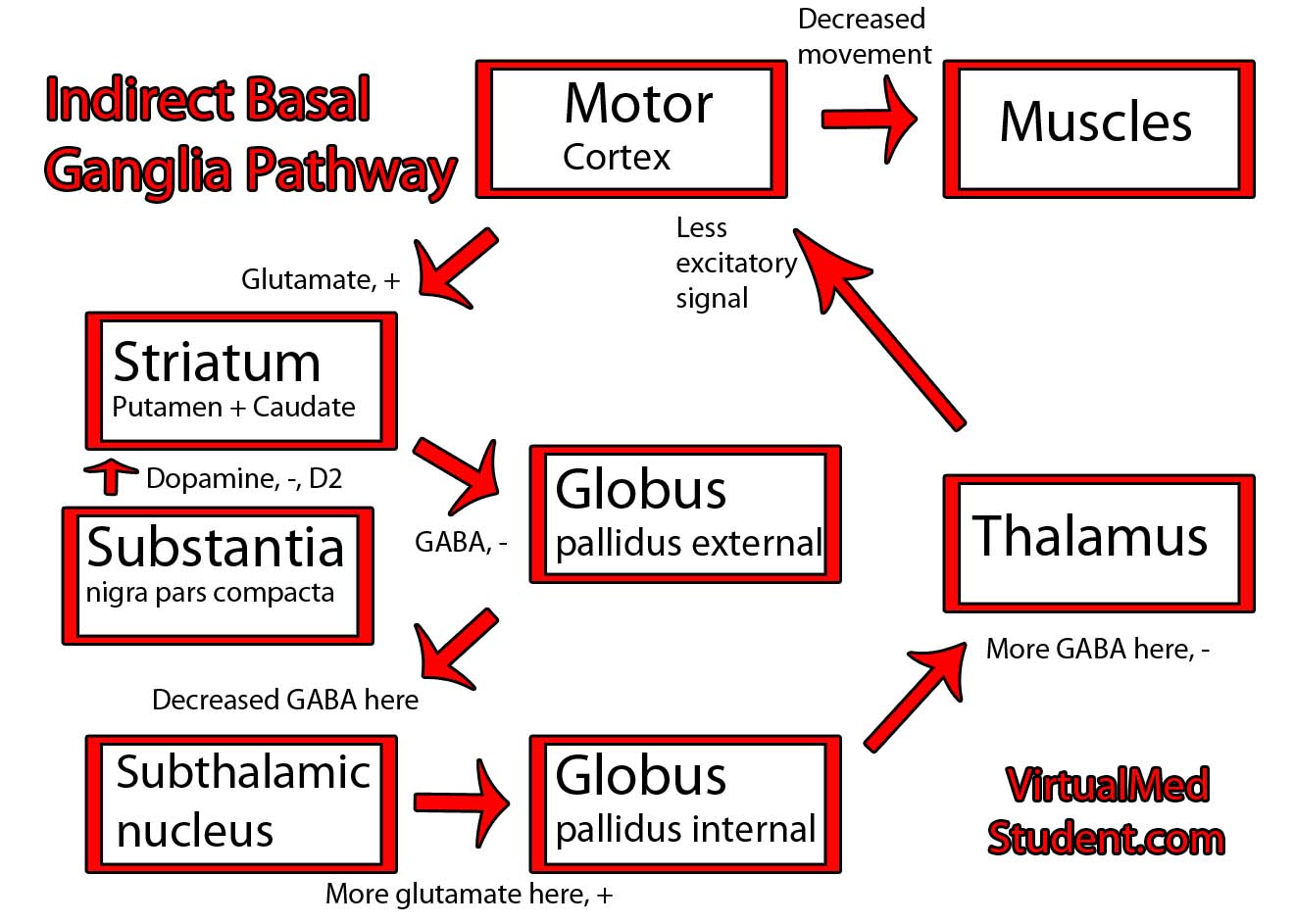
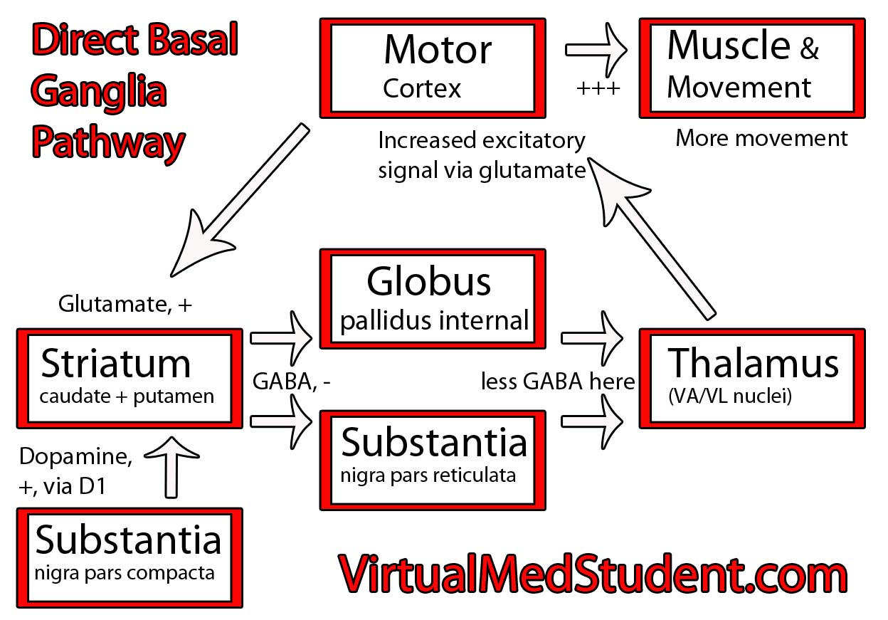
The substantia nigra is part of a system of connected neurons known collectively as the basal ganglia. This system is very important in the control of movement. The substantia nigra contains neurons that secrete a molecule known as dopamine. The loss of dopamine’s effect on the basal ganglia leads to the signs and symptoms of Parkinsonism.

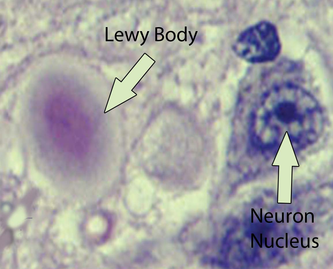
Visible abnormalities are also seen in the neurons of patients with Parkinson’s disease. These abnormalities are called Lewy bodies. They are collections of abnormal protein (specifically, a protein known as α-synuclein) that clump together to form a redish-pink cytoplasmic inclusion in the substantia nigra neurons.

Lewy body
Interestingly, it is not known what causes most cases of Parkinson’s disease. However, there are some known causes, almost all of which involve insults to the substantia nigra.

For example, toxins such as carbon disulfide, manganese, and certain street drugs have been known to kill substantia nigra cells resulting in Parkinson’s.

Another toxin known as MPTP (1-methyl-4-phenyl-1,2,3,6-tetrahydropyridine, trying saying that four times fast!), which was produced accidentally by drug chemists trying to make a synthetic opiod, is extremely toxic to substantia nigra cells. It caused irreversible Parkinson’s disease in those who were unfortunate enough to ingest it!

Finally, the genetics of Parkinson’s disease are not well known. There appears to be multiple genetic causes of the disease. One such genetic mutation involves the gene for α-synuclein, the main component of Lewy bodies. Other genetic mutations may also play a role in Parkinson’s, but they appear to contribute to only a small percentage of cases.

Diagnosis

Parkinson’s disease is a clinical diagnosis. This means that there are currently no special laboratory tests that can reliably detect the disease. Instead, the disorder is diagnosed based on the clinical symptoms discussed below. A true pathological diagnosis can only be made at autopsy by looking at the neurons of the substantia nigra under a microscope.

Signs and Symptoms

Parkinson’s disease has a number of symptoms that commonly begin in the 6th and 7th decade’s of life. Most of these are related to difficulties with movement. The classic description of Parkinson’s involves several signs and symptoms: resting tremor (4-7 Hz frequency), postural instability, bradykinesia (slow movements), rigidity (of the cog-wheeling type), and masked facies.

In addition, late in the disease course other symptoms can become apparent. Roughly 10 to 15% of patients develop dementia. This is believed to be due to the development of Lewy bodies in the cerebral cortex. The dementia of Parkinson’s disease is likely on a continuum with another form of dementia known as Lewy body dementia.

This YouTube video illustrates the common signs and symptoms seen in Parkinson’s disease:

Treatment

The mainstay of treatment for Parkinson’s disease is replacing or augmenting dopamine levels in the brain. A molecule known as levodopa (L-dopa), which is a precursor of dopamine is commonly used. The reason L-dopa is given instead of dopamine is because it crosses the blood brain barrier more readily.

Once inside the brain, L-dopa is converted to dopamine by neuronal enzymes. Sinemet® is a common formulation of L-dopa. It also contains a molecule known as carbidopa. Carbidopa inhibits the breakdown of L-dopa in the body so that more of it can reach the brain. L-dopa is most effective at improving tremor and bradykinesia.

Other medications are designed to mimic the actions of dopamine in the brain. These medications are known as dopamine agonists. They bind to dopamine receptors and cause the same types of cellular reactions that dopamine normally would. The two common dopamine agonists in use today are ropinirole and pramipexole.

Treating Parkinson’s –
(1) Levodopa/carbidopa
(2) MAO-B inhibitors
(3) COMT inhibitors
(4) Dopamine agonists
(5) Anticholinergics
(6) Glutamate antagonists
(7) Deep brain stimulation
Other medications used to treat Parkinson’s disease attempt to indirectly increase the amount of dopamine.

One category of medications that does this is known as MAO-B inhibitors. MAO, or monoamine oxidase, is an enzyme that normally breaks down monoamines like dopamine. Therefore, inhibitors of this enzyme decrease the break down of dopamine allowing it to remain in the brain longer. Selegiline and rasagiline are example of MAO-B inhibitors.

Another class of medications that perform a similar function to the MAOIs are the COMT inhibitors. COMT (catechol-O-methyl transferase) is an enzyme that breaks down dopamine. Entacapone is a COMT inhibitor that may increase the amount of dopamine in brain tissue.

Medications that interfere with another neurotransmitter known as acetylcholine are also sometimes used. These anticholinergic medications, namely benztropine (Cogentin) and trihexylphenidyl (Artane), are most useful for tremor reduction. Since acetylcholine is present in many parts of the body, the anticholinergics tend to have many side effects (the mnemonic "dry as a bone (dry mouth), mad as a hatter (delirium), blind as a bat (pupil dilation), and hot as a hare (fever)" is commonly used for symptoms pertaining to anticholinergic medications).

Finally, medications that antagonize the neurotransmitter glutamate are less commonly used. Amantadine is the name of one medication in this category. It helps mostly with decreasing, or smoothing out, fluctuations in movement.

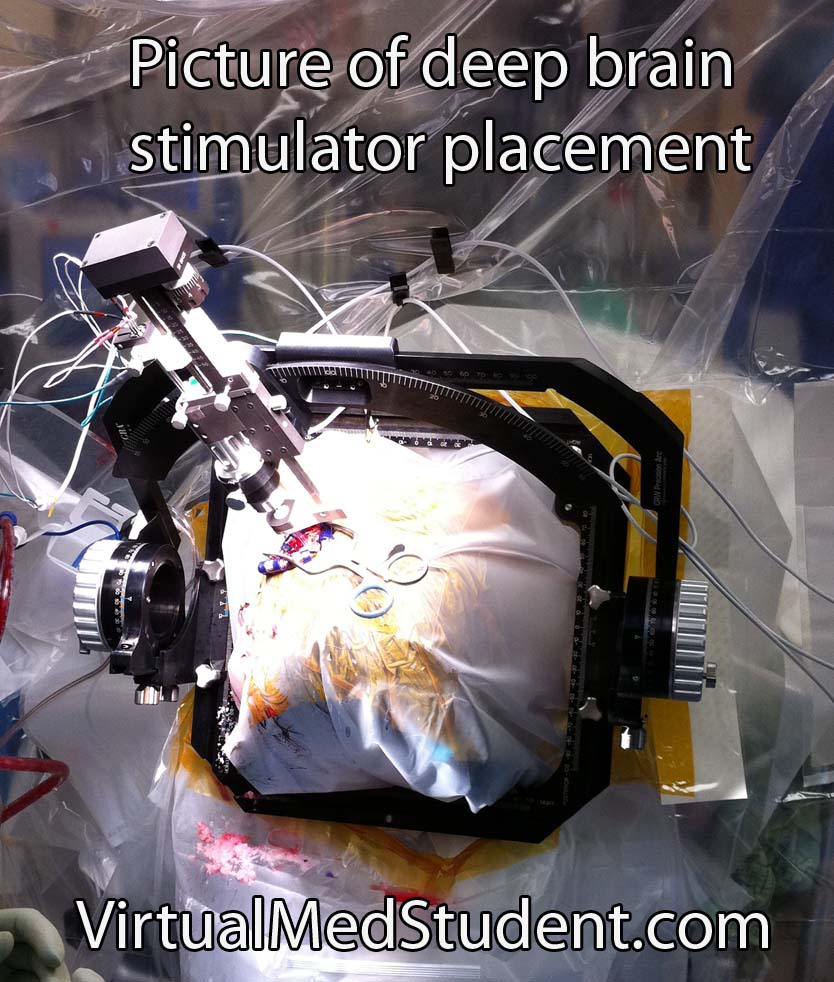
If medical therapy fails to control symptoms then surgical interventions may be used. In the past a procedure known as pallidotomy was used. This involved making a tiny incision in part of the basal ganglia. This procedure has been replaced by deep brain stimulation (DBS). During DBS surgery small electrodes are implanted in certain areas of the basal ganglia.

Deep Brain Stimulation

Overview

Parkinson’s disease is characterized by postural instability, resting tremor, slow movements, and rigidity. It’s caused by loss of dopaminergic neurons in the substantia nigra of the brain. Why these neurons die is not entirely understood. Diagnosis is based on clinical signs and symptoms. Treatment is by replacing or augmenting dopamine in the brain.

Related Articles

References and Resources

The Indirect Basal Ganglia Pathway

The basal ganglia represent a system of several discrete collections of neurons within the brain. These collections of neurons interact closely with the part of the cerebral cortex that initiates movement. The basal ganglia fine tune the starting and stopping of movements.

The term "basal ganglia" encompasses several separate, but interrelated neuron populations. The putamen, caudate, globus pallidus internus (GPi), globus pallidus externus (GPe), substantia nigra (SN), and subthalamic nucleus (STN) are all discrete neuron populations that, as a whole, compose the "basal ganglia". These named populations of neurons work together to achieve a common goal. The term "striatum" includes the caudate and putamen only, and the term "lentiform nuclei" includes the putamen and globus pallidus.

The basal ganglia modulate movement through a complex loop of both inhibitory and excitatory signals. When you decide to move, your frontal lobes send an excitatory signal via the neurotransmitter glutamate to the striatum (FYI: striatum = caudate and putamen).

In the indirect basal ganglia pathway the striatum then sends an inhibitory signal via the neurotransmitter GABA to the external segment of the globus pallidus. This is different from the direct pathway where the striatum sends a signal to the internal segment of the globus pallidus.

The external segment of the globus pallidus normally indirectly (more on this in the next few paragraphs) inhibits its internal counterpart. Thus, when the striatum inhibits the external segment, it is, in effect, releasing the internal segment from inhibition (that sure seems like a lot of double negatives!).

At this point, the internal segment of the globus pallidus is able to send its inhibitory signals to the thalamus, which causes thalamic neurons to stop sending excitatory signals to the motor cortex. The cortex is then unable to send an impulse down the spinal cord and, ta-da, the net result is a decrease in movement.

It would be easier to understand if the external segment of the globus pallidus “talked” directly to the internal segment, but that is not how it works. The message is relayed through another nucleus known as the subthalamic nucleus.

The subthalamic nucleus is usually inhibited by the external segment of the globus pallidus. Therefore, when the striatum inhibits the external globus pallidus, it causes the cells in the subthalamic nucleus to become more active (ie: the subthalamic nucleus is released from the inhibitory effects of the external globus pallidus).

The subthalamic nucleus, in turn, is able to send an excitatory signal to the neurons in the internal segment of the globus pallidus. The cells in GPi then become more active, which means that they suppress the activity of the thalamus more robustly. The thalamus is then unable to send its normal excitatory messages to the motor cortex. End result? Decreased movement!


Basal Ganglia Indirect Pathway Schematic


So how does dopamine act on the indirect pathway? Dopamine is secreted by the substantia nigra and binds to D2 receptors (these are different than the D1 receptors of the direct pathway) in the striatum. This causes striatal neurons to decrease their inhibitory message to the external segment of the globus pallidus. The external segment of the globus pallidus is then free to carry out its “normal” job and suppress the excitatory actions of the subthalamic nucleus on its internal counterpart. Less excitation going to the internal globus pallidus translates to less inhibition of the thalamus, and ultimately more excitation of the cortex!

Therefore, if you’ve managed to work through these complicated systems, you’ll realize that dopamine causes increased movement by activating the direct pathway and inhibiting the indirect pathway.

Overall, the indirect basal ganglia pathway has the exact opposite effect of the direct pathway. The indirect pathway serves as a negative modulator of movement and the direct pathway serves as a positive modulator of movement. Now that is some complicated sh**t!

Importance in Disease

Diseases of the basal ganglia cause unwanted movements, or a failure to initiate movement.

The classic basal ganglia disease is Parkinson’s disease, which has elements of both unwanted movement (resting tremor) and difficulty initiating movement (bradykinesia). Other diseases such as hemiballismus, in which the affected person violently flings an extremity, can occur when there is damage to the subthalamic nucleus.

Additionally, in Huntington’s disease the GABA and enkephalin projections from the caudate nucleus to the external globus pallidus are affected. This is believed to be responsible for many of the movement abnormalities seen in patients with this disease.

Overview

The indirect basal ganglia pathway fine tunes motor movements. It involves both excitatory and inhibitory signals through the striatum, globus pallidus, substantia nigra, thalamus, and motor cortex. Diseases such as Parkinson’s disease, hemiballismus, and Huntington’s disease may occur when there is damage to one of the components of the basal ganglia.

References and Resources

  • Baehr M, Frotscher M. Fourth Edition. Stuttgart: Thieme, 2005.
  • Bickley LS, Szilagyi PG. Bates’ Guide to Physical Examination and History Taking. Ninth Edition. New York: Lippincott Williams and Wilkins, 2007.
  • Nolte J. The Human Brain: An Introduction to its Functional Anatomy. Sixth Edition. Philadelphia: Mosby, 2008.
  • Purves D, Augustine GJ, Fitzpatrick D, et al. Neuroscience. Fourth Edition. Sinauer Associates, Inc., 2007.
  • Related Articles

The Direct Basal Ganglia Pathway

The basal ganglia are a complex system of several discrete collections of neurons within the brain. These collections of neurons interact closely with the part of the cerebral cortex that initiates movement. As such, the basal ganglia are important in fine-tuning the starting and stopping of movements.

The term "basal ganglia" encompasses several separate, but interrelated neuron populations. The putamen, caudate, globus pallidus internus (GPi), globus pallidus externus (GPe), substantia nigra (SN), and subthalamic nucleus (STN) are all discrete neuron populations that, as a whole, compose the "basal ganglia". These named populations of neurons work together to achieve a common goal. The term "striatum" includes the caudate and putamen only, and the term "lentiform nuclei" includes the putamen and globus pallidus.

The basal ganglia modulate movement through a complex loop of inhibitory and excitatory signals. When you decide to move, your frontal lobes send an excitatory signal via the neurotransmitter glutamate to the striatum (ie: caudate and putamen).

The neurons in the striatum then send an inhibitory signal to the globus pallidus internus (GPi) and the substantia nigra pars reticulata (SNpr). As a result, GPi and SNpr are no longer able to inhibit the thalamus, which is their normal resting function.

The thalamus now finds itself disinhibited and is able to send a message back to the cerebral cortex saying it is ok to allow the desired movement to occur. The motor cortex then sends a message down the spinal cord causing the desired movement.

So where does the neurotransmitter dopamine enter the picture? Dopamine is secreted by a different part of the substantia nigra known as the pars compacta. These neurons secrete dopamine onto specific cells in the striatum. The dopamine interacts with the D1 (dopamine-1) receptors on these cells causing them to become more active (ie: dopamine has a stimulatory effect via the D1 receptor).

Basal Ganglia Direct Pathway

The overall effect is that dopamine activates the striatum (ie: caudate and putamen), which, as we discussed above, inhibits the internal segment of the globus pallidus. The GPi is then unable to inhibit the thalamus, which in turn allows the thalamus to stimulate the cortex. Huh??? Simply stated, dopamine causes an increased propensity towards movement. Dopamine has a similar role, but via a different mechanism in the indirect pathway, which is discussed in detail in another article.

Importance in Disease

When the basal ganglia malfunction it causes unwanted movements or a failure to initiate movements. The classic basal ganglia disease is Parkinson’s disease, which has elements of both unwanted movements (resting tremors) and difficulty initiating movement (bradykinesia). Other diseases such as hemiballismus, in which the affected person violently flings an extremity, can occur when there is damage to the subthalamic nucleus (the subthalamic nucleus is discussed in more detail on the indirect pathway article).

Overview

The direct basal ganglia pathway fine-tunes motor movements. It involves both excitatory and inhibitory signals through the striatum, globus pallidus, substantia nigra, thalamus, and motor cortex. Diseases such as Parkinson’s disease and hemiballismus may occur when there is damage to one of the components of the basal ganglia.

Related Articles

References and Resources

  • Baehr M, Frotscher M. Duus’ Topical Diagnosis in Neurology: Anatomy, Physiology, Signs, Symptoms. Fourth Edition. Stuttgart: Thieme, 2005.
  • Bickley LS, Szilagyi PG. Bate’s Guide to Physical Examination and History Taking. Ninth Edition. New York: Lippincott Williams and Wilkins, 2007.
  • Nolte J. The Human Brain: An Introduction to its Functional Anatomy. Sixth Edition. Philadelphia: Mosby, 2008.
  • Purves D, Augustine GJ, Fitzpatrick D, et al. Neuroscience. Fourth Edition. Sinauer Associates, Inc., 2007.