Atlas Fractures: The Weight of the World On Its Shoulders

The atlas, or the first cervical vertebra (C1), is a ring shaped structure. It forms joints with the base of the skull above and the axis (ie: the second cervical vertebrae) below. It also has two foramen transversarium, which are holes that allow the passage of the vertebral arteries on either side of the spinal cord.

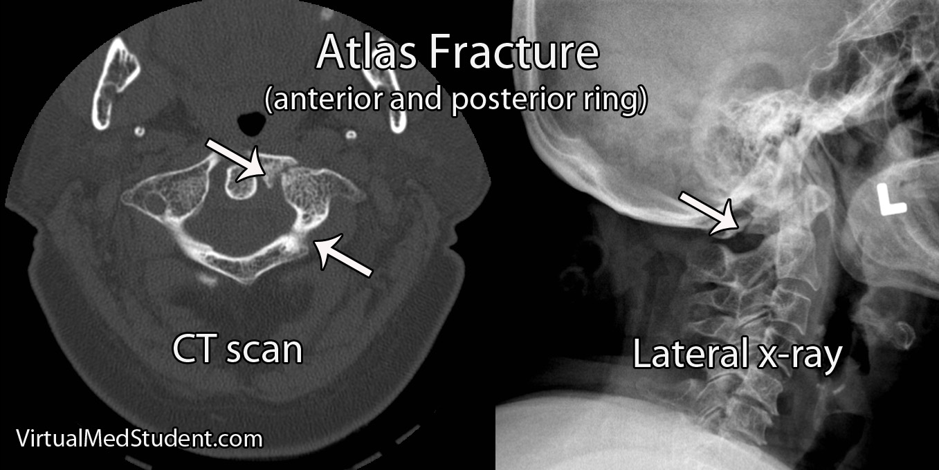
Fractures of the atlas occur most commonly with forceful axial loading of the head (ie: a downward force applied to the top of the head). Pressure on the top of the head causes the skull to push down on the atlas, which results in a break(s) of its ring-like structure. Specific fracture types such as a break in the front of the ring, the back of the ring, or one side of the ring versus the other, are dependent on additional force vectors at the time of loading (ie: flexion, extension, lateral bending, etc.).

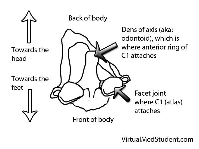
Fractures of the atlas must also include a discussion of biomechanical stability, which is usually determined by the integrity of the transverse ligament. The transverse ligament attaches the dens (odontoid) of the axis to the anterior ring of the atlas.

Fractures of the atlas with co-existent rupture of the transverse ligament lead to instability of the joint between C1 and C2. In other words, the ring of C1 may be able to move forward relative to the dens of C2. Transverse ligament injury is more common when axial loading is combined with extension of the head.

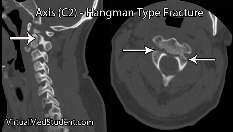
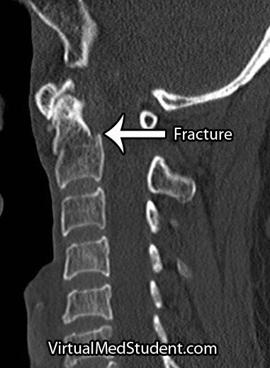
Not surprisingly, fractures of the atlas often co-exist with fractures of other cervical spine vertebrae. The most common combination is with a fracture of the axis, occurring in up to 40% of cases.

Signs and Symptoms

Patient’s with isolated atlas fractures usually have neck pain and muscle spasms. Frequently they have no injury to the spinal cord because the ring splays outwards as it fractures.

It is important to rule out injuries to the vertebral arteries, which run in bony holes (ie: foramen transversarium) on the sides of the atlas. When injured, the vertebral arteries can cause strokes in the brainstem and cerebellum, which can be life threatening.

Since the atlas is so close to the brainstem, patients may have co-existent injury to the lower cranial nerves. Specifically, injury to the 12th nerve can cause problems with tongue movements, injury to the 11th nerve can cause weakness with shoulder shrug and the ability to turn the head to the side, and injuries to the 9th and 10th cranial nerves can cause problems with swallowing and paralysis of the larynx leading to difficulty with speech.

Co-existent head and brain trauma, which can cause a constellation of different signs and symptoms depending on severity can also occcur.

Diagnosis

Diagnosis of an atlas fracture is made using x-rays, CT scans, and MRIs. X-rays should include anterior-posterior views, open mouth odontoid views, and lateral views of the cervical spine. If there is no evidence of neurological injury, flexion-extension x-rays may also be obtained to assess for stability of the C1-C2 joint.

The bony injury associated with atlas fractures is categorized according to the Jefferson or Landell and Van Peteghem classification systems. The Landells classification has three types, whereas the Jefferson classification has four types:

Landell and Van Peteghem Classification
Type 1 Fracture of either the anterior or posterior ring, but not both (posterior ring fractures are most common type)
Type 2 Fractures of both the anterior and posterior ring
Type 3 Fracture of the lateral mass(es)

Jefferson Classification
Type 1 Fracture of the posterior ring only
Type 2 Fracture of the anterior ring only
Type 3 Fracture of the anterior and posterior rings on both sides; this is the classic "burst", or traditional “Jefferson” fracture
Type 4 Fracture of the lateral mass(es)

Atlas fracture

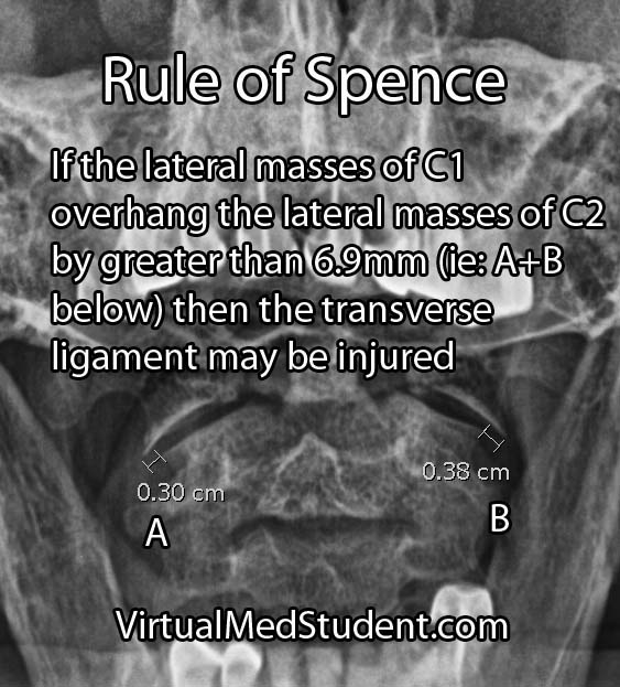
An important part of diagnosing atlas fractures involves assessing the integrity of the transverse ligament, which is best done using MRI. However, if an MRI cannot be performed then open mouth odontoid, flexion-extension x-rays, and CT scans can provide some information regarding transverse ligament injury.

The rule of Spence is one way of assessing the integrity of the transverse ligament on an open mouth odontoid x-ray. The rule states that if the right and left lateral masses of C1 overhang the lateral masses of C2 by greater than a total distance of 6.9mm than the likelihood of co-existent transverse ligament injury is high. The rule of Spence is not fool proof and should be supplemented with MRI and/or flexion-extension films whenever possible.


Atlantodental Interval
Another method for assessing transverse ligament injury is using the "atlantodental" interval (see image to the left). This is the distance between the anterior arch of C1 and the odontoid process (aka: dens) of C2.

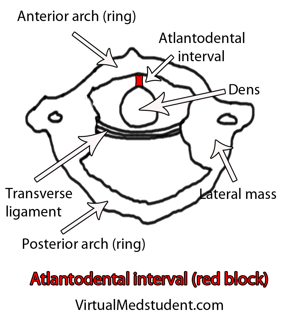
This interval is usually quite small, typically less than 3mm in adults and 5mm in children. If the interval is greater than this, then co-existent transverse ligament injury should be suspected.

Treatment

Treatment of isolated atlas fractures is usually with cervical immobilization. This may be with a halo or with a rigid cervical collar such as a cervical-occipital-mandibular-immobilizer (SOMI).

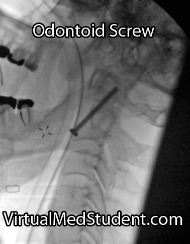
Atlas fractures that have co-existent transverse ligament rupture often require an operation to stablize the bones of the spine. This is usually in the form of fusing the atlas or occiput (back of the head) to the second cervical vertebrae.

If other injuries (ie: fractures of C2) are present and/or there is significant ligamentous injury then open surgical fusion of the bones may be necessary to re-create stability of the craniocervical junction.

Overview

Atlas fractures occur in response to vertical compression of the head on the upper cervical spine. Fractures of the anterior, posterior, or both rings of C1 may be present. Biomechanical stability is typically determined by assessing the integrity of the transverse ligament. Patients with isolated C1 fractures usually complain of neck pain, and rarely have injury to the spinal cord. Diagnosis is based on CT, x-ray, and MRI findings. Treatment is with rigid external immobilization or operative spinal fusion.

Related Articles

References and Resources

The Hangman’s Fracture

The axis, or second cervical vertebrae (C2), is unique amongst the vertebrae of the spine. It contains a body, which has an elongated structure that extends towards the head known as the dens (aka: odontoid). It also contains a ring-like structure that extends around the spinal cord, which is composed of the pedicles, pars interarticularis, and lamina. It forms joints with the atlas (ie: first cervical vertebrae) above it, and the third cervical vertebrae below it. It also has two foramen transversarium on either side, which are conduits for the vertebral arteries.

A hangman’s fracture is a break in both pedicles and/or pars interarticulari. The terms pedicle and pars interarticularis are not as well defined at C2 as they are for the other vertebrae, and thus have to be more clearly delineated before we discuss the details of a hangman’s fracture.

The pars interarticulari of C2 are the narrow pieces of bone that sit between the superior articulating facets (ie: the portion of bone that allows C2 to form a joint with C1) and the inferior articulating facets (ie: the portion of bone that allows C2 to form a joint with C3) of C2. The pedicles lie more anterior and are defined as the bony "bridges" that lie underneath the superior articulating facet and just medial (ie: closer to the spinal cord) to the transverse foramen, which house the vertebral arteries.

So now let’s get to the good stuff… Why do these fractures happen? Extension of the neck! This is why they are colloquially termed "Hangman’s" fractures; in the old days when a person was hanged the noose would pull the mandible upwards and cause the neck to violently extend. The end result was a tremendous amount of force on the pars interarticulari and pedicles of C2 leading to a fracture.

Since hangings are infrequent in today’s society, a more common cause of hyperextension of the neck is a head hitting the steering wheel or windshield of a car.

The hyperextension also often causes significant anterior ligamentous injury. The anterior longitudinal ligament (ie: the ligament that runs down the front of the spine) and the annulus fibrosis of the C2 disc are often ruptured. These findings are consistent with a hyperextension injury as the ligaments in the front get stretched to the point of rupture.

Signs and Symptoms

Surprisingly, hangman’s fractures rarely cause neurological injury. Most patients are neurologically intact meaning that there is no injury to the spinal cord and/or nerves at the level of the fracture. Typically there is neck pain, which is the most common symptom.

It is important to realize that many patients with hangman’s fractures will also have co-existent head trauma, and roughly a third of patients will have additional spine fractures. So keep a look out for associated injuries!!!

Diagnosis

The diagnosis of a hangman’s fracture can be made using x-rays and CT scans. MRI scans are also frequently ordered to determine the extent of co-existent ligamentous and soft tissue injury. A CT angiogram or MR angiogram should also be done to assess for co-existent vertebral artery injury.

Hangman's fracture

There are several different grading systems for hangman’s fractures. They include the Effendi, Francis, and Levine and Edwards classifications.

The Effendi system is based on the orientation of the fracture, as well as the degree of angulation and dislocation between C2 on C3.

The Francis system also takes into account the angulation and displacement between the bodies of C2 and C3, which is measured between the inferior endplate of C2 and the superior endplate of C3.

Perhaps the easiest to implement clinically is the Levine and Edwards classification. A type I Levine fracture is a non-displaced, non-angulated fracture. Type II fractures come in two flavors: type II is a fracture that is significantly angulated (ie: > 11 degrees) and displaced (ie: greater than 3mm) and a type IIa fracture is angulated (ie: greater than 11 degrees), but not significantly displaced. Type III fractures are fracture-dislocations of C2 on C3.

The Levine and Edwards’ Classification System
for Hangman’s Fractures

Type Angulation Displacement Treatment
Type I Minimal Minimal Rigid orthotic
Type II Greater than 11 degrees Greater than 3mm Traction ± rigid orthotic ± surgery
Type IIa Greater than 11 degrees Minimal Traction ± rigid orthotic ± surgery
Type III Minimal to severe Significant (fracture/dislocation) Traction + surgery

Treatment

Most isolated hangman’s fractures can be treated with external immobilization in a rigid cervical collar (ie: Miami J or Philadelphia collar) or in a halo immobilization device.

However, if there is significant ligamentous disruption, severe angulation and/or dislocation, or the inability to obtain adequate alignment of the spine in an immobilization device (ie: rigid collar or halo) then internal surgical fixation and fusion should be performed.

The need for surgery depends on the severity of the fracture and/or the integrity of the associated ligaments and discs.

A surgical approach from the front (aka: an "anterior" approach) may be performed to fuse the C2 and C3 vertebrae by removing the disc material between them. This approach is most often done in the presence of anterior longitudinal ligament rupture and/or intervertebral disc protrusion (ie: a "traumatic" disc).

Surgery from behind may also be used (aka: a "posterior" approach). Usually, the 1st through 3rd cervical vertebrae are incorporated into the fusion process, but some surgeons may opt to fuse to the base of the skull in cases of more severe injuries.

Overview

Hangman’s fractures occur after violent extension of the neck. The pedicles or pars interarticulari are fractured on both sides of the C2 ring. Neurological injury is rare in isolated hangman’s fractures, but frequently there are associated injuries to other bones in the cervical spine, as well as injuries to the brain and face. Diagnosis is made with x-rays, CT scans, and MRI. Treatment is with rigid immobilization of the cervical spine and/or surgical fixation depending on the extent of injury.

Related Articles

References and Resources

Latin for Toothlike: Fractures of the Odontoid Process

The odontoid process (also know as the "dens") is the finger of bone that sticks up from the second cervical vertebrae (ie: the axis).

It articulates via numerous ligaments to the anterior arch/ring of the first cervical vertebrae (ie: the atlas) to form a joint. This joint is what allows you to rotate your head from side to side as if you were nodding “no”.

Axis (C2)

Fractures of the odontoid typically occur after traumatic events. In younger, otherwise healthy individuals tremendous force is necessary to fracture the odontoid. Breaks are typically seen after car, motorcycle, or sporting accidents. In older, osteoporotic people simple ground level falls can result in a fracture. Less commonly, fractures of the odontoid may be caused by tumor chipping away at the underlying bone (a so called “pathologic” fracture).

Since the odontoid is a relatively long piece of bone it can fracture at one of several distinct sites. The most commonly used system (Anderson and D’Alonzo) categorizes fractures into one of three types:

  • Type 1 – a fracture at the tip of the odontoid.
  • Type 2 – a fracture at the base of the odontoid.
  • Type 3 – a fracture involving the body of the C2 vertebrae, which includes the odontoid within it.

Odontoid fractures

This grading system is important because it helps predict both stability of the C1-C2 (ie: atlanto-axial) joint and guides potential treatment options.

Signs and Symptoms

Roughly 80% of patients with odontoid fractures do not have any neurological injury to their spinal cord. The remaining patients can exhibit anything from quadriplegia to mild sensory disturbances. Patients with severe cervical spinal cord injury usually are unable to breath (secondary to diaphragm paralysis) and frequently die at the scene of the accident.

Many patients with odontoid fractures will have significant neck pain that radiates up into the scalp. This is usually caused by neck muscles spasming secondary to the injury.

Diagnosis

CT of odontoid fracture

CT scans, x-rays, and MRIs are all useful in diagnosing and properly treating odontoid fractures.

CT scans of the cervical spine provide excellent bony detail, and also help illustrate any additional fractures that may be present.

MRI of the cervical spine is useful for assessing any co-existent ligamentous injury. If ligamentous injury is present it drastically alters treatment decisions.

Treatment

Treatment of odontoid fractures is based on both bony and ligamentous injury. The goal of treatment is to stabilize the spine either by allowing the bone to heal on its own, or by fusing the spine artificially using rods, screws, and/or wires.

Placement of an odontoid screw is one method of fixing non-displaced type II fractures. However there are numerous contraindications for odontoid screw placement. For example severe angulation of the fractured segment precludes placement of a screw; barrel chested anatomy prevents an adequate angle for screw trajectory in the operating room. In addition, if the transverse ligament is disrupted, bony fixation with an odontoid screw alone will not stabilize the joint.

On the other hand, odontoid screws are beneficial because they typically stabilize the fracture with minimal restriction of neck motion.

Approaching the spine from behind is another option to stabilize odontoid fractures. Fusing the atlas (C1) to C2 or C3 is sometimes used if odontoid screw placement cannot be performed and the injury is deemed unstable. It is important to note that posterior approaches can restrict motion, especially in the high cervical spine.

Non-surgical options must ensure that the patient has minimal to no movement of the neck in order to give the bone an adequate chance to heal on its own. Rigid collars or halos are used to prevent neck motion.

Overview

Odontoid fractures come in three flavors depending on the location of the fracture. Symptoms can be anything from mild neck discomfort to quadriplegia, although neurological injury is surprisingly uncommon. Diagnosis is based on CT and MRI. Treatment is with cervical immobilization for an extended period of time, or surgical fusion. Treatment decisions are based on the degree of both bony and ligamentous injury, as well as the patient’s overall health status.

Related Articles

References and Resources