Spondylolisthesis: Slip, Ouch, and Wiltse

Spondylolisthesis occurs when one vertebrae "slips" forward relative to the one below it. Spondylolisthesis is most frequently used to describe this process in the lumbar spine, but it may occur in the cervical or thoracic spine as well.

Spondylolisthesis has traditionally been divided into categories based on the underlying pathology causing the slip (ie: the Wiltse classification). These categories include: congenital, isthmic, degenerative, traumatic, pathologic, and post-surgical (aka: iatrogenic).

Regardless of the category, the end result is the same: one vertebrae slips forward relative to the one below it. Let’s talk about each category in more detail…

Congenital spondylolisthesis occurs in children. It is usually caused by malformation of the superior articulating facet of the first sacral vertebrae (S1). The facet is a portion of bone that forms a joint with the vertebrae above or below it. Therefore, in congenital spondylolisthesis the malformed S1 facet joint is unable to form a stable joint with the lowest lumbar vertebrae (L5). The end result is that the L5 vertebrae is able to slip forward relative to the sacrum.

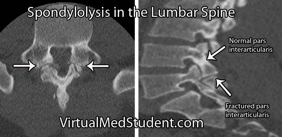
Isthmic spondylolisthesis occurs when there is a break in the bone that connects the facet of one vertebrae to the facet of the vertebrae immediately above (or below) it. This connecting bone is known as the pars interarticularis. The defect in the pars interarticularis is known formally as spondylolysis (see image below). Spondylolysis does not necessarily cause spondylolisthesis, but when it does it is classified as "isthmic". This type of spondylolisthesis is more common in those of Inuit heritage, as well as in athletes who have had repetitive hyperextension of the lumbar spine (ie: gymnastics, tennis, football, etc).

Traumatic spondylolisthesis is exactly what it sounds like. Injury to the facet joints, pars interarticularis, or ligaments of the spine can allow one vertebrae to slip forward/dislocate relative to the one above and/or below it.
CT lumbar spondy
Pathologic spondylolisthesis occurs when the slip is caused by some underlying pathology in the bone. This category is broad and may include tumors, osteoporosis, osteopetrosis, Paget’s disease, or other diseases of bone formation or metabolism.

Post surgical (also known as iatrogenic) spondylolisthesis occurs after surgery. It most frequently develops after a laminectomy is performed. If too much bone is removed during the laminectomy it may cause instability and allow a slip to occur.

The last category, degenerative spondylolisthesis, affects people over the age of forty. It is unclear exactly what causes this type of spondylolisthesis, but it is believed to be secondary to a combination of arthritic changes in the facet joints, degenerative disc disease, and bony remodeling of the pars interarticularis. Degenerative spondylolisthesis, unlike isthmic or congenital, usually results in the fourth lumbar vertebrae (L4) slipping over the fifth lumbar vertebrae (L5).

How Do Patient’s with Spondylolisthesis Present?

Spondylolisthesis typically presents with a combination of low back pain with or without radiculopathy (ie: discomfort down the legs). The radiculopathic symptoms are due to narrowing of the intervertebral foramen and pinching of the nerves as they exit the spinal column.

Neurological symptoms such as weakness, bowel or bladder problems, or sexual dysfunction are uncommon, but can also occur. These symptoms become more common as the severity of the slip increases.

Many patient’s with spondylolisthesis also have significant hamstring tightness (ie: unable to touch their toes). Additionally, many patients will walk or stand with their torso or knees in a flexed position.

Diagnosis and Severity of Slip

Diagnosis of spondylolisthesis is based on x-rays, CT scans, and MRI imaging. X-rays and CT scans are best at delineating the bony anatomy. MRIs are frequently ordered to assess the amount of nerve compression (especially in patients with signs of neurologic dysfunction).

The severity of the slip is based on the Meyerding classification system. Type one slips occur when less than 25% of the diameter of the vertebral body slips forward relative to the one below it. Type two slips are between 26% and 50%. Type three slips are between 51% and 75% and type four slips are between 76% and 100%. If a slip is more than 100% it is referred to as a grade five slip or spondyloptosis.
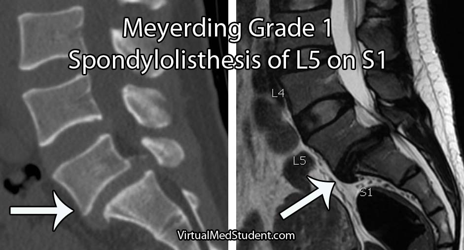
Grade 1 Spondylolisthesis L5-S1

Fix Me Doc!

Initial treatment for low grade slips presenting with back pain and mild radiculopathy is usually conservative. Physical therapy, joint injections, ibuprofen (or other non steroidal anti-inflammatory medications), and bracing can be used for several months. Physical therapy should focus on lumbar flexion exercises with avoidance of lumbar extension. If symptoms continue or worsen then surgery is indicated (talk to your local spine surgeon).

Patient’s who fail conservative treatment, have high grade slips (ie: greater than 50%), or have weakness, bowel/bladder issues, or sexual dysfunction should be offered surgery. The primary goal of surgery is to prevent further slippage by fusing the vertebrae together; realigning the spine is a secondary goal, but is not absolutely necessary. In fact, attempts to realign high grade slips have been shown to increase the risk of neurological injury.

The type of surgery offered to correct spondylolisthesis may be from the front (ie: anterior), from the back (ie: posterior), or a combination of both. As of now, there is no “right” surgical approach.

The Play by Play…

Spondylolisthesis is a fancy term that describes one vertebrae slipping forward relative to the one below. There are six categories based on the underlying pathology causing the slip. These categories are congenital, isthmic, degenerative, traumatic, pathologic, and post surgical/iatrogenic. Symptoms are typically low back pain with a component of radiculopathy. Weakness, bowel/bladder or sexual dysfunction is actually uncommon unless high grade slips are present. The Meyerding system is used to determine the severity of the slip. Treatment for low grade slips is usually conservative, but surgery may be necessary for high grade slips, patients that fail conservative management, or those with hard signs of neurological dysfunction.

Additional Pathology You Might Fancy…

References and Resources

Spondylolysis and the Scotty Dog

Spondylolysis is a fancy term for a defect in a portion of a vertebrae known as the pars interarticularis. The pars interarticularis is a section of bone that connects the superior articulating facet and the inferior articulating facet of a given vertebral body. The facets (which are joints) connect adjacent vertebrae together.

The CT scan below shows the relevant anatomy of a normal pars interarticularis and one that exhibits spondylolysis. Spondylolysis occurs about 85% of the time at the fifth (L5) lumbar segment, followed in frequency by the fourth lumbar vertebrae (L4), but it can occur elsewhere in the spine.

In spondylolysis there is a fracture/defect in the pars. Fractures of the pars interarticularis typically occur after repeated extension of the lower back. Repetitive extension causes significant pressure on the pars, which in susceptible individuals can lead to weakening of the bone.

Spondylolysis is actually quite common in gymnasts, football, and tennis players because of the repetitive back extension required of these athletes.

The importance of spondylolysis is not so much the pars defect itself, but the fact that these defects can cause one vertebrae to "slip" over the top of the one below it. When this occurs a new name is given to the condition – spondylolisthesis.

Signs and Symptoms

CT lumbar spondy
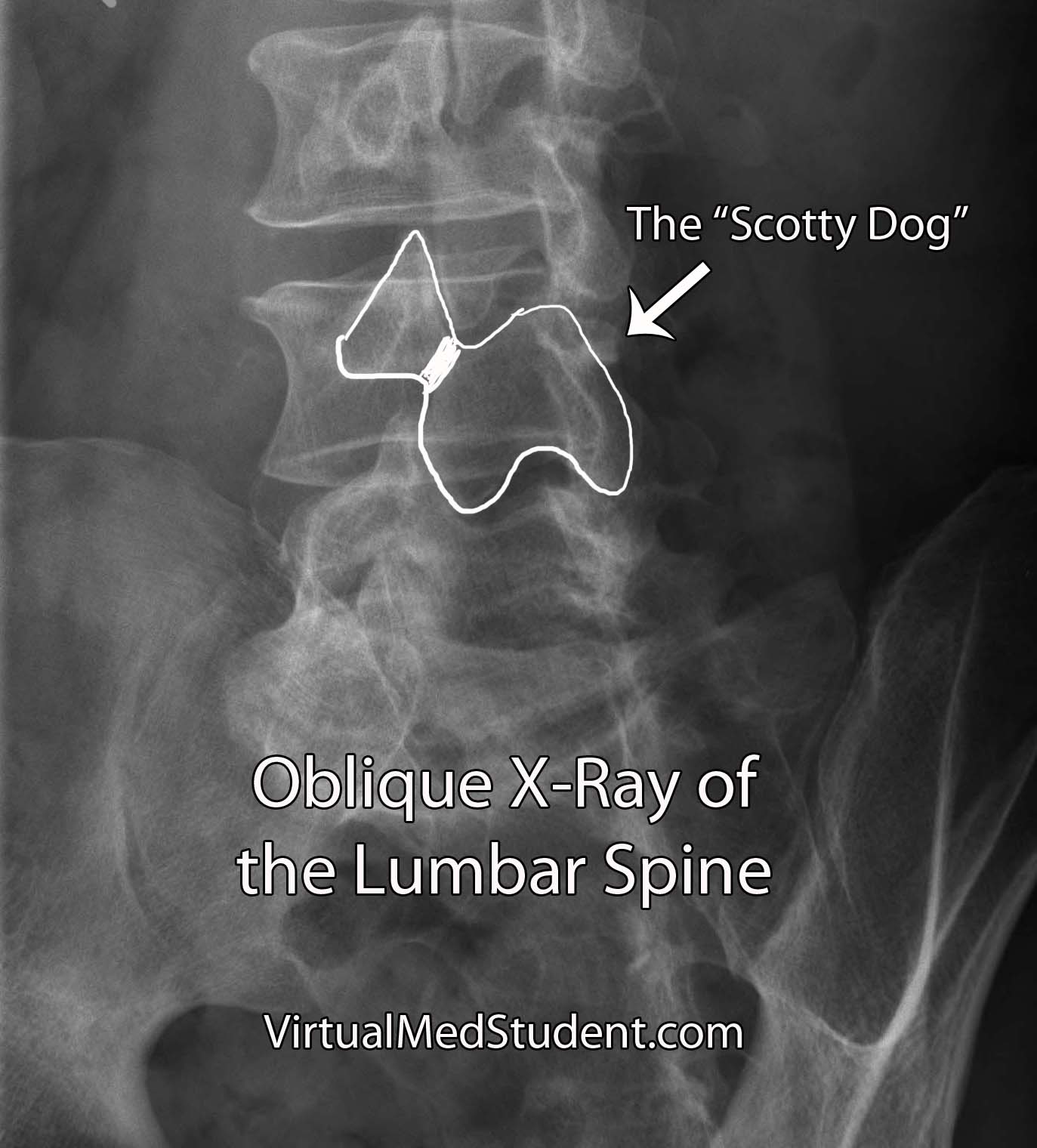
Xray Scotty Dog
The most common symptom of spondylolysis is low back pain. In fact, one of the most common causes of low back pain in individuals less than 25 years old is spondylolysis, especially in athletes. It is also important to note that spondylolysis is commonly asymptomatic.

Diagnosing Spondylolysis

Diagnosis is made by looking at an x-ray or CT scan of the spine. The classic description is a defect through the neck of the “Scotty dog”, which is seen best on an oblique x-ray of the lumbar spine.

What to do About It

Treatment starts with avoidance of the motions that caused the fracture. Occasionally a brace is used to prevent extension of the spine.

Anti-inflammatory medications like ibuprofen are commonly used if pain is significant. After a period of rest, physical therapy can be useful to strengthen muscles and prevent recurrence.

Less commonly surgical repair of the pars defect may be attempted. Surgical candidates are patients who continue to have back pain despite conservative treatment. If the patient receives significant pain relief from pre-operative bupivacaine injection into the defect, then they should be considered excellent surgical candidates.

Overview

Spondylolysis is a defect in the bony pars interarticularis of the vertebrae. It commonly occurs in the lumbar spine after repetitive extension motions. Low back pain is the most common symptom. In the absence of complications like spondylolisthesis treatment is typically conservative and involves bracing, physical therapy, and anti-inflammatories. Diagnosis is made by x-ray and/or CT scan.

For Your Reading Pleasure

If You Need Even More Info

Spinal Shock and Neurogenic Shock: The Battle Begins

Endless hours of studying, relentless exams, and the never-ending confusion between two perplexing phenomena: spinal shock and neurogenic shock. So, buckle up (so you don’t end up in spinal shock) and prepare for the journey through the world of spinal and neurogenic shock as we break down their differences.

The Tale of Two Shocks

Picture this: you’re in the ER, and a patient comes in with a recent spinal cord injury. You rack your brain to differentiate between spinal shock and neurogenic shock, but all you can remember is the cranial nerves mnemonic and your chief resident is about to pimp the you know what out of you… Let’s make it easy!

  • Spinal Shock: Think of it as your body’s initial reaction to “breaking up” with your spinal cord. It’s a temporary and unexpected “break-up shock” that leaves your reflexes, motor function, and sensation feeling lost and numb (physically, not emotionally) below the level of injury.
  • Neurogenic Shock: Now, imagine your body losing its balance between the sympathetic and parasympathetic nervous systems after a spinal cord injury. It’s like a tug-of-war, but the parasympathetic system wins, causing blood vessels to dilate and blood pressure to drop. Your heart, not knowing how to cope, slows down in response (bradycardia). Neurogenic shock occurs because the descending sympathetic fibers of the cord are injured, whereas the parasympathetic supply to the body provided by the vagus nerve (ie: “wandering nerve”) off of the brainstem is still intact and is now un-inhibited.

Diagnostic Dilemmas: The Hints are There You Just Have to Look

When you’re trying to diagnose spinal or neurogenic shock, look for these clues:

  • Spinal Shock: Your patient’s reflexes have gone on vacation, and they’re not telling you when they’ll be back. The muscles are flaccid, and sensations are playing hide-and-seek. But fear not, because those reflexes will eventually return. The delayed plantar response and bulbocavernosus reflex (don’t ask us how someone figured this one out!) are two of the earlier reflexes to come back from vacation. There is a lot of debate about how to define spinal shock and how long spinal shock lasts. However, you cannot prognosticate about the severity of cord injury until at least one reflex has returned.
  • Neurogenic Shock: Here, your patient’s blood pressure is lower than your motivation on a Monday morning, and their heart rate is slower than a sloth doing yoga. The skin may be warm and dry, resembling a cozy blanket you wish you were under instead of being in the ER.

Treatments: The Cures

Now that you’ve (hopefully) identified which shock you’re dealing with, it’s time to strategize and take action:

  • Spinal Shock: When faced with this shock, channel your inner superhero and protect the injured spinal cord at all costs! Immobilize the spine, maintain blood pressure, and ensure proper oxygenation to minimize further damage. Neurosurgical consultation is often indicated if the spinal column is unstable and requires surgical fixation.
  • Neurogenic Shock: Roll up your sleeves and get ready for some serious hemodynamic management. Rehydrate your patient with IV fluids, bring out the vasopressors to constrict those dilated blood vessels, and, if necessary, consider a temporary pacemaker to speed up the slow-motion heart rate.

Remember that both neurogenic and spinal shock are often occuring at the same time! Additionally, remember that neurogenic shock can also co-exist with other types of shock like hypovolemic shock in polytrauma patients.

As future health practitioners, you’ll face many confusing and challenging scenarios, like differentiating between spinal shock and neurogenic shock. But remember, amidst the stress, it’s essential to find some humor and light-heartedness. After all, laughter is the best medicine, and knowing the difference between these two conditions will not only help your patients, but also save you from those embarrassing moments during rounds. So, hold your head high, and step into the world of medicine with a smile on your face and the ability to distinguish between spinal and neurogenic shock in your ever-expanding medical knowledge toolbox.

More Fun Spinal Cord Pathology

References and Resources

Denis’ Three Column Spine: A Simple Way to Think About Spinal Stability

The three column model of the spine was first introduced by Dr. Francis Denis in his aptly named paper, "The Three Column Spine and its Significance in the Classification of Acute Thoracolumbar Spinal Injuries". This paper, published in 1983, proposed a new biomechanical model for spinal stability that challenged Dr. Frank Holdsworth’s two column model from the 1960s. Although replaced by more modern grading scales (ie: the TLICS model) the three column spine is a simple way to think about spinal biomechanics.

Denis’ three column model proposes that the thoracolumbar spine can be divided into three columns. The first column includes the anterior longitudinal ligament (ALL) up to the first half of the bony vertebral body. The second column includes the second half (ie: more posterior half) of the vertebral body, up to, and including the posterior longitudinal ligament (PLL). The third column includes the pedicles, spinal cord/thecal sac, lamina, transverse processes, facet joints, spinous process, and the posterior ligaments (ie: supraspinous, interspinous, and ligamentum flavum).

The purpose of Denis’ model was to delineate which injuries to the thoracolumbar spine were considered "unstable". This delineation was important because it determined which patient’s required operative treatment of their spine.

Simply stated, an unstable spine was present if two or more of the columns were involved in the injury. However, it is important to note that every rule can be broken, so not all injuries follow this rule. Part of the "art" of practicing spine surgery is determining which two-column injuries can be left alone and managed non-operatively, and which truly require operative intervention.

Types of Injuries

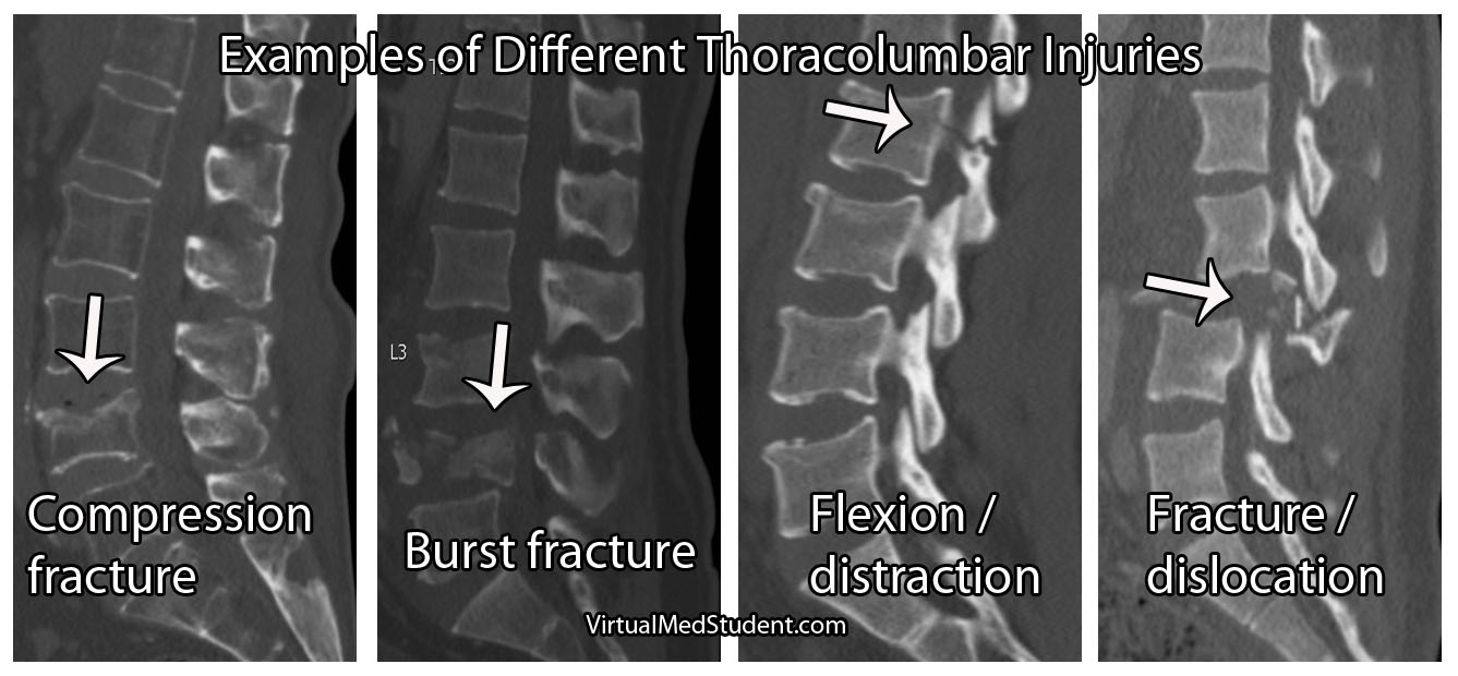
Based on Denis’ model he classified specific types of injuries. Injuries to the anterior column only were called "compression fractures". Damage to the anterior and middle columns were known as "burst fractures". Injury to the middle and posterior column were known as "flexion-distraction" injuries, or more colloquially as "seat-belt type" injuries. Finally, damage to all three columns were classified as "fracture-dislocation" injuries.

Classification of Thoracolumbar Injuries Using the Three Column Model

Columns Name Stability
Anterior Compression fracture Stable
Anterior and Middle Burst fracture Unstable
Middle and Posterior Flexion-distraction injury (aka: seat-belt type) Unstable
Anterior, middle, and posterior Fracture-dislocation Unstable

Examples of Thoracolumbar Spine Fractures

Clinical Significance

In very simple terms, unstable fractures require definitive treatment. Treatment for unstable spine injuries is usually operative, although rigid immobilization with bracing is sometimes used in certain circumstances.

The treatment of each fracture type is beyond the scope of this article (and discussed in more detail elsewhere on this site), but suffice it to stay that for the most part (and again, every rule was made to be broken) two or three column injures = unstable = surgery.

Overview

Denis’ three column model of the spine revolutionized the way we think about spinal biomechanics and stability. He divided the spine into three columns. Although it has been largely replaced by more modern grading scales such as the thoracolumbar injury classification and severity score (TLICS) it is still a nice way to “think” about spinal integrity.

Other Common Spine Injuries…

References and Resources

Cervical Myelopathy: A Squished Spinal Cord

Cervical myelopathy refers to a constellation of signs and symptoms that occur when the spinal cord is squeezed and unable to function properly. Myelopathy is most commonly caused by cervical stenosis, which is a general term used to describe a decrease in the cross sectional area of the spinal canal

Stenosis comes in two flavors: congenital or acquired. In congenital cervical stenosis the spinal canal is developmentally narrower than normal. Congenital stenosis is not considered hereditary, but is present from birth, and is a result of “abnormal” development.

Acquired stenosis is more common, and typically occurs in people over the age of 50. This type of stenosis is caused by a combination of factors and pathologic changes.

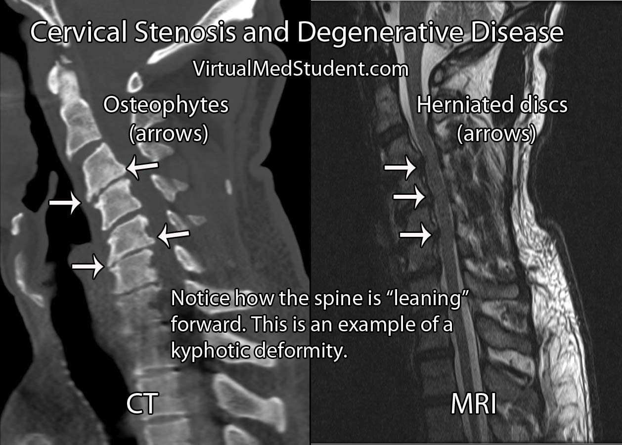
Degenerative cervical disease is the most common cause of acquired cervical stenosis and myelopathy. It has several distinct features, which include: loss of disc height, disc herniation, osteophyte formation, and resultant deformity (ie: kyphosis, or a forward leaning cervical spine). Let’s discuss each of these features in more detail…

The first feature in acquired degenerative stenosis is a loss of intervertebral disc elasticity and height. You can think of the intervertebral discs like shock absorbers for the spinal column. In the cervical spine, a healthy disc is slightly taller at its front most portion and slightly shorter at its back most portion. This gives the normal cervical spine a lordotic curvature.

As someone ages, the discs tend to dry out and the height differential between the front and back of the disc is lost. This can cause the cervical spine to bow forward resulting in a loss of lordosis. The new forward lean to the spine is called a kyphotic deformity. This deformity can cause the cord to stretch over the bones of the spine resulting in myelopathy.

The second factor that contributes to acquired degenerative stenosis is when the back most section of the disc weakens and pooches out towards the spinal cord. This is known as a disc herniation. Disc herniations narrow the cross sectional diameter of the spinal canal and contribute to stenosis.

Finally, as the discs age and dry out they lose their elasticity, and therefore lose their ability to absorb shock. As a result, the bones and ligaments take on more of the burden. They attempt to fight back by forming bony growths known as osteophytes. The osteophytes further narrow the spinal canal and contribute to stenosis.

Let’s bring it all together… Acquired degenerative cervical stenosis occurs when a combination of kyphotic deformity, disc herniations, and osteophytic growths narrow the spinal canal and cause compression of the spinal cord.

Other less common causes of cervical stenosis include ossification of the posterior longitudinal ligament. Additionally, traumatic injuries of the cervical spine can cause immediate cervical stenosis and cord injury. And even less commonly, tumors in the cervical spine can push on the cord and cause stenosis.

It is important to note that cervical stenosis does not always cause myelopathy. In fact, some patients can have a very "tight" cervical cord, but have no signs or symptoms of cord compression.

Signs and Symptoms

Patient’s that are myelopathic present with a constellation of signs and symptoms depending on how "squished" their cord is. Myelopathic patients present with a combination of gait dysfunction, clumsiness when using their hands, weakness, abnormally brisk reflexes, spasticity, and Lhermitte sign.

Signs and Symptoms
of Myelopathy:

– Gait dysfunction
– Clumsiness/weakness
– Brisk reflexes

– Spasticity
– Sensation problems
– Lhermitte’s sign

Reflexes are typically brisk, especially in the legs. Examples of brisk reflexes include clonus (a sustained contraction that causes the foot to "beat" several times), upgoing plantar response (ie: the big toes extend upwards, as opposed to curling down when the sole of the foot is stroked), and Hoffmann’s sign (ie: the thumb curls in when the tip of the index or middle finger is flicked).

Patients who have myelopathy tend to walk with a broad based gait as if steadying themselves. They often complain of feeling like they are walking “drunk”. Clumsiness is also a common symptom of myelopathy. Patients will typically complain of problems writing, using a utensil, holding a coffee cup, or performing other fine motor movements.

Finally, a phenomenon known as Lhermitte’s sign can occur. Lhermitte’s sign occurs when someone flexes their head forward and a shock like sensation runs down the neck and back into the extremities. It is a result of compression on the back most portion of the spinal cord, which is an area that relays sensation to the brain.

The Nurick grading system (table below) was designed to assess the severity of myelopathy. It is useful when guiding treatment decisions. The Japanese Orthopedic Association (JOA) score, European Myelopathy Score, Myelopathic Disability Index, and Ranawat Classification are other myelopathy grading scales that are commonly used in clinical practice.

The Nurick Grading System for Myelopathy
Grade 0 Nerve root impingement may be present (ie: radiculopathy), but no cord symptoms are present
Grade 1 Able to walk normally, but may have signs (but not symptoms) of cord compression
Grade 2 Some difficulty with walking, but still able to work
Grade 3 Unable to walk to the point of not being able to work
Grade 4 Can only walk when assisted
Grade 5 Confined to a wheelchair or bed bound

Diagnosis and Classification

Diagnosis of cervical stenosis is based on imaging (ie: CT, MRI, etc). Remember that stenosis does not necessarily translate into myelopathy! Patient’s should be symptomatic as well! A stenotic spine is radiographically present when the diameter of the spinal canal in the antero-posterior plane is less than 10mm.

An alternative way of diagnosing cervical stenosis is using Pavlov’s ratio. This is defined as the ratio of the canal diameter to the diameter of the adjacent vertebral body; any ratio less than 0.8 is worrisome.

Treatment

Treatment of cervical stenosis depends on several factors. First and foremost are symptoms. If a patient is not myelopathic, then frequently the best course of action is to monitor the patient until signs or symptoms of myelopathy appear.

If symptoms are present, the discussion becomes whether or not to reconstruct and decompress the front part of the spine (ie: an anterior approach), the back part of the spine (ie: a posterior approach), or both.

Anterior approaches come in one of two flavors. The first is removal of the disc with placement of a bone graft; this is formally known as a cervical discectomy and fusion. When the bones of the spine are causing the compression a more extensive surgery known as a corpectomy is performed; in this procedure the entire vertebral body is removed and a cage is placed in its place to reconstruct the spine.

Posterior approaches include laminoplasty and laminectomies with or without instrumented fusion. Laminoplasty involves splaying open the lamina so that the spinal cord can drift back. Laminectomies involve un-roofing the bone behind the spinal cord. Laminectomies are frequently done with an instrumented fusion in which rods and screws are placed to help the patient maintain a normal cervical alignment (ie: lordosis).

Surgical Treatment of Myelopathy
Deformity Compression Surgical Approach
Kyphotic Anterior and only 3 spinal levels or less involved Anterior (anterior cervical discectomies and/or corpectomies)
Kyphotic Anterior and posterior Combined anterior (anterior cervical discectomies and/or corpectomies) and posterior (laminectomies with or without instrumented fusion)
Lordotic Anterior and only 3 spinal levels or less involved Anterior (anterior cervical discectomies and/or corpectomies)
Lordotic Posterior Posterior (laminoplasty or laminectomies with or without posterior instrumented fusion)
Lordotic Anterior and posterior Combined anterior (anterior cervical discectomies and/or corpectomies) and posterior (laminectomies with or without instrumented fusion)

The choice of surgical options is highly dependent on the degree of deformity, instability, and where specifically (ie: what levels) in the spine the cord compression is located.

Ultimately, the surgical treatment of cervical stenosis and myelopathy must be tailored to each individual patient based on many factors.

Overview

Cervical stenosis is abnormal narrowing of the spinal canal. It has numerous causes, of which the most common one is acquired degenerative stenosis. Stenosis can cause compression of the spinal cord, which can cause signs and symptoms of myelopathy. Treatment for symptomatic patients involves decompressing the spinal cord via either an anterior, posterior, or combined approach.

Related Articles

References and Resources

Latin for Toothlike: Fractures of the Odontoid Process

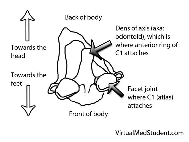
The odontoid process (also know as the "dens") is the finger of bone that sticks up from the second cervical vertebrae (ie: the axis).

It articulates via numerous ligaments to the anterior arch/ring of the first cervical vertebrae (ie: the atlas) to form a joint. This joint is what allows you to rotate your head from side to side as if you were nodding “no”.

Axis (C2)

Fractures of the odontoid typically occur after traumatic events. In younger, otherwise healthy individuals tremendous force is necessary to fracture the odontoid. Breaks are typically seen after car, motorcycle, or sporting accidents. In older, osteoporotic people simple ground level falls can result in a fracture. Less commonly, fractures of the odontoid may be caused by tumor chipping away at the underlying bone (a so called “pathologic” fracture).

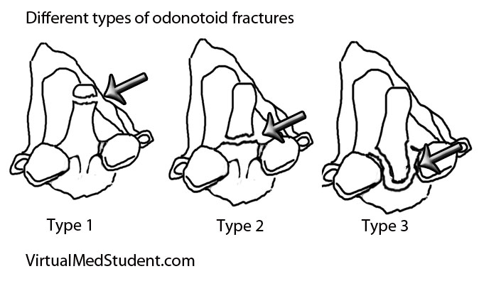
Since the odontoid is a relatively long piece of bone it can fracture at one of several distinct sites. The most commonly used system (Anderson and D’Alonzo) categorizes fractures into one of three types:

  • Type 1 – a fracture at the tip of the odontoid.
  • Type 2 – a fracture at the base of the odontoid.
  • Type 3 – a fracture involving the body of the C2 vertebrae, which includes the odontoid within it.

Odontoid fractures

This grading system is important because it helps predict both stability of the C1-C2 (ie: atlanto-axial) joint and guides potential treatment options.

Signs and Symptoms

Roughly 80% of patients with odontoid fractures do not have any neurological injury to their spinal cord. The remaining patients can exhibit anything from quadriplegia to mild sensory disturbances. Patients with severe cervical spinal cord injury usually are unable to breath (secondary to diaphragm paralysis) and frequently die at the scene of the accident.

Many patients with odontoid fractures will have significant neck pain that radiates up into the scalp. This is usually caused by neck muscles spasming secondary to the injury.

Diagnosis

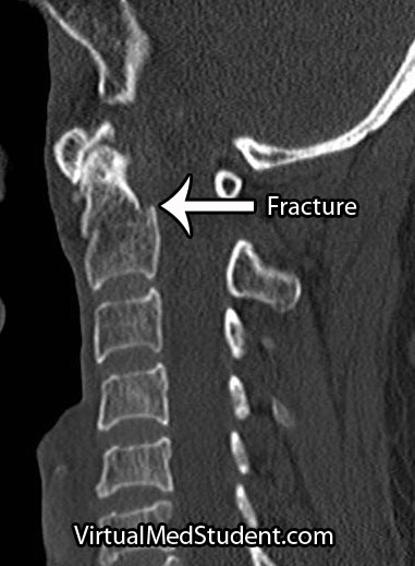
CT of odontoid fracture

CT scans, x-rays, and MRIs are all useful in diagnosing and properly treating odontoid fractures.

CT scans of the cervical spine provide excellent bony detail, and also help illustrate any additional fractures that may be present.

MRI of the cervical spine is useful for assessing any co-existent ligamentous injury. If ligamentous injury is present it drastically alters treatment decisions.

Treatment

Treatment of odontoid fractures is based on both bony and ligamentous injury. The goal of treatment is to stabilize the spine either by allowing the bone to heal on its own, or by fusing the spine artificially using rods, screws, and/or wires.

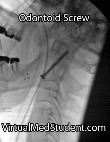
Placement of an odontoid screw is one method of fixing non-displaced type II fractures. However there are numerous contraindications for odontoid screw placement. For example severe angulation of the fractured segment precludes placement of a screw; barrel chested anatomy prevents an adequate angle for screw trajectory in the operating room. In addition, if the transverse ligament is disrupted, bony fixation with an odontoid screw alone will not stabilize the joint.

On the other hand, odontoid screws are beneficial because they typically stabilize the fracture with minimal restriction of neck motion.

Approaching the spine from behind is another option to stabilize odontoid fractures. Fusing the atlas (C1) to C2 or C3 is sometimes used if odontoid screw placement cannot be performed and the injury is deemed unstable. It is important to note that posterior approaches can restrict motion, especially in the high cervical spine.

Non-surgical options must ensure that the patient has minimal to no movement of the neck in order to give the bone an adequate chance to heal on its own. Rigid collars or halos are used to prevent neck motion.

Overview

Odontoid fractures come in three flavors depending on the location of the fracture. Symptoms can be anything from mild neck discomfort to quadriplegia, although neurological injury is surprisingly uncommon. Diagnosis is based on CT and MRI. Treatment is with cervical immobilization for an extended period of time, or surgical fusion. Treatment decisions are based on the degree of both bony and ligamentous injury, as well as the patient’s overall health status.

Related Articles

References and Resources