Ependymoma: Myxopapillary, Anaplastic, and Perivascular Pseudorosettes

Ependymomas are tumors that develop from cells known as ependymal cells (duh!). Ependymal cells are a type of glial cell that line the ventricles (ie: fluid filled cavities) of the brain and central canal of the spinal cord.

Normal ependyma have cilia and microvilli on the side of the cell that faces cerebrospinal fluid (ie: the "apical" side). Cilia are hair like extensions that are believed to "beat" cerebrospinal fluid around the ventricles. Microvilli are folds in the cellular membrane that are thought to aid in the reabsorption of cerebrospinal fluid.

Unlike other epithelial cells in the body, of which ependyma are considered a subgroup, they do not rest on a basement membrane. Instead their basal surfaces (the surface not in contact with cerebrospinal fluid) intertwine with the overlying brain tissue.

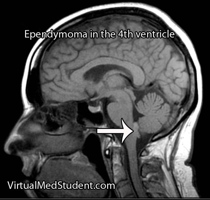
Like any other cell in the body, ependymal cells can decide to turn naughty and form a tumor. Ependymomas can occur anywhere there are ependymal cells, and therefore develop in both the brain and spinal cord. Intracranial ependymomas are more common in younger age groups, whereas spinal forms are more common in older individuals. Of those that form within the confines of the skull, the most common location is in the fourth ventricle near the brainstem.

There are three "grades" of ependymoma. There are two subsets of grade one: myxopapillary and subependymomas. The second grade of ependymoma has four distinct variants. They are cellular, papillary, clear cell, and tanycytic. The third grade is also referred to as "anaplastic" ependymoma. Regardless of the grade, each type has its own distinct characteristics when viewed under the pathology microscope.

Surgical specimens of ependymomas are often "stained" by pathologists to help aid in diagnosis, and more importantly, distinguish them from other tumor types. Ependymomas stain positive for the glial fibrillary acidic protein (GFAP), as well as phosphotungstic acid hematoxylin (PTAH).

Ependymomas may have perivascular pseudorosettes, which helps support the diagnosis. Pseudorosettes may not be apparent in tumors with dense cellularity such as anaplastic ependymomas.

In addition, ependymomas can spread throughout the cerebrospinal fluid space. For example, a tumor that arises in the fourth ventricle may "drop" tumor cells down into the spinal cord forming a secondary tumor. These secondary tumors are referred to as "drop mets".

Signs and Symptoms

The signs and symptoms depend on the location of the ependymoma.

The most common symptom of intracranial ependymoma is headache associated with nausea and/or vomiting. These symptoms occur when the ependymoma blocks the flow of cerebrospinal fluid, which causes a condition known as non-communicative hydrocephalus.

You can think of non-communicative hydrocephalus as a clog in a pipe. Everything upstream of the clog starts to back up, which eventually leads to increasing pressures. When this increased pressure occurs in the ventricular system of the brain it causes worsening headaches, nausea, and vomiting. This is especially true if the ependymoma is in the fourth ventricle of the brain, which even without tumor, is an anatomically narrow "pipe" to begin with.

Additionally, if the tumor pushes on brainstem structures a patient may present with dysfunction of the nerves that go to the various muscles of the head and face. The most commonly involved nerves are the facial nerve, which can cause weakness of the face, as well as the abducens nerve, which can cause weakness of the eye.

Tumors located in the spinal cord cause weakness and sensory disturbances.

Diagnosis

Ependymoma

MRI scans can be very useful and can support (but not prove) the diagnosis of ependymoma, especially when the tumor is in a common anatomical location.

If there is a high index of suspicion for ependymoma then the entire neuro-axis, meaning the brain and entire spinal column, should be imaged using MRI. This will detect “drop” mets, which, if present, further support the diagnosis.

Diagnosis can only be officially made when a sample of tumor (either surgical or at autopsy) is seen under the pathology microscope.

Treatment

Treatment of ependymoma is with surgical resection followed by radiation therapy. Patient outcome is most effective if the entire tumor can be removed during surgery. This is known as "gross total resection". However, the extent of surgical resection should always be weighed against the risk of harming the patient, especially if the tumor has invaded vital structures like the brainstem.

Fortunately, ependymomas are very radio-sensitive, which means that they respond well to getting zapped with radiation. Chemotherapy is not typically helpful except in very young children where the effects of radiation can be devastating.

Overview

Ependymomas arise from the cells that line the ventricular system of the brain and spinal cord. There are different subtypes depending on what it looks like under the pathology microscope. Diagnosis is based on pathological analysis and characteristic MRI findings. Treatment is with surgery and radiation.

Other Diseases You Should Know About…

References and Resources

Diffuse Axonal Injury: Shearing the Cables

Diffuse axonal injury is a form of traumatic brain injury that occurs under conditions of rapid acceleration and deceleration of the head. This frequently occurs after high impact injuries such as motorcycle crashes or high speed car accidents.

Let’s discuss a few basic brain terms before we dive into why diffuse axonal injury happens. Brain tissue is composed of neurons and glia. The neurons communicate with one another through long extensions known as axons. You can think of an axon as the telephone wire that connects one phone to another. Axons compose the bulk of what is known as “white” matter in the brain; on the other hand, “gray” matter represents clumps of neurons.

Both gray and white matter have different densities associated with them. Because of this, rapid accelerations or decelerations of the head cause the gray matter to move at a greater relative velocity compared to the white matter. If these accelerations are severe enough stretch and shear injury occurs; the end result is that the “wires” get disconnected. This, in a nutshell, is diffuse axonal injury.

Signs and Symptoms

Depending on the severity of diffuse axonal injury patients may present with different signs and symptoms. In fact, diffuse axonal injury is frequently diagnosed when patients “fail” to wake up after a traumatic event despite adequate resuscitative treatment.

In its most severe form, diffuse axonal injury results in coma. Less severe injuries may cause long term cognitive issues and personality changes.

Diagnosis

Diagnosis of diffuse axonal injury is made with an MRI scan. CT scans of the head are frequently ordered early to rule out other treatable causes for the decreased mental status often seen in patients with DAI (ie: epidural, subdural, and intraparenchymal hematomas).

The MRI will reveal restricted diffusion on diffusion weighted (DWI) and apparent diffusion coefficient (ADC) maps of the brain. Gradient echo imaging (GRE) will often show small spots of low intensity consistent with shear/stretch injury.

MRI of diffuse axonal injury

Treatment

Unfortunately, there is no effective treatment for diffuse axonal injury. There is currently no way to “re-wire” the axons. All care for diffuse axonal injury at this point is supportive.

Overview

Diffuse axonal injury occurs after rapid changes in acceleration of the head. The axons, or “wires”, between neurons get stretched, sheared and, effectively disconnected. Coma is the common presenting sign of severe axonal injury. Diagnosis is made with MR imaging. Treatment is supportive since there is currently no way to re-wire the damaged connections.

Other Stuff You Might Want to Learn About

References and Resources

  • Hijaz TA, Cento EA, Walker MT. Imaging of head trauma. J Trauma. 2010 Dec;69(6):1610-8.
  • Andriessen TM, Jacobs B, Vos PE. Clinical characteristics and pathophysiological mechanisms of focal and diffuse traumatic brain injury. J Cell Mol Med. 2010 Oct;14(10):2381-92.
  • Meythaler JM, Peduzzi JD, Eleftheriou E. Current concepts: diffuse axonal injury-associated traumatic brain injury. Arch Phys Med Rehabil. 2001 Oct;82(10):1461-71.
  • Smith DH, Meaney DF, Shull WH. Diffuse axonal injury in head trauma. J Head Trauma Rehabil. 2003 Jul-Aug;18(4):307-16.
  • Osborn AG. Osborn’s Brain: Second Edition. Elselvier, 2017.

Burst Fractures: Axial Loading Leading to Ouch!

Burst fractures are a specific type of spine fracture in which the body of a given vertebrae “bursts” into pieces. By definition a burst fracture involves the entire vertebral body. The image below is an example of a normal lumbar spine with the vertebral bodies outlined.

Burst fractures most commonly occur at the junction between the thoracic and lumbar spine. This junction is an area where the rigid thoracic spine transitions to the more mobile lumbar spine, and hence is an intrinsic point of weakness. This is why most burst fractures occur between the T10 through L2 vertebrae.

CT vertebral body

Axial loading of the spine is what causes burst fractures. They typically occur after a traumatic events like car accidents or falls from significant heights. Elderly individuals, and those with poor bone quality, may suffer burst fractures after minor trauma such as falling from a chair.

Signs and Symptoms

Burst fractures invariably present with back pain at the site of the fracture. Depending on the exact location signs and symptoms of nerve root compression or lower spinal cord injury may occur.

If the nerves that dangle in the lumbar spine (aka: the cauda equina) get compressed by the fragments of bone then weakness, numbness, tingling, and even bowel and bladder problems may occur.

Burst fractures between T10 and L1 can cause damage to the end of the spinal cord (the spinal cord ends at L1 or L2 in most individuals), which can lead to lower extremity weakness, or even paralysis, as well as bowel and bladder dysfunction.

Diagnosis

Diagnosis of a burst fracture is made using a combination of x-rays, CT scans, and MRIs. These three imaging modalities serve different functions when evaluating the severity of a burst fracture.

X-rays are usually the first imaging ordered in patients with suspected spine fractures. If the plain x-rays show a burst fracture then CT scanning is usually done to further assess the degree of bony injury (see image below for an example of an L2 burst fracture).

MRI is used to detect ligamentous injury. The degree of ligamentous injury indicates a higher degree of instability; information about ligament integrity helps determine treatment options.

Burst Fracture Lumbar Spine

Treatment

Treatment of burst fractures is highly dependent on the severity of the burst fracture. Treatment is either conservative with immobilization in a brace (ie: a "TLSO" or thoracolumbar sacral orthotic brace) or surgical fixation.

Burst fracture after instrumentation
As a rough rule of thumb patients with any of the following criteria should be strongly considered for surgical correction:

  • Greater than 50% vertebral body height loss.
  • Greater than 25 to 40 degrees of kyphosis.
  • Greater than 50% spinal canal compromise.
  • Significant posterior ligamentous injury.
  • Any neurological signs or symptoms referable to the injury.
  • If the patient fails conservative therapy with a brace.

Surgical correction can be achieved in a variety of ways and is often related to surgeon preference. Some surgeons will remove a significant portion of the fractured vertebral body and place a “cage” in the area, a procedure known as a “corpectomy”. This, combined with rods and screws from posteriorly provides the greatest stability, but has a higher risk of nerve injury. Not uncommonly, the fractured vertebral body is left alone and rods and screws are placed from behind only. This is especially true if the fractured level shows minimal spinal canal compromise.

Overview

Burst fractures of the thoracolumbar spine typically occur after high impact axial loading. They usually occur between T10 and L2, but can be seen anywhere in the spine. Patients will almost invariably have pain at the fracture site and may or may not have neurological signs and symptoms depending on the severity of the fracture. Diagnosis is made with CT, plain x-rays, and MRI. Treatment is highly dependent on the individual fracture and ranges from bracing to surgical fixation.

Related Articles

References and Resources

Atlas Fractures: The Weight of the World On Its Shoulders

The atlas, or the first cervical vertebra (C1), is a ring shaped structure. It forms joints with the base of the skull above and the axis (ie: the second cervical vertebrae) below. It also has two foramen transversarium, which are holes that allow the passage of the vertebral arteries on either side of the spinal cord.

Fractures of the atlas occur most commonly with forceful axial loading of the head (ie: a downward force applied to the top of the head). Pressure on the top of the head causes the skull to push down on the atlas, which results in a break(s) of its ring-like structure. Specific fracture types such as a break in the front of the ring, the back of the ring, or one side of the ring versus the other, are dependent on additional force vectors at the time of loading (ie: flexion, extension, lateral bending, etc.).

Fractures of the atlas must also include a discussion of biomechanical stability, which is usually determined by the integrity of the transverse ligament. The transverse ligament attaches the dens (odontoid) of the axis to the anterior ring of the atlas.

Fractures of the atlas with co-existent rupture of the transverse ligament lead to instability of the joint between C1 and C2. In other words, the ring of C1 may be able to move forward relative to the dens of C2. Transverse ligament injury is more common when axial loading is combined with extension of the head.

Not surprisingly, fractures of the atlas often co-exist with fractures of other cervical spine vertebrae. The most common combination is with a fracture of the axis, occurring in up to 40% of cases.

Signs and Symptoms

Patient’s with isolated atlas fractures usually have neck pain and muscle spasms. Frequently they have no injury to the spinal cord because the ring splays outwards as it fractures.

It is important to rule out injuries to the vertebral arteries, which run in bony holes (ie: foramen transversarium) on the sides of the atlas. When injured, the vertebral arteries can cause strokes in the brainstem and cerebellum, which can be life threatening.

Since the atlas is so close to the brainstem, patients may have co-existent injury to the lower cranial nerves. Specifically, injury to the 12th nerve can cause problems with tongue movements, injury to the 11th nerve can cause weakness with shoulder shrug and the ability to turn the head to the side, and injuries to the 9th and 10th cranial nerves can cause problems with swallowing and paralysis of the larynx leading to difficulty with speech.

Co-existent head and brain trauma, which can cause a constellation of different signs and symptoms depending on severity can also occcur.

Diagnosis

Diagnosis of an atlas fracture is made using x-rays, CT scans, and MRIs. X-rays should include anterior-posterior views, open mouth odontoid views, and lateral views of the cervical spine. If there is no evidence of neurological injury, flexion-extension x-rays may also be obtained to assess for stability of the C1-C2 joint.

The bony injury associated with atlas fractures is categorized according to the Jefferson or Landell and Van Peteghem classification systems. The Landells classification has three types, whereas the Jefferson classification has four types:

Landell and Van Peteghem Classification
Type 1 Fracture of either the anterior or posterior ring, but not both (posterior ring fractures are most common type)
Type 2 Fractures of both the anterior and posterior ring
Type 3 Fracture of the lateral mass(es)

Jefferson Classification
Type 1 Fracture of the posterior ring only
Type 2 Fracture of the anterior ring only
Type 3 Fracture of the anterior and posterior rings on both sides; this is the classic "burst", or traditional “Jefferson” fracture
Type 4 Fracture of the lateral mass(es)

Atlas fracture

An important part of diagnosing atlas fractures involves assessing the integrity of the transverse ligament, which is best done using MRI. However, if an MRI cannot be performed then open mouth odontoid, flexion-extension x-rays, and CT scans can provide some information regarding transverse ligament injury.

The rule of Spence is one way of assessing the integrity of the transverse ligament on an open mouth odontoid x-ray. The rule states that if the right and left lateral masses of C1 overhang the lateral masses of C2 by greater than a total distance of 6.9mm than the likelihood of co-existent transverse ligament injury is high. The rule of Spence is not fool proof and should be supplemented with MRI and/or flexion-extension films whenever possible.


Atlantodental Interval
Another method for assessing transverse ligament injury is using the "atlantodental" interval (see image to the left). This is the distance between the anterior arch of C1 and the odontoid process (aka: dens) of C2.

This interval is usually quite small, typically less than 3mm in adults and 5mm in children. If the interval is greater than this, then co-existent transverse ligament injury should be suspected.

Treatment

Treatment of isolated atlas fractures is usually with cervical immobilization. This may be with a halo or with a rigid cervical collar such as a cervical-occipital-mandibular-immobilizer (SOMI).

Atlas fractures that have co-existent transverse ligament rupture often require an operation to stablize the bones of the spine. This is usually in the form of fusing the atlas or occiput (back of the head) to the second cervical vertebrae.

If other injuries (ie: fractures of C2) are present and/or there is significant ligamentous injury then open surgical fusion of the bones may be necessary to re-create stability of the craniocervical junction.

Overview

Atlas fractures occur in response to vertical compression of the head on the upper cervical spine. Fractures of the anterior, posterior, or both rings of C1 may be present. Biomechanical stability is typically determined by assessing the integrity of the transverse ligament. Patients with isolated C1 fractures usually complain of neck pain, and rarely have injury to the spinal cord. Diagnosis is based on CT, x-ray, and MRI findings. Treatment is with rigid external immobilization or operative spinal fusion.

Related Articles

References and Resources