A Tale of Tails: Malpractice Tail and Its Importance

In the vibrant tapestry of medical life, malpractice insurance is that necessary gray thread that weaves its way through, dull yet indispensable. Among its variants, there’s one that leaves even the brightest minds scratching their heads: tail insurance. Like a well-tailored suit, it follows you wherever you go, long after you’ve hung up your white coat for the day or said goodbye to your employer. Let’s journey into the world of medical malpractice tail insurance and the mishmash of occurrences it covers (or doesn’t cover).

What’s the Deal with the Tail?

You might wonder why anyone would want to insure their tail. But for healthcare professionals, the ‘tail’ is not some newfound evolutionary feature but refers to an extended reporting period (ERP). This allows you to report claims for alleged incidents that occurred during your policy term, even if they are filed after the policy ended. Think of it as a way to cover your professional backside.

It’s All About the Occurrences

In the medical malpractice world, policies are typically classified into “claims-made” or “occurrence-based”:

Claims-Made: This policy is like that friend who’s only there when you’re currently hanging out. It covers any incidents reported while your policy is active, even if the actual event happened earlier. But once your policy ends, it’s a ghost, leaving you high and dry unless you’ve invested in your trusty tail.

Occurrence-Based: This is the loyal friend who’s got your back no matter what. It covers incidents that happened during the policy period, regardless of when the claim is filed. The best part? No tail needed. It’s the gift that keeps on giving, long after the policy has ended.

Why Buy the Tail Coverage?

Now, you might be thinking, “Why get tail coverage if I can get an occurrence-based policy?” Excellent question, young Padawan! The thing is, occurrence-based policies can be pricier than their claims-made counterparts. They’re like that all-inclusive vacation deal – sounds excellent but check your wallet first. Tail coverage is a one-time purchase that bridges the gap when you switch jobs, retire, or if your policy lapses, ensuring you’re not left staring at a lawsuit with nothing but your stethoscope for defense.

Shopping for the Tail

Before you purchase tail coverage, it’s essential to know that not all tails are created equal (ask any dog, they’ll tell you). Some tails have a time limit, and some cover unlimited time. There are even ‘free tail’ provisions under certain conditions, like death, disability, or retirement. If you are switching jobs and need tail coverage you can sometimes negotiate that your new employer pays for the tail coverage (assuming you are that good and they need you that badly).

In the whimsical world of medicine, tail insurance is like that quirky sidekick in a sitcom: it’s there when things get messy, keeping you safe from the past’s boomerang. While it might seem like just another financial headache, a good tail coverage can be your best friend in a world where past mistakes may come knocking at the most inconvenient times. So, remember, in the great medical sitcom of life, make sure you’ve got a great ‘tail’ to tell!

Other Enjoyable Articles

Amyotrophic Lateral Sclerosis (Lou Gehrig’s Disease)

The pathology of amyotrophic lateral sclerosis (ALS) is not well known. There are familial forms, but they account for only 10% of cases. The familial form of amyotrophic lateral sclerosis is genetically heterogenous. This means that there are genes on different chromosomes that have been implicated in the disease process.

One of the most well studied is a gene known as superoxide dismutase (SOD) on chromosome 21. There are over 100 mutations of this gene. The defective protein that occurs from these mutations is believed to cause oxidative injury and/or toxic protein aggregates that damage motor neurons.

The remaining 90% of cases are sporadic. There is still debate about what causes most cases. Studies looking at cytoskeletal abnormalities, inflammation, and excitotoxicity from overactive glutamate stimulation have all been proposed as contributing factors.

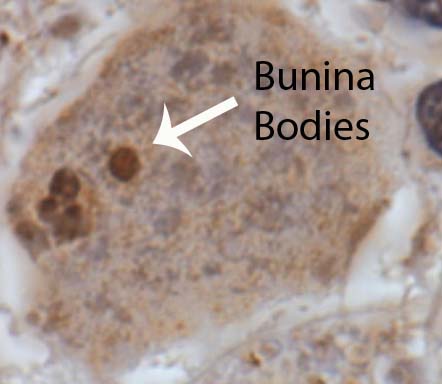
Bunina Bodies
Regardless of the cause, amyotrophic lateral sclerosis causes the death of motor neurons in both the brain and spinal cord. These motor neurons are commonly referred to as upper motor neurons (if in the brain) and lower motor neurons (if in the brainstem or spinal cord).

The dying neurons are replaced with proliferating astrocytes in a process known as gliosis. This proliferation of glial cells leads to "glial scarring".

In addition, amyotrophic lateral sclerosis neurons have intracellular inclusions when viewed under the microscope. These inclusions are composed of different abnormal protein molecules that can be phosphorylated or ubiquinated. A specific type of inclusion known as a Bunina body is commonly seen.

Signs and Symptoms

Damage to the upper and lower motor neurons gives ALS its characteristic clinical presentation. Patients present with upper motor neuron findings: spastic weakness and hyper-reflexia (increased reflexes). Lower motor neuron death results in a flaccid weakness, atrophy of the muscles, muscular fasciculations (ie: abnormal twitching), and hypo-reflexia (decreased reflexes). Thus patients with ALS have a unique mix of both spastic and flaccid weakness, muscular atrophy, and hyper-reflexia (hyperactive reflexes usually predominate).

There are also many sub-categories of ALS. For example, "ALS Plus Syndrome" is characterized by the classic motor findings of amyotrophic lateral sclerosis plus dementia, Parkinsonism, autonomic nervous system instability, and sensory disturbances. Another sub-category known as bulbar palsy occurs when the symptoms of ALS predominately affect the face/cranial musculature. Cranial muscular weakness can cause difficulty swallowing (ie: dysphagia) and speaking (ie: dysarthria).

Diagnosis

Amyotrophic lateral sclerosis, for the most part, remains a clinical diagnosis. It is based upon finding the classic motor neuron findings on physical exam with symptoms that worsen over time. Unfortunately, there is still no laboratory test that definitively diagnoses the disease.

The El Escorial World Federation of Neurology criteria help guide the clinical diagnosis. In order to qualify for a diagnosis of amyotrophic lateral sclerosis a patient must meet the following criteria: evidence of lower motor neuron pathology on physical exam, electrophysiological, or neuropathological testing, and progressive worsening / spread of symptoms, as well as evidence of upper motor neuron disease on physical exam. In addition to the above, there must also be no evidence to support another possible explanatory diagnosis for the symptoms.

There are adjunctive tests that can support the diagnosis of amyotrophic lateral sclerosis. These include electromyography (EMG). EMG looks at the electrical activity produced in muscles. In amyotrophic lateral sclerosis, the EMG will generally show findings of denervation (ie: muscles that do not have nerves connecting to them). Fasciculation potentials are often seen on the EMG as well. Interestingly, nerve conduction studies are often normal in amyotrophic lateral sclerosis.

Finally, blood testing can help support the diagnosis. For example, creatine kinase (a marker of muscle damage) may be elevated, which is a result of denervation.

Other laboratory tests are often sent. Lyme disease antibodies, anti-nuclear antibodies, HIV, vitamin B12, thyroid function tests, calcium, and phosphorus are often sent to rule out other causes for the symptoms.

Treatment

Treatment for amyotrophic lateral sclerosis currently consists of a medication known as riluzole. This medication has been shown to improve survival rates.

In addition, supportive care is extremely important. Most patients eventually require tracheostomy and gastric tube placement in order to breath and eat. Motorized wheel chairs can help patients maintain some independence.

Regardless of treatment, the prognosis is extremely guarded. Most patients are deceased within 5 years of their diagnosis. Death is usually related to pulmonary complications like pneumonia.

Overview

Amyotrophic lateral sclerosis is a neurodegenerative disease that results in death of upper and lower motor neurons. Signs and symptoms are a bizarre mix of flaccid weakness with increased reflexes. Diagnosis is based on physical exam findings and symptoms that worsen over time. In addition, there should be no evidence that other causes are responsible. Treatment is with a medication known as riluzole, which has been shown to slow the progression of disease. Supportive treatment is extremely important for quality of life.

Learn More…

References and Resources

  • Brooks BR. Managing amyotrophic lateral sclerosis: slowing disease progression and improving patient quality of life. Ann Neurol. 2009 Jan;65 Suppl 1:S17-23.
  • Baehr M, Frotscher M. Duus’ Topical Diagnosis in Neurology: Anatomy, Physiology, Signs, Symptoms. Fourth Edition. Stuttgart: Thieme, 2005.
  • Kumar V, Abbas AK, Fausto N. Robbins and Cotran Pathologic Basis of Disease. Seventh Edition. Philadelphia: Elsevier Saunders, 2004.
  • Simon RP, Aminoff MJ, Greenberg DA. Clinical Neurology, Seventh Edition (LANGE Clinical Medicine). Seventh Edition. New York: McGraw Hill, 2009.
  • Boillée S, Vande Velde C, Cleveland DW. ALS: a disease of motor neurons and their nonneuronal neighbors. Neuron. 2006 Oct 5;52(1):39-59.
  • Brooks BR. El Escorial World Federation of Neurology criteria for the diagnosis of amyotrophic lateral sclerosis. J Neurol Sci. 1994 Jul;124 Suppl:96-107.

Sir William Ogilvie and His Syndrome: Colonic Pseudo-Obstruction

Ogilvie syndrome, which is also known as acute colonic pseudo-obstruction, is a pathological dilation of the colon (ie: “large” intestine) in the absence of a mechanical lesion responsible for the dilation. This differs from colon obstruction caused by tumors, volvulus, strictures, or infection (ie: "mechanical" causes).

The underlying reason why Ogilvie syndrome occurs is not fully understood. The current hypothesis is that the autonomic nervous system, which is partially responsible for gut motility, stops working properly.

Therefore, in order to understand Ogilvie syndrome, we have to understand a little about the autonomic nervous system. The autonomic nervous system is broken up into two parts: sympathetic and parasympathetic. The sympathetic nervous system is responsible for the “fight or flight” response and the parasympathetic system is responsible for the “rest and digest” response. When it comes to the colon, the parasympathetic system increases gut motility and the sympathetic system decreases motility.

Ok, so what is happening in colonic pseudo-obstruction? It is believed that there is an over-abundance of sympathetic input to the colon. This causes a failure of the gut to contract (aka: peristalsis) and therefore feces is not pushed through the GI tract.

If the colon stops contracting then it starts to expand as mucous and stool accumulate. For unknown reasons, the most common part of the colon that is involved in Ogilvie syndrome is the cecum (first portion of the large colon) and the ascending colon.

Signs and Symptoms

Acute colonic pseudo-obstruction causes belly pain, nausea, and vomiting. In addition, since the colon is not contracting, many patients have obstipation (ie: a fancy term for intractable constipation).

If medical attention is not sought soon, the colon may perforate, which can cause peritonitis (infection of the abdominal cavity). Fever, chills, and septic shock may occur in the setting of colonic perforation and peritonitis.

Diagnosis

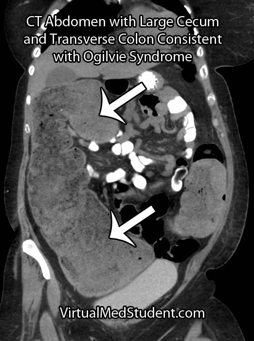
Ogilvie Syndrome CT Abdomen
Diagnosis is made when there is evidence of an enlarged colon with no mechanical obstruction. In essence, diagnosing Ogilvie syndrome is more about ruling out other causes of colon obstruction.

CT scans, abdominal x-rays, barium swallow studies, and colonoscopies are commonly used as adjunctive means for diagnosing pseudo-obstruction.

Treatment

Treatment of Ogilvie syndrome is primarily conservative. This means making the patient NPO (ie: nothing by mouth) and placing a nasogastric tube (NGT) to suction. The NGT helps "decompress" the colon from above until the pseudo-obstruction resolves.

If conservative measures fail then a medication known as neostigmine can be given. Neostigmine is a parasympathetic "mimic". It blocks the breakdown of acetylcholine by inhibiting an enzyme known as acetylcholine esterase. Acetylcholine is the main neurotransmitter of the parasympathetic nervous system. Therefore, if there is more acetylcholine, then more gut motility occurs.

Another medication known as methylnaltrexone may also be useful in cases of acute colonic pseudo-obstruction caused by opioids.

In cases that are refractory to conservative and medical therapies the use of colonoscopic decompression is extremely effective. Patient’s who have evidence of colonic perforation require prompt surgical exploration.

Overview

Ogilvie syndrome, or acute colonic pseudo-obstruction, is a pathologic dilation of the colon that occurs in the absence of a mechanical cause. It is believed to be the result of autonomic nervous system dysfunction. Diagnosis is made with x-rays and CT scans of the abdomen. Treatment is with nasogastric decompression, neostigmine, methylnaltrexone, colonoscopic decompression, or surgery in advanced cases.

References and Resources

  • Hsu HL, Wu YM, Liu KL. Ogilvie syndrome: acute pseudo-obstruction of the colon. CMAJ. 2011 February 22; 183(3): E162.
  • Eisen GM, Baron TH, Dominitz JA, et al. Acute colonic pseudo-obstruction. Gastrointest Endosc. 2002 Dec;56(6):789-92.
  • Saunders MD. Acute colonic pseudo-obstruction. Gastrointest Endosc Clin N Am. 2007 Apr;17(2):341-60, vi-vii.
  • Saunders MD, Kimmey MB. Colonic pseudo-obstruction: the dilated colon in the ICU. Semin Gastrointest Dis. 2003 Jan;14(1):20-7.
  • Ponec RJ, Saunders MD, Kimmey MB. Neostigmine for the treatment of acute colonic pseudo-obstruction. N Engl J Med. 1999 Jul 15;341(3):137-41.
  • Laine L. Management of Acute Colonic Pseudo-Obstruction. N Engl J Med 1999; 341:192-193.
  • Thomas J, Karver S, Cooney GA, et al. Methylnaltrexone for opioid-induced constipation in advanced illness. N Engl J Med. 2008 May 29;358(22):2332-43.

Carbs, Fats, and Proteins: How the Body Burns Fuel

Introduction

In its simplest terms, metabolic biochemistry is “fuel in, energy out”. The energy gained from burning fuel (ie: food) is used to drive all the processes going on in your body. These include the building of proteins, DNA (your genetic material), and fat, as well as mechanical things like muscle contraction.

Fuel for the human body takes three basic forms: carbohydrates (sugars), protein, and fat. Humans are capable of burning all three of these fuels, but do so at different times, rates, and under different circumstances. Using an extreme example, under starvation conditions the body burns its fat stores. Once fat stores are depleted the body begins digesting non-essential proteins and then essential proteins, which ultimately leads to organ damage and death. Thankfully, the body is relatively efficient and uses the best available fuel first before it has to tap into essential reserves.

Carbohydrates

Let’s start by discussing carbohydrates. Carbohydrates, or “carbs”, are simply sugar molecules linked to one another in varying arrangements. For example, starch, the most important carbohydrate in the human diet, is nothing more than numerous glucose molecules linked together in a long strand. Potatoes are an excellent example.

Another example of a carbohydrate is glycogen. Glycogen is how humans store excess glucose (a single sugar molecule) for later use. Unlike starch, which is a long chain of individual glucose molecules, glycogen is a highly branched structure that allows the body to rapidly cleave off individual sugar molecules to be burned for energy.

Carbohydrates can be further broken down into 2 categories: simple and complex. We’ve all heard of the term “complex carbohydrates”, which is a fancy way of saying multiple sugar molecules linked together in a complicated way. Contrarily, a simple carbohydrate is merely a few (usually 1 to 3) sugar molecules linked together.

Why make the distinction between simple and complex carbs? For starters, simple carbohydrates are rapidly absorbed by the gut and enter the bloodstream very quickly. Candy bars are a great example! If you need a quick boost of energy unwrap a Snickers®!

The problem is that since simple carbs enter the bloodstream so rapidly they get metabolized quickly. This causes you to lose that energy boost fast, which is why you often feel “de-energized” an hour or so after eating "junk food". In contrast, complex carbs get degraded by the gut much less rapidly, and therefore slowly trickle into the bloodstream. This gives you a more sustained, but less pronounced energy boost. Whole grains are a great example of complex carbs.

Why all the hub-bub about carbohydrates? Because they are, for the most part, the first energy source that is utilized during exercise. This forms the basis behind “carbo loading”, or eating a meal rich in carbohydrates the night before, or morning of, a planned work out. During exercise, the body will then utilize the individual sugar molecules in the carbohydrates to provide energy for your muscles and brain. Once you run out of sugar (or the form that humans store it in, glycogen) your body turns to the other fuel sources, namely, protein and fat.

Fats

The next fuel that gets burned is fat. All human beings have a certain percentage of body weight that is fat. From an evolutionary stand point this is advantageous. During times of drought or famine there was not enough food to provide reliable carbohydrate or protein, and thus humans survived by “burning” their fat stores. In biochemical terms, fat is nothing more than long chains of carbon atoms linked together. Suffice it to say that it is the carbon in the fat that gets utilized to form energy that your muscles and other body tissues use.

Why not burn fat first? Because fat is not as efficient an energy provider as sugar. This is the reason that endurance athletes, a few hours into a work out, hit the proverbial “wall”. The wall represents the point where they have burned up all the carbohydrate in their body, and are now running on fat reserves. The decreased amount of energy gained per unit of fat, when compared to what you get with carbs, results in a relative feeling of fatigue.

These principles can also be used as a weight loss system. Using the basics of carbohydrate and fat metabolism it makes sense that people have difficulty losing weight when they exercise vigorously for only half an hour. This is because the quick vigorous exercise burns mostly carbohydrate stores in the liver (ie: glycogen); the body never touches its fat reserves!

In contrast, running a marathon (or a nice long walk or jog in the park) causes the body to tap into its fat reserves. This is also the idea behind exercising early in the morning before having breakfast. In the morning your body has been burning carbohydrates to keep all your organs functioning; therefore, in the morning your body has less carbohydrate available to burn because it was slowly getting eaten away during sleep. If you exercise at this point you’ll have to tap into your fat stores earlier than you normally would.

Proteins

The third and final fuel is protein. The body rarely burns protein as its sole fuel source, and when it does it is usually under conditions of starvation. Interestingly, when no carbohydrate is present in the diet, the body will use the amino acid backbones of protein to form glucose (a carbohydrate) in order to supply the brain with adequate energy.

It was once thought that protein provided the energy that athletes used during exercise. This was the basis behind the “steak-and-eggs” breakfast prior to an athletic event. This has fallen out of favor as biochemists (and athletes) now realize that the body prefers to burn carbohydrates, then fat, and finally protein if all else fails.

Overview

The three main fuel sources in humans are carbohydrates, fats, and proteins. They are used preferentially under different conditions. In general, the body burns carbohydrates, then fats, and then proteins, in that order.

It is important to realize that energy metabolism is not an "all-or-none" phenomenon. The body is constantly fine tuning the exact blend of carbohydrate, fat, and protein metabolism to ensure the appropriate supply of energy to the bodies tissues.

References and Resources

  • Champe PC. Lippincott’s Illustrated Reviews: Biochemistry. Second Edition. Lippincott-Ravens Publishers, 1992.
  • Nelson DL, Cox MM. Lehninger Principles of Biochemistry. Fifth Edition. New York: Worth Publishers, 2008.
  • Summerbell CD, Cameron C, Glasziou PP. WITHDRAWN: Advice on low-fat diets for obesity. Cochrane Database Syst Rev. 2008 Jul 16;(3):CD003640.
  • Elliott SA, Truby H, Lee A. Associations of body mass index and waist circumference with: energy intake and percentage energy from macronutrients, in a cohort of Australian Children. Nutr J. 2011 May 26;10(1):58. [Epub ahead of print]