What Do New Orleans and Canada Have in Common? Head CT Rules

Background

CT scans have become the sine-qua-non of assessing traumatic brain injury. CT scans of the head are fast and relatively inexpensive. In addition, they are able to pick up injuries that require emergent intervention.

CT scanners are so ubiquitous, at least in the United States, that it is easy to order a scan regardless of whether or not it is clinically indicated. The “scan everybody with a head bonk” mentality is dangerous for several reason. First, it exposes the patient to unnecessary radiation. Second, the term “relatively” inexpensive is exactly that, “relative”. CT scans are still costly by comparison, and every unnecessary scan only adds to the economic health care crisis.

So who should we scan? The literature on who to scan is based heavily on the Glasgow Coma Scale (GCS). This scale divides patients with head trauma into three categories: mildly injured, moderately injured, or severely injured.

The Glasgow Coma Score is based on three behavioral components: eye opening, verbal performance, and motor responsiveness. Scores range from 3 to 15, with 15 being a "normal" score and 3 being completely comatose (or even dead!). Patients with a score of 8 or less are considered severely injured. Those with a score of 9 to 12 are considered moderately injured, and those with a score of 13 to 15 are considered mildly injured.

Patients with moderate to severe GCS scores should always be scanned. These people are at high risk for clinically important brain injury.

That brings us to the next question… What should we do with all the mild head bonks? The mildly injured patient usually looks good clinically (ie: a normal neurological examination), but may still harbor intracranial nastiness! So how do we determine which mild injuries to scan and which ones to send home?

The New Orleans Criteria

New Orleans Criteria:
Scan if GCS 15 and
any of the following
are present…

– Headache
– Vomiting
– Age 60 or older
– Short term memory
problems
– Seizure
– Intoxicated
– Visible injury above
clavicles
The answer lies in two commonly used guidelines. The first set of guidelines is known as the "New Orleans Criteria for Minor Head Injury". The New Orleans Criteria state that anyone with a normal GCS should be scanned if any of the following criteria are present: headache, vomiting, 60 years of age or older, short term memory problems, seizure, intoxication (ie: alcohol or drugs), or visible injuries above the clavicles.

The Canadian Head CT Rules

The second set of guidelines is known as the "Canadian Head CT Rules in Minor Head Injury". This set of guidelines states that any patient with a mild GCS score (ie: a 13, 14 or 15) are at high risk for neurosurgical intervention if the following factors are present: GCS score of less than 15 for longer than 2 hours after the injury, open or depressed skull fracture, signs of basilar skull fracture on physical examination, greater than two episodes of vomiting, and age greater than 65. In addition, patients at risk for brain injury (although not necessarily requiring surgical intervention) include those with amnesia longer than 30 minutes from the injury, or those involved in a dangerous mechanism of injury.

Comparing the Two Criteria

Canadian Head CT Rules:
Scan if GCS 13, 14, or 15 and any
of the following are present…

– GCS < 15 at 2 hours
– Open/depressed skull fracture
– Vomiting > 2 times
– Signs of basilar skull fracture
– Age 65 or older
– Dangerous mechanism
– Antegrade amnesia > 30 minutes
The two sets of criteria for scanning are surprisingly different. I would argue that the New Orleans Criteria are more "loose" compared to the Canadian Rules. For example, most people have a "headache" after traumatic injury, and based on the New Orleans criteria this alone would be enough to warrant a scan. In a head-to-head comparison both criteria were very sensitive at picking up clinically important head injuries, but the Canadian Rules were more specific.

Overview

Deciding who to scan after mild head injuries has been studied extensively. Currently, two common sets of criteria are used to decide who gets a CT scan. Both sets of criteria are sensitive in picking up clinically significant head injury, but the Canadian Head CT Rules are more specific than the New Orleans Criteria and may help further reduce unnecessary scanning.

References and Resources

  • Washington CW, Grubb RL Jr. Are routine repeat imaging and intensive care unit admission necessary in mild traumatic brain injury? J Neurosurg. 2012 Mar;116(3):549-57.
  • Stiell IG, Clement CM, Rowe BH, et al. Comparison of the Canadian CT Head Rule and the New Orleans Criteria in patients with minor head injury. JAMA. 2005 Sep 28;294(12):1511-8.
  • Teasdale G, Jennett B. Assessment of coma and impaired consciousness. A practical scale. Lancet. 1974 Jul 13;2(7872):81-4. This is the original GCS paper.
  • Bouida W, Marghli S, Souissi S, et al. Prediction value of the Canadian CT head rule and the New Orleans criteria for positive head CT scan and acute neurosurgical procedures in minor head trauma: a multicenter external validation study. Ann Emerg Med. 2013 May;61(5):521-7.
Burst Fractures: Axial Loading Leading to Ouch!

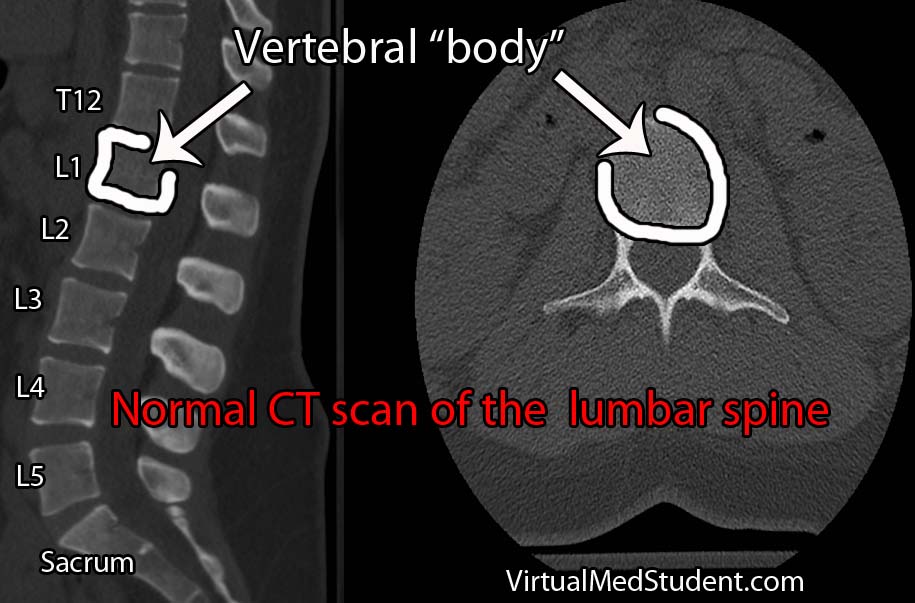
Burst fractures are a specific type of spine fracture in which the body of a given vertebrae “bursts” into pieces. By definition a burst fracture involves the entire vertebral body. The image below is an example of a normal lumbar spine with the vertebral bodies outlined.

Burst fractures most commonly occur at the junction between the thoracic and lumbar spine. This junction is an area where the rigid thoracic spine transitions to the more mobile lumbar spine, and hence is an intrinsic point of weakness. This is why most burst fractures occur between the T10 through L2 vertebrae.

CT vertebral body

Axial loading of the spine is what causes burst fractures. They typically occur after a traumatic events like car accidents or falls from significant heights. Elderly individuals, and those with poor bone quality, may suffer burst fractures after minor trauma such as falling from a chair.

Signs and Symptoms

Burst fractures invariably present with back pain at the site of the fracture. Depending on the exact location signs and symptoms of nerve root compression or lower spinal cord injury may occur.

If the nerves that dangle in the lumbar spine (aka: the cauda equina) get compressed by the fragments of bone then weakness, numbness, tingling, and even bowel and bladder problems may occur.

Burst fractures between T10 and L1 can cause damage to the end of the spinal cord (the spinal cord ends at L1 or L2 in most individuals), which can lead to lower extremity weakness, or even paralysis, as well as bowel and bladder dysfunction.

Diagnosis

Diagnosis of a burst fracture is made using a combination of x-rays, CT scans, and MRIs. These three imaging modalities serve different functions when evaluating the severity of a burst fracture.

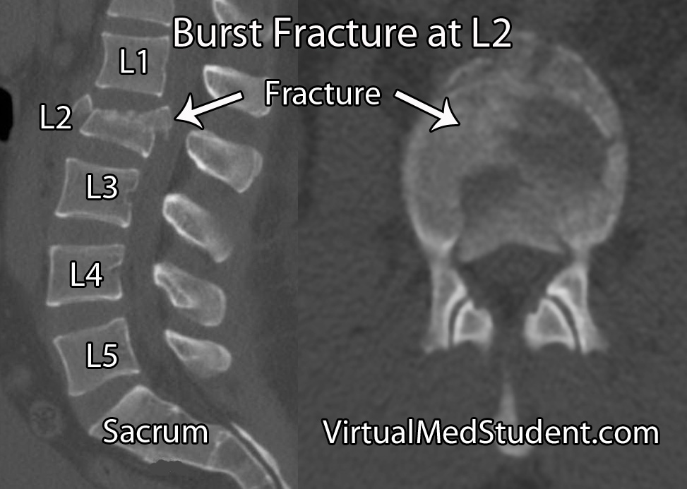
X-rays are usually the first imaging ordered in patients with suspected spine fractures. If the plain x-rays show a burst fracture then CT scanning is usually done to further assess the degree of bony injury (see image below for an example of an L2 burst fracture).

MRI is used to detect ligamentous injury. The degree of ligamentous injury indicates a higher degree of instability; information about ligament integrity helps determine treatment options.

Burst Fracture Lumbar Spine

Treatment

Treatment of burst fractures is highly dependent on the severity of the burst fracture. Treatment is either conservative with immobilization in a brace (ie: a "TLSO" or thoracolumbar sacral orthotic brace) or surgical fixation.

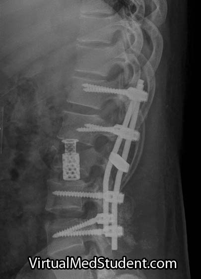
Burst fracture after instrumentation
As a rough rule of thumb patients with any of the following criteria should be strongly considered for surgical correction:

  • Greater than 50% vertebral body height loss.
  • Greater than 25 to 40 degrees of kyphosis.
  • Greater than 50% spinal canal compromise.
  • Significant posterior ligamentous injury.
  • Any neurological signs or symptoms referable to the injury.
  • If the patient fails conservative therapy with a brace.

Surgical correction can be achieved in a variety of ways and is often related to surgeon preference. Some surgeons will remove a significant portion of the fractured vertebral body and place a “cage” in the area, a procedure known as a “corpectomy”. This, combined with rods and screws from posteriorly provides the greatest stability, but has a higher risk of nerve injury. Not uncommonly, the fractured vertebral body is left alone and rods and screws are placed from behind only. This is especially true if the fractured level shows minimal spinal canal compromise.

Overview

Burst fractures of the thoracolumbar spine typically occur after high impact axial loading. They usually occur between T10 and L2, but can be seen anywhere in the spine. Patients will almost invariably have pain at the fracture site and may or may not have neurological signs and symptoms depending on the severity of the fracture. Diagnosis is made with CT, plain x-rays, and MRI. Treatment is highly dependent on the individual fracture and ranges from bracing to surgical fixation.

Related Articles

References and Resources

Atlas Fractures: The Weight of the World On Its Shoulders

The atlas, or the first cervical vertebra (C1), is a ring shaped structure. It forms joints with the base of the skull above and the axis (ie: the second cervical vertebrae) below. It also has two foramen transversarium, which are holes that allow the passage of the vertebral arteries on either side of the spinal cord.

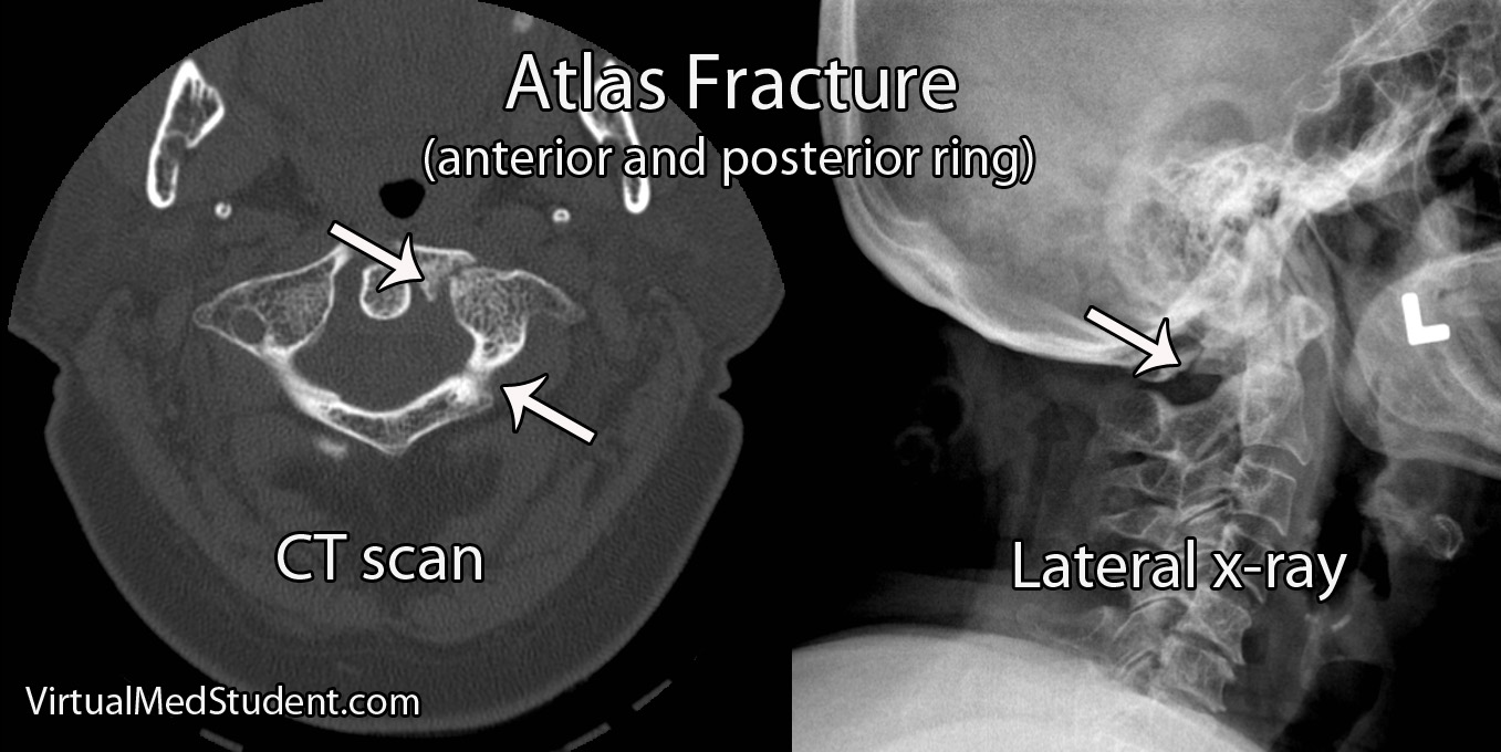
Fractures of the atlas occur most commonly with forceful axial loading of the head (ie: a downward force applied to the top of the head). Pressure on the top of the head causes the skull to push down on the atlas, which results in a break(s) of its ring-like structure. Specific fracture types such as a break in the front of the ring, the back of the ring, or one side of the ring versus the other, are dependent on additional force vectors at the time of loading (ie: flexion, extension, lateral bending, etc.).

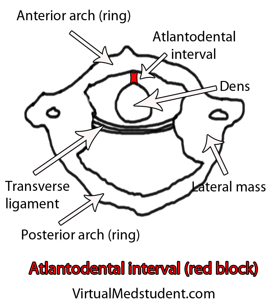
Fractures of the atlas must also include a discussion of biomechanical stability, which is usually determined by the integrity of the transverse ligament. The transverse ligament attaches the dens (odontoid) of the axis to the anterior ring of the atlas.

Fractures of the atlas with co-existent rupture of the transverse ligament lead to instability of the joint between C1 and C2. In other words, the ring of C1 may be able to move forward relative to the dens of C2. Transverse ligament injury is more common when axial loading is combined with extension of the head.

Not surprisingly, fractures of the atlas often co-exist with fractures of other cervical spine vertebrae. The most common combination is with a fracture of the axis, occurring in up to 40% of cases.

Signs and Symptoms

Patient’s with isolated atlas fractures usually have neck pain and muscle spasms. Frequently they have no injury to the spinal cord because the ring splays outwards as it fractures.

It is important to rule out injuries to the vertebral arteries, which run in bony holes (ie: foramen transversarium) on the sides of the atlas. When injured, the vertebral arteries can cause strokes in the brainstem and cerebellum, which can be life threatening.

Since the atlas is so close to the brainstem, patients may have co-existent injury to the lower cranial nerves. Specifically, injury to the 12th nerve can cause problems with tongue movements, injury to the 11th nerve can cause weakness with shoulder shrug and the ability to turn the head to the side, and injuries to the 9th and 10th cranial nerves can cause problems with swallowing and paralysis of the larynx leading to difficulty with speech.

Co-existent head and brain trauma, which can cause a constellation of different signs and symptoms depending on severity can also occcur.

Diagnosis

Diagnosis of an atlas fracture is made using x-rays, CT scans, and MRIs. X-rays should include anterior-posterior views, open mouth odontoid views, and lateral views of the cervical spine. If there is no evidence of neurological injury, flexion-extension x-rays may also be obtained to assess for stability of the C1-C2 joint.

The bony injury associated with atlas fractures is categorized according to the Jefferson or Landell and Van Peteghem classification systems. The Landells classification has three types, whereas the Jefferson classification has four types:

Landell and Van Peteghem Classification
Type 1 Fracture of either the anterior or posterior ring, but not both (posterior ring fractures are most common type)
Type 2 Fractures of both the anterior and posterior ring
Type 3 Fracture of the lateral mass(es)

Jefferson Classification
Type 1 Fracture of the posterior ring only
Type 2 Fracture of the anterior ring only
Type 3 Fracture of the anterior and posterior rings on both sides; this is the classic "burst", or traditional “Jefferson” fracture
Type 4 Fracture of the lateral mass(es)

Atlas fracture

An important part of diagnosing atlas fractures involves assessing the integrity of the transverse ligament, which is best done using MRI. However, if an MRI cannot be performed then open mouth odontoid, flexion-extension x-rays, and CT scans can provide some information regarding transverse ligament injury.

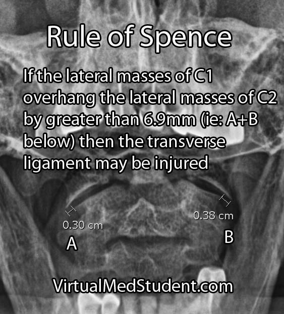
The rule of Spence is one way of assessing the integrity of the transverse ligament on an open mouth odontoid x-ray. The rule states that if the right and left lateral masses of C1 overhang the lateral masses of C2 by greater than a total distance of 6.9mm than the likelihood of co-existent transverse ligament injury is high. The rule of Spence is not fool proof and should be supplemented with MRI and/or flexion-extension films whenever possible.


Atlantodental Interval
Another method for assessing transverse ligament injury is using the "atlantodental" interval (see image to the left). This is the distance between the anterior arch of C1 and the odontoid process (aka: dens) of C2.

This interval is usually quite small, typically less than 3mm in adults and 5mm in children. If the interval is greater than this, then co-existent transverse ligament injury should be suspected.

Treatment

Treatment of isolated atlas fractures is usually with cervical immobilization. This may be with a halo or with a rigid cervical collar such as a cervical-occipital-mandibular-immobilizer (SOMI).

Atlas fractures that have co-existent transverse ligament rupture often require an operation to stablize the bones of the spine. This is usually in the form of fusing the atlas or occiput (back of the head) to the second cervical vertebrae.

If other injuries (ie: fractures of C2) are present and/or there is significant ligamentous injury then open surgical fusion of the bones may be necessary to re-create stability of the craniocervical junction.

Overview

Atlas fractures occur in response to vertical compression of the head on the upper cervical spine. Fractures of the anterior, posterior, or both rings of C1 may be present. Biomechanical stability is typically determined by assessing the integrity of the transverse ligament. Patients with isolated C1 fractures usually complain of neck pain, and rarely have injury to the spinal cord. Diagnosis is based on CT, x-ray, and MRI findings. Treatment is with rigid external immobilization or operative spinal fusion.

Related Articles

References and Resources