Basics of Pregnancy: GxPy, Naegele’s Rule, and that Bundle of Joy

This article will discuss the basics of pregnancy. In order to understand the basics we have to understand two important obstetric terms: gravity and parity.

Gravity refers to the number of times a woman has been pregnant. Parity refers to how many times the woman has given birth to an infant older than 20 weeks gestational age, or weighing greater than 500 grams.

On a medical chart a woman’s obstetric history is commonly written as “G x P y”. “G” refers to the gravity, or the number of times, “x”, a woman has been pregnant. The “P” stands for the parity, or the number of times, “y”, a woman has given birth to an infant older than 20 weeks gestation, or weighing greater than 500 grams. In other words the “y” equals the number of pregnancies that made it past 20 weeks gestation.

However, a woman’s obstetric history may also contain miscarriages, abortions, preterm births, and still births. In addition, some of her children may have died early in infancy. To account for these facts the “Gx Py” system is sometimes written as “G x P y, a, b, c, d”. The “a” refers to the number of term pregnancies; the "b" refers to the number of preterm pregnancies. The "c" refers to the number of abortions; abortions includes both spontaneous and induced, as well as ectopic pregnancies. And finally, the "d" refers to the number of living children the woman currently has.

So, as an example, a woman with two living children and one previous spontaneous abortion at twelve weeks gestation, who is currently pregnant for the third time would have an obstetric chart that reads "G 3 P 2, 2, 0, 1, 2."

Fetal Age nd Due Date

Pregnancy Wheel
There are different measures of fetal age. They include the following:

(1) Developmental age
(2) Gestational age

The developmental age is the age of the fetus measured from the time of fertilization. Since it is often difficult to determine exactly when fertilization occurred, a surrogate marker is used.

The surrogate marker is the gestational age. The gestational age is measured from the first day of the last menstrual period. Typically, the gestational age overestimates the developmental age by approximately two weeks.

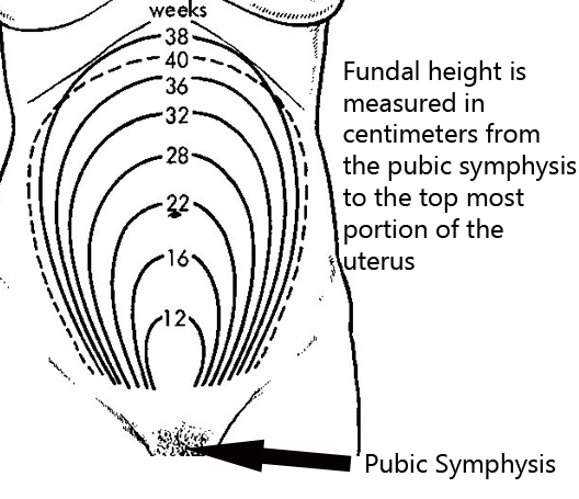
If the last menstrual period is not known there are other ways of determining the gestational age. One method is to use fundal height; fundal height is the location of the fundus (ie: the top most portion of the uterus) as felt by palpating the maternal abdomen. By 20 weeks of gestation the fundal height should be at the mother’s umbilicus (belly button). From there on, the height roughly correlates with gestational age (ie: at 28 weeks the fundal height is 28cm above the pubic symphysis).

The second method, albeit an even more imprecise one, is to use quickening. Quickening is the detection of fetal movements by the mother. It usually corresponds to a gestational age of 17 to 18 weeks. Ultrasound can also be used to determine the gestational age, and is the most reliable way, assuming the last menstrual period is not known.

In order to calculate the delivery date a simple mathematics trick known as Nagele’s rule is used. It states that the estimated delivery date is determined by adding nine months plus seven days to the first day of the last menstrual period.

Naegele’s rule -> Last menstrual period + 9 months + 7 days = expected due date

Pregnancy Tests

How exactly do you determine if someone is pregnant? The most common way is to use a urine pregnancy test. These tests detect a molecule known as beta-HCG (ie: beta human chorionic gonadotropin). Beta-HCG is a molecule secreted by the placenta. Its level doubles roughly ever 48 hours during the first few days of pregnancy. It peaks at about 10 weeks of gestation. Urine tests tend to have a high false negative rate. This means that they miss pregnancies, especially if the pregnancy has just begun.

A more sensitive test is the serum (ie: blood) pregnancy test. This test measures exactly the same molecule, beta-HCG, but it does so from a blood, rather than a urine sample. This test can pick up pregnancies earlier than a urine test can.

Related Articles

References and Resources

Bun in the Oven: Fetus Surveillance Techniques

Fetal growth is most commonly monitored by ultrasound. Most “routine” pregnancies begin with one fetal ultrasound between 18 and 20 weeks of gestation. This ultrasound can pick up important defects that may not be apparent at later gestational dates. If the 18-20 week ultrasound is normal, and the pregnancy is otherwise uncomplicated, repeat ultrasounds are not routinely performed.

Fetus

Fetal growth can also be monitored by measuring the "fundal height". The fundal height is the distance between the fundus (ie: top of the uterus), which is felt via palpation of the abdomen, and the pubic symphysis (see image to left).

A rule of the thumb is that the top of the uterus should be felt at the level of the umbilicus (ie: belly button) at approximately 20 weeks gestation. It then increases proportionally until about 36 weeks gestation. At this point the height may actually decrease as the fetal head enters the pelvis.

Fetal Maturity

There are three different ways to assess fetal maturity. They are used to determine if the fetus has adequate lung maturity to survive outside the uterus. These tests measure molecules that are present in the amniotic fluid. Therefore an invasive test known as an amniocentesis is necessary. The different methods are:

(1) Lecithin to sphingomyelin ratio
(2) Phosphatidylglycerol test
(3) Foam stability index

The first way of determining fetal lung maturity is to measure the lecithin to sphingomyelin ratio. Sphingomyelin is a specialized phospholipid that is produced in constant amounts by the fetus throughout pregnancy. Lecithin is a component of surfactant (ie: molecules that help keep the alveoli of the lung inflated) and its production increases as fetal lung maturity increases. An L:S ratio of greater than two indicates that the fetal lungs are mature enough to work outside the uterus.

The second test is known as the phosphatidylglycerol (PG) test. The presence of this molecule in the amniotic fluid indicates fetal lung maturity. PG is a component of pulmonary surfactant, which helps keep the gas exchange portions of the lung (ie: alveoli) from deflating at the end of exhalation.

The third test is the "foam stability index" test. This test measures the stability of bubbles when amniotic fluid is shaken with variable percent alcohol solutions. If the bubbles are stable this indicates fetal lung maturity.

Fetal Well-Being

Fetal well-being is assessed using several different methods. They include:

(1) Non-stress test
(2) Stress test
(3) Biophysical profile

Then on-stress test measures fetal well-being by looking at changes in the fetal heart rate during movement. A normal (aka: "reactive") non-stress test occurs when the fetus’ heart rate increases by 15 beats per minute over 15 seconds following fetal movement. If this increase in heart rate occurs twice within 20 minutes of monitoring the test is normal or “reactive”. Note: if the fetus is less than 32 weeks gestation then only a 10 beat per minute increase is needed to satisfy the non-stress test.

The stress test (aka: "contraction stress test", or "oxytocin stress test" depending on how contractions are elicited) measures the fetus’ ability to handle the "stress" of a uterine contraction. When the uterus contracts blood flow from the placenta is momentarily decreased. A healthy fetus will have no discernible change in their heart rate; however, an at risk fetus will have heart rate decelerations. Stress tests, especially oxytocin induced stress tests, commonly result in false-positives (ie: indicate fetal jeopardy when the fetus is normal); therefore, an abnormal stress test is followed by a non-stress test. If the non-stress test is normal/reactive then the stress test is often times considered a false-positive.

The biophysical profile uses five parameters for assessing fetal well-being. They include: fetal breathing movements, gross body movements, fetal muscle tone, fetal heart rate as measured by non-stress test, and qualitative amniotic fluid levels. Each of these parameters is given a score of 0 if absent/abnormal, or 2 if normal. A score of 8-10 is reassuring and indicates no problems with the fetus.

Related Articles

References and Resources

Appendicitis: A Vestigial Remnant to Belly Pain

The appendix is a small out-pouching off a part of the large intestine known as the cecum. It functions similar to normal large intestine by secreting mucous and absorbing water. Its overall importance, however, is not well understood, and it is likely a vestigial remnant from a distant ancestor. Unfortunately for some unlucky folk it can become inflamed; when this occurs it is called “appendicitis”.

Appendicitis occurs when something blocks the opening of the appendix into the cecum. There are numerous causes. The most common in younger individuals is a mass of inflammatory cells known as lymphoid hyperplasia, which can occur after a viral or bacterial infection of the gut. In older individuals, the most common cause is a small, hard piece of poop known as a “fecalith”.

When lymphoid hyperplasia or a fecalith (or any other obstructing thing) blocks the opening of the appendix, any mucous secreted by it gets trapped. When the appendix becomes distended enough, it literally chokes off its own blood supply and starts to die.

The dying appendix sets off a cascade of inflammation. Bacteria within the intestine are able to move in and wreak further havoc! The end result is a nasty inflammatory process, that if left unchecked, can lead to a very serious surgical illness.

Signs and Symptoms

Appendicitis initially presents with periumbilical pain (ie: pain around the belly button) that quickly migrates to involve the right lower quadrant of the abdomen.

The reason pain occurs in this sequence is because the initial discomfort of appendicitis is due to inflammation of the visceral peritoneum and appendix itself. The visceral peritoneum is a layer of tissue that envelopes the gut tube. This type of pain is carried back to the spinal cord by autonomic nerves, and due to their embryological origin, that pain gets referred to the midline of the abdomen (the belly button).

Over the course of the disease, the parietal peritoneum eventually becomes inflamed. This pain is carried by somatosensory nerves with a very specific dermatomal distribution, thus the pain gets localized directly to the anatomical location of the appendix – the right lower quadrant. This pain is often very well localized at an area known as McBurney’s point.

Patients often have nausea, vomiting, and a decreased appetite. In fact, if patients are hungry and want to eat, appendicitis becomes a highly unlikely diagnosis for abdominal pain. This is referred to informally as the "cheeseburger sign".

There are also several physical exam findings. Pain with palpation of the right lower quadrant associated with rebound tenderness (ie: pain that occurs when you release pressure during palpation) is frequently seen. Sometimes palpating the left lower quadrant (ie: the area on the other side of the abdomen from the appendix) will cause discomfort in the right lower quadrant. This is known as "Rovsing’s sign".

Any maneuvers that irritate the peritoneum will also cause discomfort. The first of these signs occurs when you flex the hip. This causes the iliopsoas muscle to rub up against an inflamed peritoneum. This is known as the "psoas sign". Another way to irritate the peritoneum is to internally rotate the leg while the patient’s knee and hip are flexed. This is known as the "obturator sign".

Diagnosis

Appendicitis CT Scan
Appendicitis is first and foremost a clinical diagnosis. Therefore, in patients with a history of periumbilical pain that migrates to the right lower quadrant appendicitis is the most likely diagnosis.

However, in a world with advanced imaging technologies we can quickly get pictures that support the diagnosis. Both ultrasound (frequently used in children, pregnant patients, and younger adults) or CT scans can be obtained quickly and inexpensively. The CT scan will show fat stranding and fluid around an enlarged appendix (see image to the left).

Blood tests such as a complete blood count (CBC) will show an elevated white blood cell count (ie: the cells that fight off infection and are responsible for inflammation).

Treatment

Treatment is surgical (ie: appendectomy)! Get that nasty inflamed appendix out of there STAT! In addition, patients are started on intravenous fluids and antibiotics that cover anaerobic organisms.

Commonly used antibiotics for a non-ruptured appendix are metronidazole, ampicillin/sulbactam, and ciprofloxacin. If the appendix has ruptured, broad spectrum coverage with piperacillin/tazobactam or a combination of ampicillin, ciprofloxacin, and clindamycin is started and continued for at least 5 days.

Occasionally a ruptured appendix will wall itself off into an abscess. If this is the case, the abscess must be drained with a needle. Antibiotics are started, and the patient is taken to surgery roughly six weeks later to remove the appendix after the inflammation has "calmed down".

Overview

Appendicitis occurs when something blocks the opening of the appendix into the cecum. Progressive enlargement of the appendix occurs eventually chocking off its own blood supply. Pain in the right lower quadrant of the abdomen is a common symptom of appendicitis. Diagnosis is clinical, but ultrasound and CT scanning can be helpful in elucidating unclear cases. Treatment is appendectomy (removal of the appendix), IV fluids, and antibiotics.

References and Resources

  • Merlin MA, Shah CN, Shiroff AM. Evidence-based appendicitis: the initial work-up. Postgrad Med. 2010 May;122(3):189-95.
  • Kim JK, Ryoo S, Oh HK, et al. Management of appendicitis presenting with abscess or mass. J Korean Soc Coloproctology. 2010 Dec;26(6):413-9. Epub 2010 Dec 31.
  • Lee SL, Islam S, Cassidy LD. Antibiotics and appendicitis in the pediatric population: an American Pediatric Surgical Association Outcomes and Clinical Trials Committee systematic review. J Pediatr Surg. 2010 Nov;45(11):2181-5.
  • Markides G, Subar D, Riyad K. Laparoscopic versus open appendectomy in adults with complicated appendicitis: systematic review and meta-analysis. World J Surg. 2010 Sep;34(9):2026-40.
  • Grundmann RT, Petersen M, Lippert H, et al. The acute (surgical) abdomen – epidemiology, diagnosis and general principles of management. Z Gastroenterol. 2010 Jun;48(6):696-706. Epub 2010 Jun 1.