Burrill B. Crohn and His Disease: GI Inflammation

Crohn’s disease is one of the inflammatory bowel diseases. Its pathology is related to transmural (ie: full wall thickness) inflammation of the bowel wall. Why this inflammation occurs is not particularly well understood.

Genetics appears to play an important role. Specific human leukocyte antigen genes are associated with Crohn’s (DR1/DQ5). In addition, there is a gene known as IBD1 that increases the risk of Crohn’s disease. The IBD1 gene encodes a protein called NOD2. This protein normally helps people “contain” bacteria in the gut. In some patients with Crohn’s disease the protein is mutated and does not function properly.

The immune system also plays an important role. For reasons that are actively being researched patients with inflammatory bowel disease may attack bacteria in the intestine that would otherwise be left alone. This leads to inflammation of the gut wall. Why dysregulation of the immune system occurs is not well understood, but many different types of immune cells are involved including T-cells, neutrophils, and macrophages.

The inflammation in Crohn’s disease can affect almost any location in the gastrointestinal tract from the mouth to the colon. The terminal ileum and proximal colon are the most common sites affected. Roughly 40% of cases involve the small bowel only. Pathological specimens resected from patients with the disease show noncaseating granulomas, aggregates of lymphocytes (ie: a type of white blood cell) in the bowel wall, and extension of fat along the serosal surface.

Signs and Symptoms

Symptoms usually start between the ages of 15 and 40, with a second smaller peak starting between 50 and 80. Men and women are affected equally.

Diarrhea, abdominal pain, and weight loss that are chronic, or occur repeatedly are concerning for Crohn’s. Blood in the stool is not always seen, but may be present. Since any portion of the gastrointestinal tract can be involved, some patients have oral manifestations such as aphthous ulcers (“canker sores”).

Complications can occur and include bowel wall perforation, abscess formation, and strictures (ie: narrowing) of the bowel’s diameter resulting in obstruction. Fistulas, which are abnormal connections between two unrelated organs/body parts can occur between the bowel and the skin (enterocutaneous), bowel and bladder (enterovesicular), bowel and vagina (enterovaginal), and between adjacent bowel segments (enteroenteric).

Non-intestinal manifestations are also common. They include an arthritis that migrates around the body and involves multiple joints. Uveitis is inflammation of the uvea of the eye, which can lead to a painful eye and vision problems. Ankylosing spondylitis and sacroiliitis are inflammatory conditions of the axial skeleton that can co-exist with Crohn’s. Erythema nodosum, which is inflammation of the fat cells under the skin commonly occurs around the shins in patient’s with Crohn’s. Finally, primary sclerosing cholangitis (PSC), which is inflammation of the bile ducts, both inside and outside the liver, can also occur (PSC is more strongly associated with ulcerative colitis).

Patient’s with long standing Crohn’s disease are also at risk of developing colon cancer, although the risk is less than that seen in ulcerative colitis.

Diagnosis

The diagnosis is based on the history provided by the patient and imaging studies. Imaging studies can be x-rays taken after contrast is given (barium). The x-rays may show a “string sign”. The string sign is a result of nodular outpouchings of the bowel wall (secondary to inflammation), alternating with normal areas.

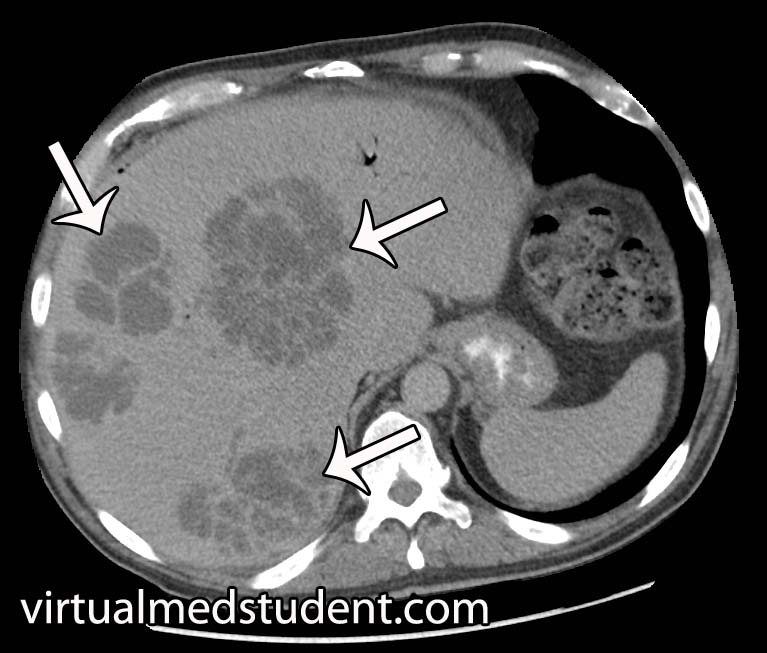
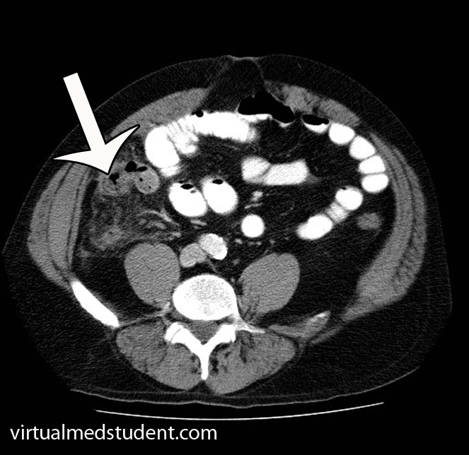
CT and MRI scans of the abdomen with contrast can show increased thickness of the bowel wall; they can also show abscess and fistulae formation, which are complications of the disease.

The "gold standard" for diagnosis is colonoscopy with biopsy of affected tissue. Skip lesions, in which areas of inflammation alternate with normal bowel, are highly suggestive of Crohn’s disease. The bowel wall will often appear “cobblestoned” and aphthous ulcers may also be present along the mucosal surface.

Treatment

Active Crohn’s disease is usually treated with one or more medications depending on the severity. Severe flairs of the disease are treated with steroids like prednisone.

Mild to moderate disease is often treated with one of the 5-aminosalicylic acid (5-ASA) derivatives. Sulfasalazine and mesalamine are two such derivatives. Unfortunately, 5-ASA medications do not appear to prevent relapses.

Some patients will not respond to prednisone or 5-ASA medications. For disease that does not respond to the above medications there are other options such as azathioprine and methotrexate.

Finally a class of medications known as “biologics” can be used. These medications dampen the immume response and therefore decrease the inflammation that occurs in autoimmune diseases. These include the anti-tumor necrosis factor medications known as infliximab and adalimumab. Vedolizumab (Entyvio®) blocks integrins on the surface of immune cells from binding to cells in the GI system. Ustekinumab (Stelara®) works on a molecule known as an interleukin, which is involved in inflammation. And last, but not least, risankizumab (Skyrizi®) also blocks a specific interleukin (number 23 to be exact), which decreases inflammation.

Overview

Crohn’s disease is characterized by inflammation of the entire bowel wall. It manifests with both gastrointestinal and extra-intestinal symptoms. It is diagnosed by history and imaging studies. Treatment is with immune dampening therapies like prednisone, 5-ASA derivatives, azathioprine, 6-mercaptopurine, methotrexate, and tumor necrosis factor inhibitors.

Other Stuff You May Want to Learn About

References and Resources

  • Gura Y, Bonen DK, Inohara N, et al. Frameshift mutation in NOD2 associated with susceptibility to Crohn’s disease. Nature 2001 May 31;411(6837):603-6.
  • Kumar V, Abbas AK, Fausto N. Robbins and Cotran Pathologic Basis of Disease. Tenth Edition. Philadelphia: Elsevier Saunders, 2004.
  • Akobeng AK, Gardener ES. Oral 5-aminosalicylic acid for maintenance of medically-induced remission in Crohn’s Disease. Cochrane Database Syst Rev. 2005 Jan 25;(1):CD003715.

Pyogenic Liver Abscesses: Pus, Needle Drainage, and Antibiotics

Pyogenic liver abscesses are localized collections of pus and bacteria. The initial infection occurs when bacteria travel through the portal vein. This most commonly occurs after bowel contents leak into the peritoneal cavity from the gut. Other causes of liver abscesses include direct spread of infection from the bile duct system, or from other bacterial infections of the blood. Rarely, penetrating injuries (gunshots, stab wounds, surgery) may directly introduce infection. There are numerous “bugs” that can cause pyogenic liver abscesses. They include, but are not limited to, streptococcus species, klebsiella pneumoniae, and staphylococcus species.

Signs and Symptoms

Similar to other infections, liver abscesses can cause fevers, chills, decreased appetite, abdominal pain, and a generalized sense of not feeling well (ie: malaise). Interestingly, hiccups may also be present if the abscesses are causing adjacent inflammation/irritation of the diaphragm.

Diagnosis

Pyogenic Liver Abscesses
The diagnosis of pyogenic liver abscesses is made with imaging. The most commonly used method is a CT scan of the abdomen with and without contrast. If present, a liver abscess will look like a collection of fluid, with or without septated dividers.

Another commonly employed imaging modality is the use of ultrasound to detect the fluid filled pockets within the liver.

However, it is important to note that imaging studies alone cannot distinguish between the different types of liver abscesses. Imaging can also not tell you what bacteria is responsible for the abscess.

Treatment

Pyogenic abscesses must be drained and treated with antibiotics. Drainage is usually done under image guidance using a needle placed through the skin into the abscess. Although occasionally direct surgical evacuation of the abscess is necessary.

Draining the abscess is important for two reasons. First, it decreases the size of the abscess allowing antibiotic therapy to work more effectively. And secondly, it provides abscess fluid that can be sent to the lab for bacterial culture and gram stain.

The results of the culture help guide subsequent antibiotic therapy. Commonly used antibiotics include piperacillin-tazobactam (Zosyn®), vancomycin, metronidazole (Flagyl®), and ceftriaxone; once cultures confirm the causative bug antibiotic therapy can be narrowed.

Overview

Pyogenic liver abscesses are collections of pus and bacteria. They occur most commonly after the spilling of gut bacteria into the peritoneal space (ie: peritonitis). There are numerous causative bacteria. Symptoms include fever, chills, decreased appetite, and abdominal discomfort. Treatment is with drainage and antibiotics.

References and Resources

  • Hasper D, Schefold JC, Baumgart DC. Management of severe abdominal infections. Recent Pat Antiinfect Drug Discov. 2009 Jan;4(1):57-65.
  • Benedetti NJ, Desser TS, Jeffrey RB. Imaging of hepatic infections. Ultrasound Q. 2008 Dec;24(4):267-78.
  • Mortelé KJ, Segatto E, Ros PR. The infected liver: radiologic-pathologic correlation. Radiographics. 2004 Jul-Aug;24(4):937-55.
  • Kurland JE, Brann OS. Pyogenic and amebic liver abscesses. Curr Gastroenterol Rep. 2004 Aug;6(4):273-9.

Cholelithiasis (Gallstones): Female, Fat, Fertile, and Forty

Gallstones (aka: cholelithiasis) are stones that form – you guessed it – in the gallbladder. In order to understand gallstones we have to understand how bile is formed. The gallbladder functions as a bile storage bin that releases its contents in response to signals from the intestine and stomach (ie: usually after a meal rich in fats). Bile is formed in the liver and is a unique mix of bile acids, cholesterol, bilirubin, water, phospholipids, and other electrolytes. The most important component is the bile acids. They help keep bile in its liquid state.

With a basic understanding of bile we can now address the problem of gallstone formation. There are different types of gallstones, and the most common type are cholesterol stones. When there is too much cholesterol in bile, the bile acids can no longer keep it soluble. The result? The excess cholesterol precipitates out and forms a stone.

The second type of gallstones are pigmented stones, which come in two flavors: brown and black. Brown stones form as a result of biliary tract infection. Black stones occur when the body breaks down red blood cells excessively, such as in hemolytic anemias. Black stones form from the excess bilirubin (a breakdown product of red blood cells), which gets sent to the liver and ultimately ends up in bile. An excessive amount will do exactly like cholesterol does, and precipitate to form a stone.

Signs and Symptoms

The common risk factors associated with gallstone formation are the “4 Fs”:

– Female
– Fat (overweight/obese, to use the politically "correct" term)
– Fertile
– Forty

Gallstones tend to develop in this demographic more often than in other people. Any one of these factors puts you at increased risk of developing gallstones. Other common risk factors include the following: oral contraceptive use, rapid weight loss, hemolytic diseases/disorders, small bowel resection, total parenteral nutrition (ie: nutrition given through an IV), Crohn’s disease, cystic fibrosis, and Native American ethnicity.

Gallstones by themselves are usually not symptomatic. In fact, many people are walking around with gallstones and don’t even know they have them! However, in a subset of the population symptoms develop. The main symptom associated with gallstones is biliary colic, which is pain that occurs after a meal (especially meals rich in fats). In addition, nausea, vomiting, and fatty food intolerances may also occur.

Complications

Biliary Tract
There are many potential complications of gallstones. The most common complication is acute cholecystitis. In acute cholecystitis a gallstone lodges in the cystic duct (see image) during contraction of the gallbladder. This leads to inflammation of the gallbladder wall. If left untreated, acute cholecystitis can lead to infection of the gallbladder.

The second complication of gallstones is choledocholithiasis. This is a fancy term for a gallstone lodged in the common bile duct. This can lead to jaundice, pain, pancreatitis, and potentially a very dangerous condition known as acute cholangitis. Acute cholangitis is infection of the biliary tree and can occur secondary to an obstructing stone in the common bile duct.

To review, the common complications of gallstones are:

(1) Biliary colic
(2) Acute cholecystitis
(3) Choledocholithiasis
(4) Acute cholangitis
(5) Pancreatitis

Diagnosis

Since most gallstones are cholesterol, they are radiolucent, meaning that they are not detectable by x-rays. The test that is often done first with any suspected biliary pathology is a right upper quadrant ultrasound. If stones are present they will cast a “shadow” that can be picked up on the ultrasound. Often times additional studies are performed if another diagnosis is being considered. Some clinicians will get hepatitis studies to rule out liver damage. Pancreatic function is also commonly tested.

Treatment

Most gallstones do not require treatment because they are asymptomatic. However, if symptoms develop a trial of lifestyle modification is often tried. For example, avoiding fatty foods can limit biliary colic.

The definitive treatment for symptomatic gallstones, or if any of the complications above develop, is a cholecystectomy. Cholecystectomy is the medical term for removal of the gallbladder. It is done either "open" (ie: with a large incision) or laparoscopically depending on the patient and surgeon’s preferences.

Some patients may not be able to undergo surgery. In these patients a medication known as ursodiol may be given. It is a bile acid mimic that helps liquify bile.

Overview

Cholelithiasis (gallstones) is caused by precipitation of cholesterol or bilirubin. Gallstones are usually asymptomatic, although they can sometimes lead to biliary colic, acute cholecystitis, acute cholangitis, choledocholithiasis, and pancreatitis. Treatment, when symptomatic, is usually lifestyle modifications; if dangerous symptoms or complications occur treatment is with cholecystectomy (ie: removal of the gallbladder).

References and Resources

  • Venneman NG, van Erpecum KJ. Pathogenesis of gallstones. Gastroenterol Clin North Am. 2010 Jun;39(2):171-83, vii.
  • van Erpecum KJ. Biliary lipids, water and cholesterol gallstones. Biol Cell. 2005 Nov;97(11):815-22.
  • Stinton LM, Myers RP, Shaffer EA. Epidemiology of gallstones. Gastroenterol Clin North Am. 2010 Jun;39(2):157-69, vii.
  • Gurusamy KS, Davidson BR. Surgical treatment of gallstones. Gastroenterol Clin North Am. 2010 Jun;39(2):229-44, viii.
  • O’Neill DE, Saunders MD. Endoscopic ultrasonography in diseases of the gallbladder. Gastroenterol Clin North Am. 2010 Jun;39(2):289-305, ix.
  • Flynn JA. Oxford American Handbook of Clinical Medicine (Oxford American Handbooks of Medicine). First Edition. Oxford University Press, 2007.
  • Kumar V, Abbas AK, Fausto N. Robbins and Cotran Pathologic Basis of Disease. Seventh Edition. Philadelphia: Elsevier Saunders, 2004.
  • Blackbourne LH. Surgical Recall, North American Edition (Recall Series). Ninth Edition. Philadelphia: Lippincott Williams and Wilkins, 2009.

Barrett’s Esophagus: Epithelial Metaplasia

The mucosal layer of the human esophagus is lined with cells known as stratified squamous epithelium. This epithelium is ideal for areas with lots of abrasion and use (ie: swallowing food and drink) because it is easily shed and replaced.

However, in Barrett’s esophagus the squamous epithelium is replaced by another cell type known as columnar epithelium interspersed with goblet cells (ie: cells that secrete mucous). This type of epithelium normally lines the intestine. This abnormal change in esophageal cell type is called "metaplasia".

Long standing acid reflux is responsible for the metaplasia that occurs in the esophagus. Poorly controlled reflux allows stomach acid to regurgitate into the lower esophagus. After many years of this the esophagus responds by changing its lining to be more like an intestinal cell type; this is referred to as “intestinal metaplasia”. The intestinal cell types, including goblet cells, secrete mucous and bicarbonate ions to neutralize the refluxed acid. This change is believed to help prevent damage to the esophagus.

Why is Barrett’s esophagus a bad thing? Occasionally, the metaplastic cells undergo a process known as dysplasia in which they assume pre-cancerous characteristics. Many patients do not progress past low grade dysplasia, and therefore their risk of developing esophageal cancer is minimal. However, a small percentage of patients will go on to develop high grade dysplasia, which puts them at significant risk for developing esophageal cancer.

Signs and Symptoms

Barrett’s esophagus does not cause any symptoms per se. Rather the symptoms are usually related to gastroesophageal reflux disease (GERD). Most patients with GERD have “heart burn” symptoms, especially after eating spicy foods. Heart burn refers to a burning pain behind the sternum. In addition, patients with GERD frequently have nausea and mild amounts of regurgitation.

Interestingly, patients who develop Barrett’s esophagus often have improvement in GERD symptoms. This is because the intestinal type cells are able to neutralize the acid more effectively.

Diagnosis

Diagnosis of Barrett’s esophagus usually occurs after years of uncontrolled GERD symptoms. Patient’s with severe reflux can undergo upper endoscopic visualization of the esophagus (aka: swallowing a "roto-rooter" with a camera on the end of it). At this time biopsies can be taken to look for metaplasia and dysplasia. A pathologist reviews the biopsy samples and is able to tell if Barrett’s esophagus is present.

Treatment

Treatment is highly variable and depends on the severity of dysplasia seen. For simple metaplasia and low grade dysplasia treating the acid reflux with medications such as H2-blockers (ie: ranitidine, famotidine, etc.) and proton pump inhibitors (omeprazole, lansoprazole, etc.) can often reverse the changes.

In patients with high grade dysplasia numerous options exist including surgery, radiofrequency ablation of the esophageal lining, and close endoscopic monitoring with frequent repeat biopsies. Treatment is highly tailored to the individual patient.

Overview

Barrett’s esophagus is intestinal metaplasia of the cells lining the esophagus. It normally occurs after years of uncontrolled acid reflux. The metaplastic cells can undergo a pre-cancerous change, which can ultimately lead to esophageal cancer in a certain subset of individuals. Treatment is highly individualized and is dictated by the degree of dysplasia and risk of developing esophageal cancer.

References and Resources

  • Shaheen NJ, Sharma P, Overholt BF, et al. Radiofrequency Ablation in Barrett’s Esophagus with Dysplasia. N Engl J Med 360:22, p2277-88. May 28, 2009.
  • Flynn JA. Oxford American Handbook of Clinical Medicine (Oxford American Handbooks of Medicine). First Edition. Oxford University Press, 2007.
  • Kumar V, Abbas AK, Fausto N. Robbins and Cotran Pathologic Basis of Disease. Seventh Edition. Philadelphia: Elsevier Saunders, 2004.
  • Blackbourne LH. Surgical Recall, Fifth North American Edition (Recall Series). Fifth Edition. Philadelphia: Lippincott Williams and Wilkins, 2009.

Appendicitis: A Vestigial Remnant to Belly Pain

The appendix is a small out-pouching off a part of the large intestine known as the cecum. It functions similar to normal large intestine by secreting mucous and absorbing water. Its overall importance, however, is not well understood, and it is likely a vestigial remnant from a distant ancestor. Unfortunately for some unlucky folk it can become inflamed; when this occurs it is called “appendicitis”.

Appendicitis occurs when something blocks the opening of the appendix into the cecum. There are numerous causes. The most common in younger individuals is a mass of inflammatory cells known as lymphoid hyperplasia, which can occur after a viral or bacterial infection of the gut. In older individuals, the most common cause is a small, hard piece of poop known as a “fecalith”.

When lymphoid hyperplasia or a fecalith (or any other obstructing thing) blocks the opening of the appendix, any mucous secreted by it gets trapped. When the appendix becomes distended enough, it literally chokes off its own blood supply and starts to die.

The dying appendix sets off a cascade of inflammation. Bacteria within the intestine are able to move in and wreak further havoc! The end result is a nasty inflammatory process, that if left unchecked, can lead to a very serious surgical illness.

Signs and Symptoms

Appendicitis initially presents with periumbilical pain (ie: pain around the belly button) that quickly migrates to involve the right lower quadrant of the abdomen.

The reason pain occurs in this sequence is because the initial discomfort of appendicitis is due to inflammation of the visceral peritoneum and appendix itself. The visceral peritoneum is a layer of tissue that envelopes the gut tube. This type of pain is carried back to the spinal cord by autonomic nerves, and due to their embryological origin, that pain gets referred to the midline of the abdomen (the belly button).

Over the course of the disease, the parietal peritoneum eventually becomes inflamed. This pain is carried by somatosensory nerves with a very specific dermatomal distribution, thus the pain gets localized directly to the anatomical location of the appendix – the right lower quadrant. This pain is often very well localized at an area known as McBurney’s point.

Patients often have nausea, vomiting, and a decreased appetite. In fact, if patients are hungry and want to eat, appendicitis becomes a highly unlikely diagnosis for abdominal pain. This is referred to informally as the "cheeseburger sign".

There are also several physical exam findings. Pain with palpation of the right lower quadrant associated with rebound tenderness (ie: pain that occurs when you release pressure during palpation) is frequently seen. Sometimes palpating the left lower quadrant (ie: the area on the other side of the abdomen from the appendix) will cause discomfort in the right lower quadrant. This is known as "Rovsing’s sign".

Any maneuvers that irritate the peritoneum will also cause discomfort. The first of these signs occurs when you flex the hip. This causes the iliopsoas muscle to rub up against an inflamed peritoneum. This is known as the "psoas sign". Another way to irritate the peritoneum is to internally rotate the leg while the patient’s knee and hip are flexed. This is known as the "obturator sign".

Diagnosis

Appendicitis CT Scan
Appendicitis is first and foremost a clinical diagnosis. Therefore, in patients with a history of periumbilical pain that migrates to the right lower quadrant appendicitis is the most likely diagnosis.

However, in a world with advanced imaging technologies we can quickly get pictures that support the diagnosis. Both ultrasound (frequently used in children, pregnant patients, and younger adults) or CT scans can be obtained quickly and inexpensively. The CT scan will show fat stranding and fluid around an enlarged appendix (see image to the left).

Blood tests such as a complete blood count (CBC) will show an elevated white blood cell count (ie: the cells that fight off infection and are responsible for inflammation).

Treatment

Treatment is surgical (ie: appendectomy)! Get that nasty inflamed appendix out of there STAT! In addition, patients are started on intravenous fluids and antibiotics that cover anaerobic organisms.

Commonly used antibiotics for a non-ruptured appendix are metronidazole, ampicillin/sulbactam, and ciprofloxacin. If the appendix has ruptured, broad spectrum coverage with piperacillin/tazobactam or a combination of ampicillin, ciprofloxacin, and clindamycin is started and continued for at least 5 days.

Occasionally a ruptured appendix will wall itself off into an abscess. If this is the case, the abscess must be drained with a needle. Antibiotics are started, and the patient is taken to surgery roughly six weeks later to remove the appendix after the inflammation has "calmed down".

Overview

Appendicitis occurs when something blocks the opening of the appendix into the cecum. Progressive enlargement of the appendix occurs eventually chocking off its own blood supply. Pain in the right lower quadrant of the abdomen is a common symptom of appendicitis. Diagnosis is clinical, but ultrasound and CT scanning can be helpful in elucidating unclear cases. Treatment is appendectomy (removal of the appendix), IV fluids, and antibiotics.

References and Resources

  • Merlin MA, Shah CN, Shiroff AM. Evidence-based appendicitis: the initial work-up. Postgrad Med. 2010 May;122(3):189-95.
  • Kim JK, Ryoo S, Oh HK, et al. Management of appendicitis presenting with abscess or mass. J Korean Soc Coloproctology. 2010 Dec;26(6):413-9. Epub 2010 Dec 31.
  • Lee SL, Islam S, Cassidy LD. Antibiotics and appendicitis in the pediatric population: an American Pediatric Surgical Association Outcomes and Clinical Trials Committee systematic review. J Pediatr Surg. 2010 Nov;45(11):2181-5.
  • Markides G, Subar D, Riyad K. Laparoscopic versus open appendectomy in adults with complicated appendicitis: systematic review and meta-analysis. World J Surg. 2010 Sep;34(9):2026-40.
  • Grundmann RT, Petersen M, Lippert H, et al. The acute (surgical) abdomen – epidemiology, diagnosis and general principles of management. Z Gastroenterol. 2010 Jun;48(6):696-706. Epub 2010 Jun 1.