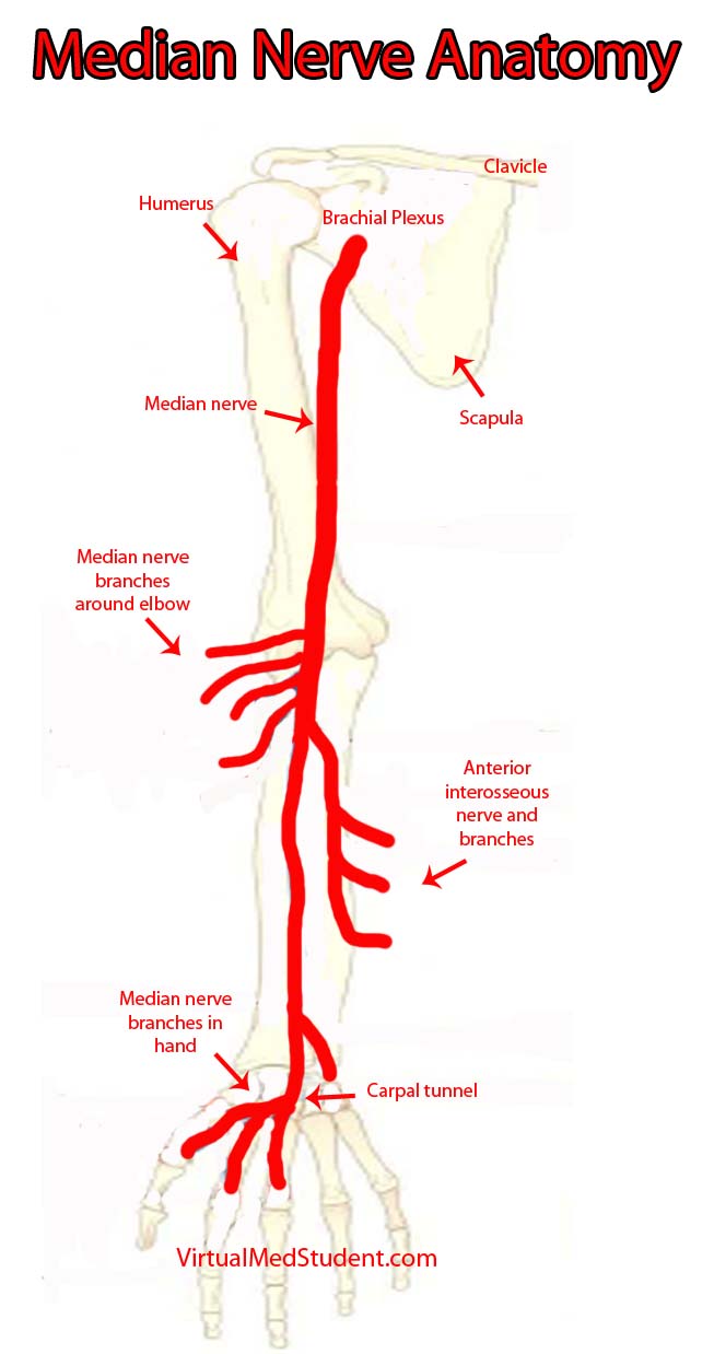
The "highways" merging into the brachial plexus are the 5th, 6th, 7th, and 8th cervical nerve roots, as well as the 1st thoracic nerve root. These nerve roots mix together to form trunks, divisions, cords, and finally branches. The median nerve is one of the final branches of the brachial plexus. It is composed of fibers from the 6th, 7th and 8th cervical nerve roots, as well as the 1st thoracic nerve root.
After branching from the brachial plexus, the median nerve courses along the front aspect of the humerus in the upper arm. At the elbow it branches to supply the pronator teres, flexor carpi radialis, palmaris longus, and flexor digitorum superficialis muscles.
In the forearm the nerve divides into a branch called the anterior interosseous nerve, which supplies the flexor pollicis longus, flexor digitorum profundus (more specifically the lateral head, the medial being supplied by the ulnar nerve), and the pronator quadratus muscles.
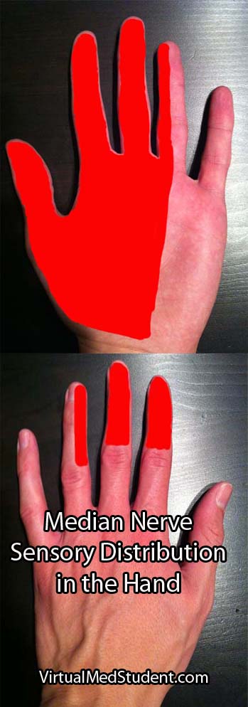
The other forearm branch travels into the hand where it supplies the abductor pollicis brevis (abductor pollicis longus is supplied by the radial nerve), flexor pollicis brevis, opponens pollicis, and the first and second lumbrical muscles. This branch also supplies the skin on the palmar side of the hand including the thumb, index, middle, and lateral half of the ring finger (see picture below).
For the most part the median nerve and its branches are involved in muscles that allow joints to flex, especially at the wrist and fingers.
| Muscles Innervated by the Median Nerve and its Branches | |
| Muscle | Action of Muscle |
| Pronator teres | Pronates the hand (ie: facing the palm towards the floor as if you were patting a dog) |
| Flexor carpi radialis | Flexion of the wrist |
| Flexor digitorum superficialis | Flexion of the proximal interphalangeal joints primarily (ie: flexion of the second knuckles of the fingers) |
| Palmaris longus | Variable, frequently not even present in some people |
| Flexor digitorum profundus (lateral half of the muscle) | Helps flex all the fingers joints (ie: as if you were squeezing something or making a fist) |
| Flexor pollicis longus | Helps flex the thumb |
| Pronator quadratus | Helps pronate the hand (as if you were "patting" a dog) |
| Opponens pollicis | Helps oppose the thumb (ie: bringing the thumb towards the little finger) |
| Lumbricals (1st and 2nd) | Help flex the metacarpophalangeal joints and extend the interphalangeal joints of the index and middle fingers |
| Abductor pollicis brevis | Helps abduct the thumb (ie: moving your thumb further from the palm) |
| Flexor pollicis brevis | Helps flex the thumb |
Importance in Disease

Patients with compression of the nerve by the pronator teres muscle have an inability to flex the index or middle fingers. This is due to dysfunction in the flexor digitorum profundus and first and second lumbricals. The ability to flex or oppose the thumb is also affected because of flexor pollicis longus and brevis dysfunction, as well as opponens pollicis muscle dysfunction. Abduction of the thumb is moderately affected because abductor pollicis brevis is affected; however, abductor pollicis longus is innervated by the radial nerve so some thumb abduction may be possible. The muscles affected above can cause the "hand of Benediction" sign when the patient is asked to make a fist. Finally, there is decreased sensation in the distribution of the median nerve in the hand (see image to right).
Compression of the anterior interosseous nerve can occur in the forearm. The flexor digitorum profundus and flexor pollicis longus muscles are affected, which causes a decreased ability to flex the thumb, index, and middle fingers. This causes an abnormal "pinch attitude" when the patient is asked to make an "OK" sign with their index finger and thumb. Sensation is normal.
The nerve can also get trapped in the carpal tunnel near the wrist. Patients typically have weakness in the abductor pollicis brevis, flexor pollicis brevis, opponens pollicis, and the first two lumbricals. This causes decreased thumb, index, and middle finger function. In addition, sensation is affected. Patients with carpal tunnel syndrome typically have pain, numbness, and tingling in their affected hand, which is worst at night. This is in contrast to pronator teres compression in which the sensation changes are not exacerbated at night.
Overview
The median nerve is one of the terminal branches of the brachial plexus. It sends branches to the main wrist and finger flexors, as well as many of the muscles that control thumb function. It provides sensation to most of the palmar side of the hand. It gives rise to three syndromes: pronator teres syndrome, anterior interosseous syndrome, and carpal tunnel syndrome depending on where along its course the nerve is affected.
Related Anatomy You Should Know About…
- An Overview of the Sciatic nerve
- Radial nerve and the Saturday Night Palsy
- Peroneal Nerve and the Dropped Foot
References and Resources
- Ducic I, Felder JM 3rd, Quadri HS. Common nerve decompressions of the upper extremity: reliable exposure using shorter incisions. Ann Plast Surg. 2012 Jun;68(6):606-9
- Colbert SH, Mackinnon SE. Nerve compressions in the upper extremity. Mo Med. 2008 Nov-Dec;105(6):527-35.
- Reddy MP. Peripheral nerve entrapment syndromes. Am Fam Physician. 1983 Nov;28(5):133-43.
- Calfee RP, Wilson JM, Wong AH. Variations in the anatomic relations of the posterior interosseous nerve associated with proximal forearm trauma. J Bone Joint Surg Am. 2011 Jan 5;93(1):81-90.
- Arle JE, Zager EL. Surgical treatment of common entrapment neuropathies in the upper limbs. Muscle Nerve. 2000 Aug;23(8):1160-74.