An Overview of the Sciatic Nerve

The sciatic nerve is a collection of nerve fibers that exit the spinal column between the 4th lumbar and 3rd sacral levels (L4-S3).

In the upper leg, the sciatic nerve gives off branches to the hamstring muscles. These muscles, which include the semimembranous, semitendinous, biceps femoris and part of the adductor magnus, are powerful flexors of the knee joint.

After sending off branches to the hamstrings, the sciatic nerve hits the back of the knee. At the popliteal fossa the sciatic splits into two distinct nerves: the common peroneal nerve and the tibial nerve.

The first nerve, the common peroneal (aka: common fibular), wraps around the outside of the knee and over the head of the fibula. It then branches into two separate nerves: the superficial peroneal and deep peroneal nerves.

The superficial peroneal nerve innervates two muscles: peroneus longus and brevis (aka: fibularis longus and brevis). These muscles allow you to evert (ie: allow you to lift your "pinky" toe higher than your "big" toe) and plantarflex (ie: help you step on the gas pedal) the foot. This nerve also provides the sensation to the outside half (ie: lateral half) of the lower leg, as well as the top of the foot.

The deep peroneal nerve innervates three muscles in the leg: tibialis anterior, extensor digitorum longus, and extensor hallucis longus. Tibialis anterior allows you to dorsiflex (ie: lift your foot off the ground) and invert (ie: bring your "big" toe higher than your "pinky" toe) your foot. Extensor digitorum longus helps you extend your toes (ie: the opposite of curling them), as well as evert, and dorsiflex your foot. The third muscle, extensor hallucis longus allows you to extend your big toe.

The deep peroneal nerve also innervates two muscles in the foot: extensor digitorum brevis (also helps to extend the toes) and extensor hallucis brevis (also helps to extend the big toe).

Peroneal Nerve and its Branches
Branch Muscles
Superficial peroneal – Peroneus longus
– Peroneus brevis
Deep peroneal
(leg branches)
– Tibialis anterior
– Extensor digitorum longus
– Extensor hallucis longus
Deep peroneal
(foot branches)
– Extensor digitorum brevis
– Extensor hallucis brevis

The other branch of the sciatic nerve, the tibial nerve, dives deep into the lower part of the leg where it acts on muscles in the calf and foot. In the calf it innervates the gastrocnemius (commonly called the "calf" muscle), soleus, popliteus, plantaris, flexor digitorum longus, flexor hallucis longus, and tibialis posterior. The gastrocnemius, soleus, and tibialis posterior are important plantarflexors of the foot (ie: allow you to "step on the gas pedal"). Flexor digitorum longus and flexor hallucis help flex (ie: curl) the toes.

The tibial nerve also sends branches into the foot. It further branches into the medial plantar and lateral plantar nerves, which innervate numerous muscles (see table below) in the foot itself.

The tibial nerve branches in the foot, namely the medial plantar and lateral plantar nerves also provide sensation to the sole of the foot.

Tibial Nerve and its Branches
Branch Muscles
Tibial nerve (leg branches) – Gastrocnemius
– Soleus
– Popliteus
– Plantaris
– Flexor digitorum longus
– Flexor hallucis longus
– Tibialis posterior

Tibial nerve (foot branches) -> medial plantar nerve

– Abductor hallucis
– Flexor digitorum brevis
– First lumbrical
Tibial nerve (foot branches) ->
lateral plantar nerve
– Flexor digiti minimi
– Adductor hallucis
– Interossei
– 2nd, 3rd, 4th lumbricals
– Abductor digiti minimi

Importance in Disease

The most well known problem of the sciatic nerve is a condition termed "sciatica". Sciatica is not a disease or disorder in itself, but rather a symptom of some underlying spine or nerve pathology. It is most often due to compression of one or more of the nerve roots that contribute axons to the sciatic nerve (L4-S3).

Compression of the nerve roots can be caused by many different pathologies, of which the most common is a herniated disc. Other causes include spinal stenosis and spondylolisthesis. Rarely, the nerve itself is compressed at some point as it travels down the leg.

A common symptom of sciatica is a severe and sharp pain that starts in the lower back and shoots down the buttock and back of the leg. If the involved nerve roots are severely compressed, weakness may also occur. This can cause a "foot drop" (ie: an inability to lift your foot off the ground).

The sciatic nerve may also get compressed as it passes through the piriformis muscle in the pelvis. When this occurs weakness of the hamstrings, lower leg, and foot muscles occurs; in addition, sensation of the outside of the lower leg, the calf, and the sole of the foot may also be affected.

Overview

The sciatic nerve is really two nerves that split at the level of the knee. The two main branches are the common peroneal (aka: common fibular) and tibial nerves, each of which branch again to innervate different muscles in the lower leg and foot. The most common pathologic problem with the sciatic nerve is a constellation of symptoms termed "sciatica". Sciatica is caused, most commonly, by compression of the nerve roots (L4-S3) in the spine that give rise to the sciatic nerve.

More Anatomy You Should Know…

References and Resources

The Median Nerve: Anatomy, Function, and Clinical Relevance

Radial Nerve Course
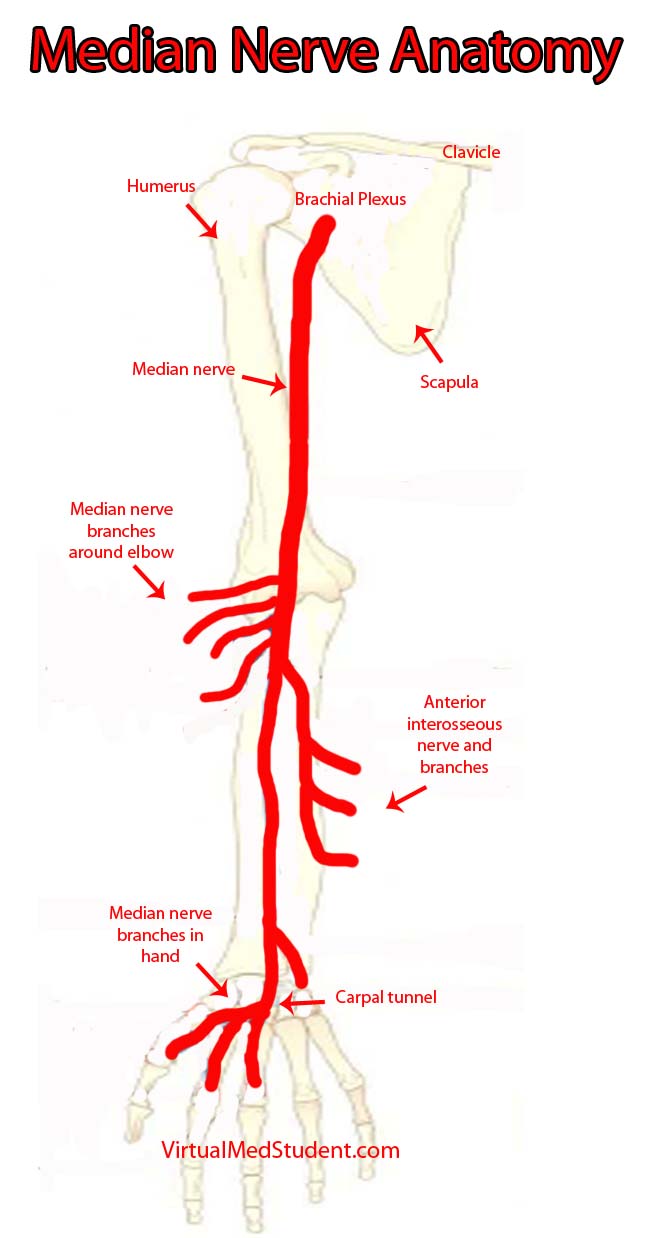
In order to appreciate the median nerve, we have to first understand the brachial plexus. The brachial plexus can be thought of as a massive system of highway intersections, in which numerous highways come together and then split apart.

The "highways" merging into the brachial plexus are the 5th, 6th, 7th, and 8th cervical nerve roots, as well as the 1st thoracic nerve root. These nerve roots mix together to form trunks, divisions, cords, and finally branches. The median nerve is one of the final branches of the brachial plexus. It is composed of fibers from the 6th, 7th and 8th cervical nerve roots, as well as the 1st thoracic nerve root.

After branching from the brachial plexus, the median nerve courses along the front aspect of the humerus in the upper arm. At the elbow it branches to supply the pronator teres, flexor carpi radialis, palmaris longus, and flexor digitorum superficialis muscles.

In the forearm the nerve divides into a branch called the anterior interosseous nerve, which supplies the flexor pollicis longus, flexor digitorum profundus (more specifically the lateral head, the medial being supplied by the ulnar nerve), and the pronator quadratus muscles.

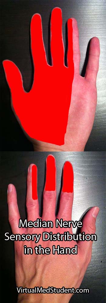
The other forearm branch travels into the hand where it supplies the abductor pollicis brevis (abductor pollicis longus is supplied by the radial nerve), flexor pollicis brevis, opponens pollicis, and the first and second lumbrical muscles. This branch also supplies the skin on the palmar side of the hand including the thumb, index, middle, and lateral half of the ring finger (see picture below).

For the most part the median nerve and its branches are involved in muscles that allow joints to flex, especially at the wrist and fingers.

Muscles Innervated by the Median Nerve and its Branches
Muscle Action of Muscle
Pronator teres Pronates the hand (ie: facing the palm towards the floor as if you were patting a dog)
Flexor carpi radialis Flexion of the wrist
Flexor digitorum superficialis Flexion of the proximal interphalangeal joints primarily (ie: flexion of the second knuckles of the fingers)
Palmaris longus Variable, frequently not even present in some people
Flexor digitorum profundus (lateral half of the muscle) Helps flex all the fingers joints (ie: as if you were squeezing something or making a fist)
Flexor pollicis longus Helps flex the thumb
Pronator quadratus Helps pronate the hand (as if you were "patting" a dog)
Opponens pollicis Helps oppose the thumb (ie: bringing the thumb towards the little finger)
Lumbricals (1st and 2nd) Help flex the metacarpophalangeal joints and extend the interphalangeal joints of the index and middle fingers
Abductor pollicis brevis Helps abduct the thumb (ie: moving your thumb further from the palm)
Flexor pollicis brevis Helps flex the thumb

Importance in Disease

Median Nerve Sensory Distribution in Hand
Damage to the median nerve occurs at one of three places along its course. It may be compressed by the two heads of the pronator teres muscle near the elbow, or at the wrist in the carpal tunnel. Additionally, the anterior interosseous nerve may become compressed in the forearm.

Patients with compression of the nerve by the pronator teres muscle have an inability to flex the index or middle fingers. This is due to dysfunction in the flexor digitorum profundus and first and second lumbricals. The ability to flex or oppose the thumb is also affected because of flexor pollicis longus and brevis dysfunction, as well as opponens pollicis muscle dysfunction. Abduction of the thumb is moderately affected because abductor pollicis brevis is affected; however, abductor pollicis longus is innervated by the radial nerve so some thumb abduction may be possible. The muscles affected above can cause the "hand of Benediction" sign when the patient is asked to make a fist. Finally, there is decreased sensation in the distribution of the median nerve in the hand (see image to right).

Compression of the anterior interosseous nerve can occur in the forearm. The flexor digitorum profundus and flexor pollicis longus muscles are affected, which causes a decreased ability to flex the thumb, index, and middle fingers. This causes an abnormal "pinch attitude" when the patient is asked to make an "OK" sign with their index finger and thumb. Sensation is normal.

The nerve can also get trapped in the carpal tunnel near the wrist. Patients typically have weakness in the abductor pollicis brevis, flexor pollicis brevis, opponens pollicis, and the first two lumbricals. This causes decreased thumb, index, and middle finger function. In addition, sensation is affected. Patients with carpal tunnel syndrome typically have pain, numbness, and tingling in their affected hand, which is worst at night. This is in contrast to pronator teres compression in which the sensation changes are not exacerbated at night.

Overview

The median nerve is one of the terminal branches of the brachial plexus. It sends branches to the main wrist and finger flexors, as well as many of the muscles that control thumb function. It provides sensation to most of the palmar side of the hand. It gives rise to three syndromes: pronator teres syndrome, anterior interosseous syndrome, and carpal tunnel syndrome depending on where along its course the nerve is affected.

Related Anatomy You Should Know About…

References and Resources

Radial Nerve and the Saturday Night Palsy

Radial Nerve Course
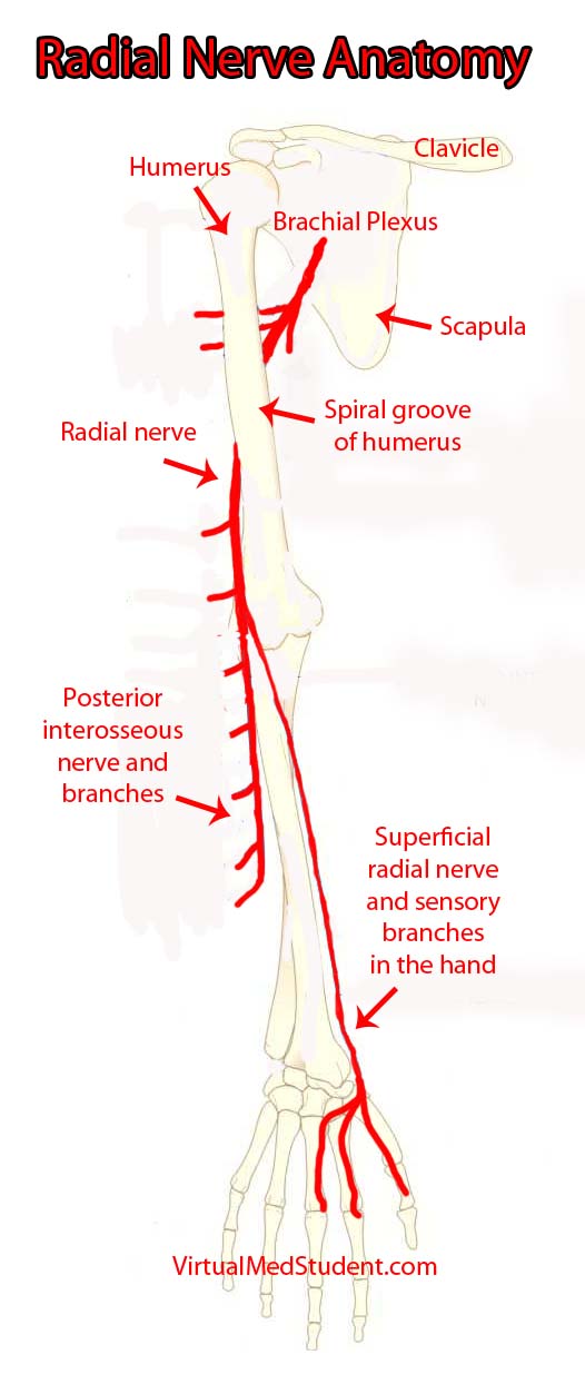
In order to appreciate the radial nerve, we have to first understand the brachial plexus. The brachial plexus can be thought of as a massive highway intersection, in which numerous highways come together and then split apart again.

The "highways" merging into the brachial plexus are the 5th, 6th, 7th, and 8th cervical nerve roots, as well as the 1st thoracic nerve root. These nerve "highways" tangle together to form trunks, divisions, cords, and then branches. The radial nerve is one of the branches of the brachial plexus; it gets its input from the 5th, 6th, 7th and 8th cervical nerve roots.

The radial nerve courses along the humerus in the upper arm. It wraps around the humerus in a spot called the spiral groove. Just before wrapping around the humerus, it sends a branch that innervates the triceps muscle (long, medial, and lateral heads) in the upper arm.

After wrapping around the spiral groove, it sends additional branches to the brachioradialis, extensor carpi radialis longus, and extensor carpi radialis brevis muscles.

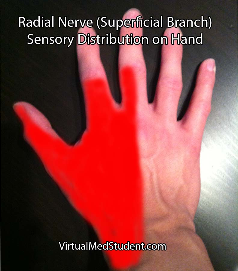
The first major branch in the forearm is known as the superficial radial nerve. This nerve courses along the medial/ulnar aspect of the forearm (ie: the ulnar or medial side of the forearm is closest to your body when your palms are facing forward) and heads straight for the hand. It relays sensory information from the lateral portion of the back of the hand.

The second major branch at the elbow can be thought of as the deep radial nerve, but it is formally known as the posterior interosseous nerve.

The posterior interosseus nerve sends branches to eight muscles in the forearm. They include the supinator (through which the nerve travels via a fibrous tunnel known as the arcade of Frohse), extensor digiti minimi, extensor carpi ulnaris, extensor digitorum, abductor pollicis longus, extensor pollicis longus, extensor pollicis brevis, and extensor indicis.

Muscles Innervated by the Radial Nerve and Its Branches
Muscle Action of Muscle
Triceps brachii – Extension of the forearm away from upper arm
Brachioradialis – Helps flex the forearm closer to the upper arm
– Helps supinate (ie: palm towards the sky)
– Helps pronate (ie: palm towards the floor)
Extensor carpi radialis
(longus and brevis)
– Helps extend the wrist
– Abducts the hand (ie: hand moves away from
the body when the palms are facing forward)
Extensor digiti minimi – Helps extend the little finger (5th digit)
Extensor carpi ulnaris – Helps extend the wrist
– Adducts the hand (ie: hand moves towards
the body when the palms are facing forward)
Supinator – Allows palms to face up towards the sky
Extensor digitorum – Helps with extension of the fingers
(specifically at the metacarpophalngeal joint)
Abductor pollicis longus – Helps abduct the thumb
Extensor pollicis
(longus and brevis)
– Help extend the thumb
Extensor indicis – Helps extend the index finger

Generally speaking, the radial nerve and its branches are involved in muscles that allow joints to extend (ie: widen or separate away from one another).

Importance in Disease

Damage to the radial nerve may take the form of compression or sheering injuries, typically after traumatic events.

The most common site of injury is at the spiral groove of the humerus. The nerve may be damaged if someone breaks their humerus, or if someone leans the back of their arm on something for an extended period of time (ie: a "Saturday night palsy" is the informal term given to a drunk who falls asleep with their arms draped over a chair… they end up waking up with a radial nerve palsy!).

Injury to the nerve at the spiral groove causes a wrist drop, in which the affected person cannot extend their wrist. The triceps are not affected because nerve branches to this muscle are proximal to the spiral groove. In addition, patients also complain of decreased sensation on the back of the hand (see image to right).

Radial Nerve Sensory Distribution in Hand
Additionally, the posterior interosseous branch of the radial nerve may get compressed as it passes through the supinator muscle in the forearm. This is referred to as posterior interosseous nerve syndrome.

The compression occurs at a fibrous portion of the supinator muscle known as the "arcade of Frohse", which causes an inability to extend the fingers at the metacarpophalangeal joints; this is due to dysfunction of the extensor digitorum muscle (extension at the interphalangeal – both distal and proximal – joints is controlled by the lumbricals and interossei muscles which are innervated by the median and ulnar nerves).

In posterior interosseous nerve syndrome it is still possible to extend the wrist because the branches of the nerve to the extensor carpi radialis muscle are unaffected (ie: they branch before the arcade). However, when the wrist is extended it deviates towards the radial side of the forearm because of the unopposed action of the extensor carpi radialis muscles (in other words, the extensor carpi ulnaris is affected and cannot keep the wrist extension neutral).

Sensation is entirely normal when the posterior interosseous nerve gets compressed because it contains no sensory fibers.

Overview

The radial nerve is a terminal branch of the brachial plexus. It sends branches to most of the extensor muscles of the arm and forearm. It also provides sensation to the back side of the hand. If injured it causes either a radial nerve palsy, or posterior interosseous syndrome, in which the affected patient has an inability to extend various joints at the wrist and/or fingers.

Related Anatomy You Should Know About…

References and Resources

  • Ducic I, Felder JM 3rd, Quadri HS. Common nerve decompressions of the upper extremity: reliable exposure using shorter incisions. Ann Plast Surg. 2012 Jun;68(6):606-9
  • Colbert SH, Mackinnon SE. Nerve compressions in the upper extremity. Mo Med. 2008 Nov-Dec;105(6):527-35.
  • Reddy MP. Peripheral nerve entrapment syndromes. Am Fam Physician. 1983 Nov;28(5):133-43.
  • Calfee RP, Wilson JM, Wong AH. Variations in the anatomic relations of the posterior interosseous nerve associated with proximal forearm trauma. J Bone Joint Surg Am. 2011 Jan 5;93(1):81-90.
  • Arle JE, Zager EL. Surgical treatment of common entrapment neuropathies in the upper limbs. Muscle Nerve. 2000 Aug;23(8):1160-74.