The Anterior Choroidal Artery: Small but Mighty

The anterior choroidal arteries are small, but vital blood vessels in the brain. They are branches of the internal carotid arteries. They arise proximal to the splitting of the internal carotid into the anterior and middle cerebral arteries.

The anterior choroidal arteries deliver blood to vital brain structures. These structures include the posterior limbs of the internal capsules, portions of the thalami, optic tracts, middle third of the cerebral peduncles, portions of the temporal lobes (ie: parts of the pyriform cortex, uncus, and amygdala), substantia nigra, portions of the globus pallidus, as well as the choroid plexus in the lateral ventricles.

The anterior choroidal artery forms connections (anastamoses) with the posterior lateral choroidal arteries. Cerebral angiograms are the best way to visualize the anterior choroidal arteries.

Importance in Disease

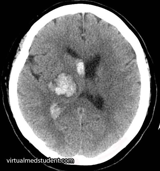
Blockage of the anterior choroidal artery can cause a stroke. The most common symptoms of an anterior choroidal stroke are hemiparesis (weakness on the opposite side of the body), hemianesthesia (decreased sensation on the opposite side of the body), and a homonymous hemianopsia (loss of a portion of the visual field of both eyes). High blood pressure is the most common underlying factor in people with anterior choroidal artery strokes.

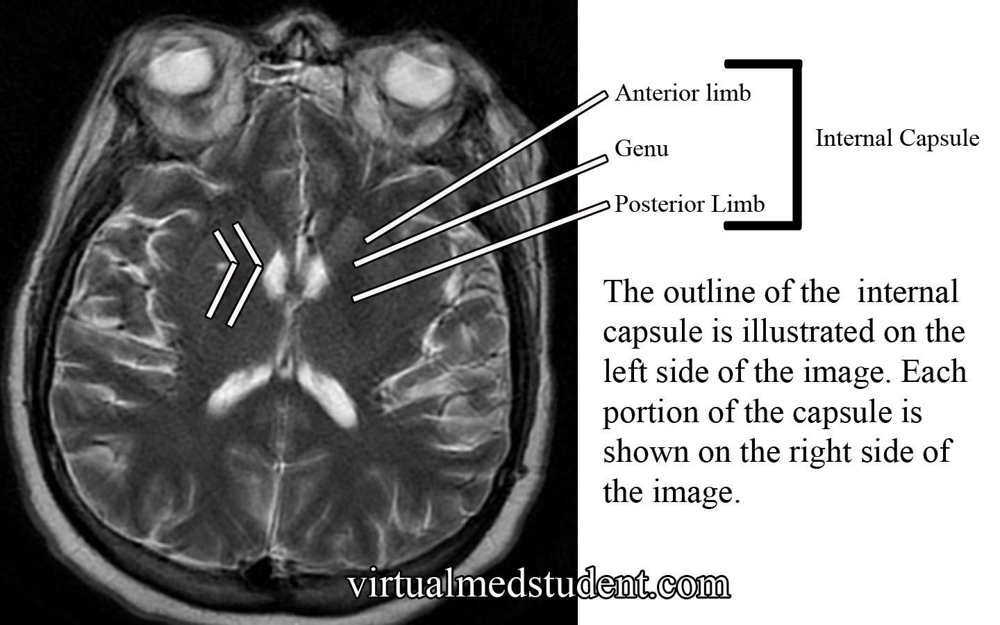
The hemiparesis is a result of damage to the posterior limb and genu of the internal capsule. The posterior limb contains the corticospinal tracts, which send information about movement from the brain to the spinal cord.

The hemianesthesia is a result of damage to the ventral posterolateral nucleus of the thalamus. This nucleus contains neurons that receive information from the spinal cord about sensation from the body. This symptom is less common than weakness, and occurs in roughly half of patients with an anterior choroidal stroke.

Cerebral angiogram showing anterior choroidal artery.

The final symptom, homonymous hemianopsia, is caused by damage to the optic tracts and lateral geniculate nucleus of the thalamus. Patients lose the ability to see objects on the left or right side (depending on which anterior choroidal artery is involved) in both eyes. This is an even more uncommon symptom, which occurs in less than 10% of patients with an anterior choroidal artery stroke.

Strokes of the anterior choroidal artery rarely cause all three symptoms. This is because the brain tissue served by the anterior choroidals also receives blood flow from other arteries.

Aneurysms of the anterior choroidal arteries are rare and will not be discussed in this article.

Overview

The anterior choroidal arteries are paired structures that arise from the internal carotid arteries. They supply blood to many important structures within the brain. Stroke is the most common pathological disease related to this blood vessel and frequently causes weakness of the opposite side of the body. High blood pressure is the most common underlying disease seen in people who have a stroke in this vascular distribution.

Other Stuff Worth Looking At…

References and Resources

  • Pezzella FR, Vadalà R. Anterior choroidal artery territory infarction. Front Neurol Neurosci. 2012;30:123-7. Epub 2012 Feb 14.
  • Bruno A, Graff-Radford NR, Biller J, et al. Anterior choroidal artery territory infarction: a small vessel disease. Stroke. 1989 May;20(5):616-9.
  • Baehr M, Frotscher M. Duus’ Topical Diagnosis in Neurology: Anatomy, Physiology, Signs, Symptoms. Fourth Edition. Stuttgart: Thieme, 2005.
  • Nolte J. The Human Brain: An Introduction to its Functional Anatomy. Sixth Edition. Philadelphia: Mosby, 2008.
  • Baskaya MK, Coscarella E, Gomez F, et al. Surgical and angiographic anatomy of the posterior communicating and anterior choroidal arteries. Neuroanatomy (2004) v3:38-42.

Denis’ Three Column Spine: A Simple Way to Think About Spinal Stability

The three column model of the spine was first introduced by Dr. Francis Denis in his aptly named paper, "The Three Column Spine and its Significance in the Classification of Acute Thoracolumbar Spinal Injuries". This paper, published in 1983, proposed a new biomechanical model for spinal stability that challenged Dr. Frank Holdsworth’s two column model from the 1960s. Although replaced by more modern grading scales (ie: the TLICS model) the three column spine is a simple way to think about spinal biomechanics.

Denis’ three column model proposes that the thoracolumbar spine can be divided into three columns. The first column includes the anterior longitudinal ligament (ALL) up to the first half of the bony vertebral body. The second column includes the second half (ie: more posterior half) of the vertebral body, up to, and including the posterior longitudinal ligament (PLL). The third column includes the pedicles, spinal cord/thecal sac, lamina, transverse processes, facet joints, spinous process, and the posterior ligaments (ie: supraspinous, interspinous, and ligamentum flavum).

The purpose of Denis’ model was to delineate which injuries to the thoracolumbar spine were considered "unstable". This delineation was important because it determined which patient’s required operative treatment of their spine.

Simply stated, an unstable spine was present if two or more of the columns were involved in the injury. However, it is important to note that every rule can be broken, so not all injuries follow this rule. Part of the "art" of practicing spine surgery is determining which two-column injuries can be left alone and managed non-operatively, and which truly require operative intervention.

Types of Injuries

Based on Denis’ model he classified specific types of injuries. Injuries to the anterior column only were called "compression fractures". Damage to the anterior and middle columns were known as "burst fractures". Injury to the middle and posterior column were known as "flexion-distraction" injuries, or more colloquially as "seat-belt type" injuries. Finally, damage to all three columns were classified as "fracture-dislocation" injuries.

Classification of Thoracolumbar Injuries Using the Three Column Model

Columns Name Stability
Anterior Compression fracture Stable
Anterior and Middle Burst fracture Unstable
Middle and Posterior Flexion-distraction injury (aka: seat-belt type) Unstable
Anterior, middle, and posterior Fracture-dislocation Unstable

Examples of Thoracolumbar Spine Fractures

Clinical Significance

In very simple terms, unstable fractures require definitive treatment. Treatment for unstable spine injuries is usually operative, although rigid immobilization with bracing is sometimes used in certain circumstances.

The treatment of each fracture type is beyond the scope of this article (and discussed in more detail elsewhere on this site), but suffice it to stay that for the most part (and again, every rule was made to be broken) two or three column injures = unstable = surgery.

Overview

Denis’ three column model of the spine revolutionized the way we think about spinal biomechanics and stability. He divided the spine into three columns. Although it has been largely replaced by more modern grading scales such as the thoracolumbar injury classification and severity score (TLICS) it is still a nice way to “think” about spinal integrity.

Other Common Spine Injuries…

References and Resources

The Internal Capsule: Some Pricey Brain Real Estate

The internal capsule is one pricey piece of brain real estate! It contains all of the pathways that allow information to be transferred between the cerebral cortex and the spinal cord, brainstem, and subcortical structures (ie: thalamus, basal ganglia). It is divided into an anterior limb, posterior limb, and genu (ie: the area where the anterior and posterior limbs meet).

The anterior limb contains axons that send information between the thalamus and the cingulate gyrus and pre-frontal cortex. It also contains axons in the frontopontine pathway (ie: axons going from the frontal cortex to a portion of the brainstem known as the pons).

The genu contains the corticobulbar tract, which originate in the motor areas of the frontal lobes and extend to the cranial nerve nuclei in the brainstem. It also contains axons that connect the motor section of the thalamus (ie: VA and VL nuclei) with the motor areas of the frontal cortex.

The posterior limb contains the corticospinal tract, which are axons that come from the motor area of the frontal cortex and travel all the way to the anterior horns of the spinal cord where α-motor neurons are located. The posterior limb also contains sensory information coming from the body via the medial lemniscus and the anterolateral (aka: spinothalamic tract) systems.

Internal Capsule MRI

The blood supply to most of the internal capsule comes from the lenticulostriate arteries. These small arteries originate from the first portion of the middle cerebral artery. Two other important arteries also supply portions of the internal capsule: the anterior choroidal artery and the recurrent artery of Heubner. The anterior choroidal artery is a branch of the internal carotid. It supplies the inferior portion of the posterior limb. The recurrent artery of Heubner is a branch of the anterior cerebral artery. It supplies the inferior portions of the anterior limb and the genu.

Anatomy of the Internal Capsule
Division Major Communication Tracts Blood Supply
Anterior limb

– Tracts between the frontal lobe and pons (brainstem)

– Tracts between the thalamus and prefrontal cortex

– Tracts between the thalamus and cingulate gyrus

– Lenticulostriate arteries (branches of the middle cerebral artery)

– Recurrent artery of Heubner (branch of the anterior cerebral artery)

Genu – Tracts between the motor cortex in the frontal lobe and the cranial nerve nuclei in the brainstem (aka: corticobulbar tract)

– Lenticulostriate arteries (branches of the middle cerebral artery)

– Recurrent artery of Heubner (branch of the anterior cerebral artery)
Posterior limb

– Tracts between the motor cortex of frontal lobe and anterior horn of spinal cord (aka: corticospinal tract)

– Medial lemniscus tract (a continuation of the dorsal columns), which carries information about light touch, vibration, and pressure sensation from the body and spinal cord.

– Anterolateral (aka: spinothalamic) tract, which carries pain and temperature information

– Lenticulostriate arteries (branches of the middle cerebral artery)

– Anterior choroidal artery (branch of the internal carotid)

Importance in Disease

Thalamic Hemorrhage
Thalamic intracerebral hematoma
compressing the posterior limb
of the internal capsule

Damage to the internal capsule can be devastating neurologically because it contains so many vital tracts.

For example, a stroke of the anterior choroidal artery can lead to posterior limb damage. This can cause paralysis of the contralateral (ie: opposite) arm and leg secondary to interruption of the corticospinal tract.

Posterior limb disruption can also cause co-existent sensory deficits including an inability to feel light touch, pain, and temperature due to damage of the spinothalamic and medial lemniscal pathways.

Hypertensive hemorrhages in the thalamus or basal ganglia can compress the adjacent fibers of the internal capsule leading to similar clinical findings. The head CT to the right shows a thalamic hemorrhage secondary to severely elevated blood pressure. The patient had compression of the posterior limb of the internal capsule. As a result she was unable to move her left arm and leg, and could not feel pain or light touch on the left side of her body.

Overview

The internal capsule is a vital structure. It contains many communication pathways between the brain’s cortex, brainstem, spinal cord, and subcortical nuclei (ie: thalamus, basal ganglia). Its blood supply comes from branches of the middle cerebral artery (ie: lenticulostriates), anterior cerebral artery (ie: recurrent artery of Heubner), and the internal carotid (ie: anterior choroidal artery). Lesions in this area caused by strokes or hypertensive hemorrhages can have devastating clinical consequences.

Other Pertinent Articles…

References and Resources

  • Greenberg MS. Handbook of Neurosurgery. 9th Edition. New York: Thieme, 2006. Chapter 25.
  • Chowdhury F, Haque M, Sarkar M, et al. White fiber dissection of brain; the internal capsule: a cadaveric study. Turk Neurosurg. 2010 Jul;20(3):314-22. doi: 10.5137/1019-5149.JTN.3052-10.2.
  • Simon RP, Aminoff MJ, Greenberg DA. Clinical Neurology, Seventh Edition (LANGE Clinical Medicine). Seventh Edition. New York: McGraw Hill, 2009.
  • Nolte J. The Human Brain: An Introduction to its Functional Anatomy. Sixth Edition. Philadelphia: Mosby, 2008.
  • Bickley LS, Szilagyi PG. Bates’ Guide to Physical Examination and History Taking. Ninth Edition. New York: Lippincott Williams and Wilkins, 2007.

Peroneal (Fibular) Nerve and the Dropped Foot

The peroneal nerve is one of the branches of the sciatic nerve. It receives most of its innervation from the L4, L5, and S1 nerves. The common peroneal nerve (aka: common fibular nerve) wraps around the outside of the knee over the head of the fibula. It then branches into two separate nerves: the superficial peroneal nerve and the deep peroneal nerve.

The superficial peroneal nerve innervates two muscles: peroneus longus and brevis (aka: fibularis longus and brevis). These muscles allow you to evert (ie: move your foot outwards) and plantarflex (ie: help you step on the gas pedal) the foot. This nerve also provides the sensation to the outside half (ie: lateral half) of the lower leg, as well as the top of most of the foot (ie: the dorsum of the foot).

The deep peroneal nerve innervates three muscles in the leg: tibialis anterior, extensor digitorum longus, and extensor hallucis longus. Tibialis anterior allows you to dorsiflex (ie: lift your foot off the ground) and invert (ie: bringing your big to closer to the middle of the body) your foot. Extensor digitorum longus helps you extend your toes (ie: the opposite of curling them), as well as evert, and dorsiflex your foot. The third muscle, extensor hallucis longus, allows you to extend your big toe.

The deep peroneal nerve also innervates two muscles in the foot: extensor digitorum brevis (also helps to extend the toes) and extensor hallucis brevis (also helps to extend the big toe).

Importance in Disease

The peroneal nerve is most frequently compressed over the fibular head. Compression typically affects the deep peroneal nerve rather than the common or superficial nerve; however, all of the nerves may be involved.

When the deep peroneal nerve is compressed, the foot is unable to dorsiflex secondary to dysfunction of the tibialis anterior muscle. Additionally, the patient is unable to extend the toes. Sensation may be decreased on a small patch of skin between the big toe and second toe; pain in the area of the lateral lower leg may also be present.

When the superficial peroneal nerve is compressed, the peroneus longus and brevis muscles are affected. Dysfunction of these muscles prevents the patient from everting their foot. Sensation over the lateral half of the lower leg and top of the foot may also be decreased.

Placing the nerve under passive, or active, stretch by placing the patient’s foot in inversion will often reproduce the symptoms. Percussing the nerve over the fibular head (Tinel’s test) can reproduce the symptoms.

It is important to distinguish a peroneal nerve palsy from a herniated L4-L5 disc. A patient with a herniated L4-L5 disc – causing an L5 radiculopathy – will not only have difficulty dorsiflexing the foot and toes (secondary to dysfunction of the L5 component of the peroneal nerve), but will also have difficulty inverting the foot (secondary to dysfunction of the L5 component of the tibial nerve).

Wait a second! This still shouldn’t make sense if you are actually thinking about it, and this is where things get tricky… Both the anterior tibialis (deep peroneal nerve innervated) and the posterior tibialis (tibial nerve innervated) help invert the foot. So how do we know that the weakness in inversion is related to an L5 disc herniation, a peroneal nerve palsy, or a tibial nerve palsy? You need to have the patient attempt to invert the foot while plantarflexing! The posterior tibialis is an inverter and plantarflexor of the foot so if dorsiflexion is weak (L5 –> deep peroneal nerve –> anterior tibialis) and the patient cannot invert (L5 –> tibial nerve –> posterior tibialis OR L5 –> deep peroneal nerve –> anterior tibialis) well while the foot is plantarflexed you probably have an L5 radiculopathy and not a peroneal nerve palsy.

To make it a bit easier clinically… A foot drop WITH inversion weakness is most likely an L5 radiculopathy (most commonly from a herniated disc) because you are getting the invertors for both the deep peroneal nerve (tibialis anterior muscle) AND the tibial nerve (tibialis posterior muscle), which both have get fibers from the L5 nerve root. However, injury to the sciatic nerve proximally in the leg could also give you this, but it would be associated with significant plantarflexion weakness too (due to gastroc weakness from S1)! So therefore a foot drop with eversion weakness and toe extension weakness is also suspicious for a peroneal nerve injury.

Overview

The peroneal nerve splits at the level of the knee from the sciatic nerve. It then further divides into the superficial and deep peroneal nerves. The superficial branch controls the evertors of the foot (peroneus longus and brevis) and provides sensation over the lateral aspect of the lower leg and top of the foot. The deep branch controls the dorsiflexors of the foot, and the extensor muscles of the toes. The common peroneal, and either of its branches, is most commonly compressed near the fibular head.

More Important Anatomy to Master…

References and Resources

  • Greenberg MS. Handbook of Neurosurgery. Sixth Edition. New York: Thieme, 2006. Chapter 25.
  • Prakash, Bhardwaj AK, Devi MN. Sciatic nerve division: a cadaver study in the Indian population and review of the literature. Singapore Med J. 2010 Sep;51(9):721-3.
  • Yuen EC, So YT. Sciatic neuropathy. Neurol Clin. 1999 Aug;17(3):617-31, viii.
  • Simon RP, Aminoff MJ, Greenberg DA. Clinical Neurology, Seventh Edition (LANGE Clinical Medicine). Seventh Edition. New York: McGraw Hill, 2009.
  • Netter FH. Atlas of Human Anatomy: with Student Consult Access (Netter Basic Science). Fifth Edition. Philadelphia: Saunders Elsevier, 2010.
  • Bickley LS, Szilagyi PG. Bates’ Guide to Physical Examination and History Taking. Ninth Edition. New York: Lippincott Williams and Wilkins, 2007.

An Overview of the Sciatic Nerve

The sciatic nerve is a collection of nerve fibers that exit the spinal column between the 4th lumbar and 3rd sacral levels (L4-S3).

In the upper leg, the sciatic nerve gives off branches to the hamstring muscles. These muscles, which include the semimembranous, semitendinous, biceps femoris and part of the adductor magnus, are powerful flexors of the knee joint.

After sending off branches to the hamstrings, the sciatic nerve hits the back of the knee. At the popliteal fossa the sciatic splits into two distinct nerves: the common peroneal nerve and the tibial nerve.

The first nerve, the common peroneal (aka: common fibular), wraps around the outside of the knee and over the head of the fibula. It then branches into two separate nerves: the superficial peroneal and deep peroneal nerves.

The superficial peroneal nerve innervates two muscles: peroneus longus and brevis (aka: fibularis longus and brevis). These muscles allow you to evert (ie: allow you to lift your "pinky" toe higher than your "big" toe) and plantarflex (ie: help you step on the gas pedal) the foot. This nerve also provides the sensation to the outside half (ie: lateral half) of the lower leg, as well as the top of the foot.

The deep peroneal nerve innervates three muscles in the leg: tibialis anterior, extensor digitorum longus, and extensor hallucis longus. Tibialis anterior allows you to dorsiflex (ie: lift your foot off the ground) and invert (ie: bring your "big" toe higher than your "pinky" toe) your foot. Extensor digitorum longus helps you extend your toes (ie: the opposite of curling them), as well as evert, and dorsiflex your foot. The third muscle, extensor hallucis longus allows you to extend your big toe.

The deep peroneal nerve also innervates two muscles in the foot: extensor digitorum brevis (also helps to extend the toes) and extensor hallucis brevis (also helps to extend the big toe).

Peroneal Nerve and its Branches
Branch Muscles
Superficial peroneal – Peroneus longus
– Peroneus brevis
Deep peroneal
(leg branches)
– Tibialis anterior
– Extensor digitorum longus
– Extensor hallucis longus
Deep peroneal
(foot branches)
– Extensor digitorum brevis
– Extensor hallucis brevis

The other branch of the sciatic nerve, the tibial nerve, dives deep into the lower part of the leg where it acts on muscles in the calf and foot. In the calf it innervates the gastrocnemius (commonly called the "calf" muscle), soleus, popliteus, plantaris, flexor digitorum longus, flexor hallucis longus, and tibialis posterior. The gastrocnemius, soleus, and tibialis posterior are important plantarflexors of the foot (ie: allow you to "step on the gas pedal"). Flexor digitorum longus and flexor hallucis help flex (ie: curl) the toes.

The tibial nerve also sends branches into the foot. It further branches into the medial plantar and lateral plantar nerves, which innervate numerous muscles (see table below) in the foot itself.

The tibial nerve branches in the foot, namely the medial plantar and lateral plantar nerves also provide sensation to the sole of the foot.

Tibial Nerve and its Branches
Branch Muscles
Tibial nerve (leg branches) – Gastrocnemius
– Soleus
– Popliteus
– Plantaris
– Flexor digitorum longus
– Flexor hallucis longus
– Tibialis posterior

Tibial nerve (foot branches) -> medial plantar nerve

– Abductor hallucis
– Flexor digitorum brevis
– First lumbrical
Tibial nerve (foot branches) ->
lateral plantar nerve
– Flexor digiti minimi
– Adductor hallucis
– Interossei
– 2nd, 3rd, 4th lumbricals
– Abductor digiti minimi

Importance in Disease

The most well known problem of the sciatic nerve is a condition termed "sciatica". Sciatica is not a disease or disorder in itself, but rather a symptom of some underlying spine or nerve pathology. It is most often due to compression of one or more of the nerve roots that contribute axons to the sciatic nerve (L4-S3).

Compression of the nerve roots can be caused by many different pathologies, of which the most common is a herniated disc. Other causes include spinal stenosis and spondylolisthesis. Rarely, the nerve itself is compressed at some point as it travels down the leg.

A common symptom of sciatica is a severe and sharp pain that starts in the lower back and shoots down the buttock and back of the leg. If the involved nerve roots are severely compressed, weakness may also occur. This can cause a "foot drop" (ie: an inability to lift your foot off the ground).

The sciatic nerve may also get compressed as it passes through the piriformis muscle in the pelvis. When this occurs weakness of the hamstrings, lower leg, and foot muscles occurs; in addition, sensation of the outside of the lower leg, the calf, and the sole of the foot may also be affected.

Overview

The sciatic nerve is really two nerves that split at the level of the knee. The two main branches are the common peroneal (aka: common fibular) and tibial nerves, each of which branch again to innervate different muscles in the lower leg and foot. The most common pathologic problem with the sciatic nerve is a constellation of symptoms termed "sciatica". Sciatica is caused, most commonly, by compression of the nerve roots (L4-S3) in the spine that give rise to the sciatic nerve.

More Anatomy You Should Know…

References and Resources

The Median Nerve: Anatomy, Function, and Clinical Relevance

Radial Nerve Course
In order to appreciate the median nerve, we have to first understand the brachial plexus. The brachial plexus can be thought of as a massive system of highway intersections, in which numerous highways come together and then split apart.

The "highways" merging into the brachial plexus are the 5th, 6th, 7th, and 8th cervical nerve roots, as well as the 1st thoracic nerve root. These nerve roots mix together to form trunks, divisions, cords, and finally branches. The median nerve is one of the final branches of the brachial plexus. It is composed of fibers from the 6th, 7th and 8th cervical nerve roots, as well as the 1st thoracic nerve root.

After branching from the brachial plexus, the median nerve courses along the front aspect of the humerus in the upper arm. At the elbow it branches to supply the pronator teres, flexor carpi radialis, palmaris longus, and flexor digitorum superficialis muscles.

In the forearm the nerve divides into a branch called the anterior interosseous nerve, which supplies the flexor pollicis longus, flexor digitorum profundus (more specifically the lateral head, the medial being supplied by the ulnar nerve), and the pronator quadratus muscles.

The other forearm branch travels into the hand where it supplies the abductor pollicis brevis (abductor pollicis longus is supplied by the radial nerve), flexor pollicis brevis, opponens pollicis, and the first and second lumbrical muscles. This branch also supplies the skin on the palmar side of the hand including the thumb, index, middle, and lateral half of the ring finger (see picture below).

For the most part the median nerve and its branches are involved in muscles that allow joints to flex, especially at the wrist and fingers.

Muscles Innervated by the Median Nerve and its Branches
Muscle Action of Muscle
Pronator teres Pronates the hand (ie: facing the palm towards the floor as if you were patting a dog)
Flexor carpi radialis Flexion of the wrist
Flexor digitorum superficialis Flexion of the proximal interphalangeal joints primarily (ie: flexion of the second knuckles of the fingers)
Palmaris longus Variable, frequently not even present in some people
Flexor digitorum profundus (lateral half of the muscle) Helps flex all the fingers joints (ie: as if you were squeezing something or making a fist)
Flexor pollicis longus Helps flex the thumb
Pronator quadratus Helps pronate the hand (as if you were "patting" a dog)
Opponens pollicis Helps oppose the thumb (ie: bringing the thumb towards the little finger)
Lumbricals (1st and 2nd) Help flex the metacarpophalangeal joints and extend the interphalangeal joints of the index and middle fingers
Abductor pollicis brevis Helps abduct the thumb (ie: moving your thumb further from the palm)
Flexor pollicis brevis Helps flex the thumb

Importance in Disease

Median Nerve Sensory Distribution in Hand
Damage to the median nerve occurs at one of three places along its course. It may be compressed by the two heads of the pronator teres muscle near the elbow, or at the wrist in the carpal tunnel. Additionally, the anterior interosseous nerve may become compressed in the forearm.

Patients with compression of the nerve by the pronator teres muscle have an inability to flex the index or middle fingers. This is due to dysfunction in the flexor digitorum profundus and first and second lumbricals. The ability to flex or oppose the thumb is also affected because of flexor pollicis longus and brevis dysfunction, as well as opponens pollicis muscle dysfunction. Abduction of the thumb is moderately affected because abductor pollicis brevis is affected; however, abductor pollicis longus is innervated by the radial nerve so some thumb abduction may be possible. The muscles affected above can cause the "hand of Benediction" sign when the patient is asked to make a fist. Finally, there is decreased sensation in the distribution of the median nerve in the hand (see image to right).

Compression of the anterior interosseous nerve can occur in the forearm. The flexor digitorum profundus and flexor pollicis longus muscles are affected, which causes a decreased ability to flex the thumb, index, and middle fingers. This causes an abnormal "pinch attitude" when the patient is asked to make an "OK" sign with their index finger and thumb. Sensation is normal.

The nerve can also get trapped in the carpal tunnel near the wrist. Patients typically have weakness in the abductor pollicis brevis, flexor pollicis brevis, opponens pollicis, and the first two lumbricals. This causes decreased thumb, index, and middle finger function. In addition, sensation is affected. Patients with carpal tunnel syndrome typically have pain, numbness, and tingling in their affected hand, which is worst at night. This is in contrast to pronator teres compression in which the sensation changes are not exacerbated at night.

Overview

The median nerve is one of the terminal branches of the brachial plexus. It sends branches to the main wrist and finger flexors, as well as many of the muscles that control thumb function. It provides sensation to most of the palmar side of the hand. It gives rise to three syndromes: pronator teres syndrome, anterior interosseous syndrome, and carpal tunnel syndrome depending on where along its course the nerve is affected.

Related Anatomy You Should Know About…

References and Resources

Radial Nerve and the Saturday Night Palsy

Radial Nerve Course
In order to appreciate the radial nerve, we have to first understand the brachial plexus. The brachial plexus can be thought of as a massive highway intersection, in which numerous highways come together and then split apart again.

The "highways" merging into the brachial plexus are the 5th, 6th, 7th, and 8th cervical nerve roots, as well as the 1st thoracic nerve root. These nerve "highways" tangle together to form trunks, divisions, cords, and then branches. The radial nerve is one of the branches of the brachial plexus; it gets its input from the 5th, 6th, 7th and 8th cervical nerve roots.

The radial nerve courses along the humerus in the upper arm. It wraps around the humerus in a spot called the spiral groove. Just before wrapping around the humerus, it sends a branch that innervates the triceps muscle (long, medial, and lateral heads) in the upper arm.

After wrapping around the spiral groove, it sends additional branches to the brachioradialis, extensor carpi radialis longus, and extensor carpi radialis brevis muscles.

The first major branch in the forearm is known as the superficial radial nerve. This nerve courses along the medial/ulnar aspect of the forearm (ie: the ulnar or medial side of the forearm is closest to your body when your palms are facing forward) and heads straight for the hand. It relays sensory information from the lateral portion of the back of the hand.

The second major branch at the elbow can be thought of as the deep radial nerve, but it is formally known as the posterior interosseous nerve.

The posterior interosseus nerve sends branches to eight muscles in the forearm. They include the supinator (through which the nerve travels via a fibrous tunnel known as the arcade of Frohse), extensor digiti minimi, extensor carpi ulnaris, extensor digitorum, abductor pollicis longus, extensor pollicis longus, extensor pollicis brevis, and extensor indicis.

Muscles Innervated by the Radial Nerve and Its Branches
Muscle Action of Muscle
Triceps brachii – Extension of the forearm away from upper arm
Brachioradialis – Helps flex the forearm closer to the upper arm
– Helps supinate (ie: palm towards the sky)
– Helps pronate (ie: palm towards the floor)
Extensor carpi radialis
(longus and brevis)
– Helps extend the wrist
– Abducts the hand (ie: hand moves away from
the body when the palms are facing forward)
Extensor digiti minimi – Helps extend the little finger (5th digit)
Extensor carpi ulnaris – Helps extend the wrist
– Adducts the hand (ie: hand moves towards
the body when the palms are facing forward)
Supinator – Allows palms to face up towards the sky
Extensor digitorum – Helps with extension of the fingers
(specifically at the metacarpophalngeal joint)
Abductor pollicis longus – Helps abduct the thumb
Extensor pollicis
(longus and brevis)
– Help extend the thumb
Extensor indicis – Helps extend the index finger

Generally speaking, the radial nerve and its branches are involved in muscles that allow joints to extend (ie: widen or separate away from one another).

Importance in Disease

Damage to the radial nerve may take the form of compression or sheering injuries, typically after traumatic events.

The most common site of injury is at the spiral groove of the humerus. The nerve may be damaged if someone breaks their humerus, or if someone leans the back of their arm on something for an extended period of time (ie: a "Saturday night palsy" is the informal term given to a drunk who falls asleep with their arms draped over a chair… they end up waking up with a radial nerve palsy!).

Injury to the nerve at the spiral groove causes a wrist drop, in which the affected person cannot extend their wrist. The triceps are not affected because nerve branches to this muscle are proximal to the spiral groove. In addition, patients also complain of decreased sensation on the back of the hand (see image to right).

Radial Nerve Sensory Distribution in Hand
Additionally, the posterior interosseous branch of the radial nerve may get compressed as it passes through the supinator muscle in the forearm. This is referred to as posterior interosseous nerve syndrome.

The compression occurs at a fibrous portion of the supinator muscle known as the "arcade of Frohse", which causes an inability to extend the fingers at the metacarpophalangeal joints; this is due to dysfunction of the extensor digitorum muscle (extension at the interphalangeal – both distal and proximal – joints is controlled by the lumbricals and interossei muscles which are innervated by the median and ulnar nerves).

In posterior interosseous nerve syndrome it is still possible to extend the wrist because the branches of the nerve to the extensor carpi radialis muscle are unaffected (ie: they branch before the arcade). However, when the wrist is extended it deviates towards the radial side of the forearm because of the unopposed action of the extensor carpi radialis muscles (in other words, the extensor carpi ulnaris is affected and cannot keep the wrist extension neutral).

Sensation is entirely normal when the posterior interosseous nerve gets compressed because it contains no sensory fibers.

Overview

The radial nerve is a terminal branch of the brachial plexus. It sends branches to most of the extensor muscles of the arm and forearm. It also provides sensation to the back side of the hand. If injured it causes either a radial nerve palsy, or posterior interosseous syndrome, in which the affected patient has an inability to extend various joints at the wrist and/or fingers.

Related Anatomy You Should Know About…

References and Resources

  • Ducic I, Felder JM 3rd, Quadri HS. Common nerve decompressions of the upper extremity: reliable exposure using shorter incisions. Ann Plast Surg. 2012 Jun;68(6):606-9
  • Colbert SH, Mackinnon SE. Nerve compressions in the upper extremity. Mo Med. 2008 Nov-Dec;105(6):527-35.
  • Reddy MP. Peripheral nerve entrapment syndromes. Am Fam Physician. 1983 Nov;28(5):133-43.
  • Calfee RP, Wilson JM, Wong AH. Variations in the anatomic relations of the posterior interosseous nerve associated with proximal forearm trauma. J Bone Joint Surg Am. 2011 Jan 5;93(1):81-90.
  • Arle JE, Zager EL. Surgical treatment of common entrapment neuropathies in the upper limbs. Muscle Nerve. 2000 Aug;23(8):1160-74.