Pyogenic Liver Abscesses: Pus, Needle Drainage, and Antibiotics

Pyogenic liver abscesses are localized collections of pus and bacteria. The initial infection occurs when bacteria travel through the portal vein. This most commonly occurs after bowel contents leak into the peritoneal cavity from the gut. Other causes of liver abscesses include direct spread of infection from the bile duct system, or from other bacterial infections of the blood. Rarely, penetrating injuries (gunshots, stab wounds, surgery) may directly introduce infection. There are numerous “bugs” that can cause pyogenic liver abscesses. They include, but are not limited to, streptococcus species, klebsiella pneumoniae, and staphylococcus species.

Signs and Symptoms

Similar to other infections, liver abscesses can cause fevers, chills, decreased appetite, abdominal pain, and a generalized sense of not feeling well (ie: malaise). Interestingly, hiccups may also be present if the abscesses are causing adjacent inflammation/irritation of the diaphragm.

Diagnosis

Pyogenic Liver Abscesses
The diagnosis of pyogenic liver abscesses is made with imaging. The most commonly used method is a CT scan of the abdomen with and without contrast. If present, a liver abscess will look like a collection of fluid, with or without septated dividers.

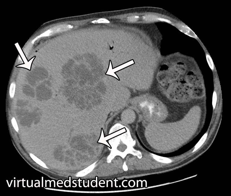
Another commonly employed imaging modality is the use of ultrasound to detect the fluid filled pockets within the liver.

However, it is important to note that imaging studies alone cannot distinguish between the different types of liver abscesses. Imaging can also not tell you what bacteria is responsible for the abscess.

Treatment

Pyogenic abscesses must be drained and treated with antibiotics. Drainage is usually done under image guidance using a needle placed through the skin into the abscess. Although occasionally direct surgical evacuation of the abscess is necessary.

Draining the abscess is important for two reasons. First, it decreases the size of the abscess allowing antibiotic therapy to work more effectively. And secondly, it provides abscess fluid that can be sent to the lab for bacterial culture and gram stain.

The results of the culture help guide subsequent antibiotic therapy. Commonly used antibiotics include piperacillin-tazobactam (Zosyn®), vancomycin, metronidazole (Flagyl®), and ceftriaxone; once cultures confirm the causative bug antibiotic therapy can be narrowed.

Overview

Pyogenic liver abscesses are collections of pus and bacteria. They occur most commonly after the spilling of gut bacteria into the peritoneal space (ie: peritonitis). There are numerous causative bacteria. Symptoms include fever, chills, decreased appetite, and abdominal discomfort. Treatment is with drainage and antibiotics.

References and Resources

  • Hasper D, Schefold JC, Baumgart DC. Management of severe abdominal infections. Recent Pat Antiinfect Drug Discov. 2009 Jan;4(1):57-65.
  • Benedetti NJ, Desser TS, Jeffrey RB. Imaging of hepatic infections. Ultrasound Q. 2008 Dec;24(4):267-78.
  • Mortelé KJ, Segatto E, Ros PR. The infected liver: radiologic-pathologic correlation. Radiographics. 2004 Jul-Aug;24(4):937-55.
  • Kurland JE, Brann OS. Pyogenic and amebic liver abscesses. Curr Gastroenterol Rep. 2004 Aug;6(4):273-9.

Giardia Lamblia (You Better Have a Toilet Nearby)

This little bastard to the left is called giardia lamblia and it is a single celled protozoan. It exits in two forms: trophozoite and cyst. The cyst form is highly resistant to destruction and can survive for weeks outside a host.

The life cycle of giardia is relatively simple. First, cysts are pooped out by a host (ie: animal or human) where they contaminate food and water supplies. They are then unknowingly ingested by a host. Once in the gut of the host, the cyst transforms into the trophozoite form. Trophozoites undergo asexual reproduction in the gut; as they reach the colon they re-encyst themselves. From there they are pooped out again to reinfect another unfortunate soul.

The trophozoite form has two nuclei (the two "eyes" in the photo to the right) and four pairs of flagella that allow it to move. The organism attaches itself to intestinal cells. How it causes symptoms is not entirely known. One possibility is that it causes inflammation of the intestinal cells resulting in a decreased ability to absorb nutrients from food.

Signs and Symptoms

Interestingly, most people infected by giardia lamblia are asymptomatic. However, in some individuals a foul smelling diarrhea results. The foul smell is due to steatorrhea (ie: undigested fat molecules). Many symptomatic people will also have belly pain, fatigue, flatulence, nausea, and possibly vomiting. All of these symptoms can give rise to weight loss.

Symptoms generally begin seven days after exposure. They generally last anywhere from two to four weeks with or without treatment. However, in a subset of patients who are untreated, symptoms may continue for months to years!

Diagnosis

Diagnosis is made most commonly by looking at the stool for proteins made by the organism (ie: giardia antigens). Also by looking at a stool sample under a microscope it is possible to directly see the trophozoites and cysts.

Treatment

Treatment is with an antibiotic known as metronidazole. This antibiotic is taken up by anaerobic bacteria and some protozoans where it is converted into toxic by products. These toxic metabolites damage DNA making the organism unable to divide; eventually, the damage causes cell death.

Overview

Giardia lamblia is a protozoan parasite that exists in two forms: trophozoite and cyst. The cyst is defecated and then ingested by another host. It turns into the trophozoite form and adheres to intestinal cells causing a foul smelling diarrhea in some individuals. Many people remain asymptomatic. Treatment is with an antibiotic known as metronidazole.

References and Resources