Enterobius Vermicularis and Itchy Butts

Enterobius vermicularis, better known as pinworm, is a nematode or roundworm. Like other worms it has a unique lifecycle that is quite interesting, albeit somewhat disgusting! It begins when an egg is ingested by a human. Eggs are usually ingested because of poor hygiene (ie: not washing your hands after doing number 2) and can sometimes be found in contaminated food. Once ingested the eggs hatch in the small intestine. From there the worms migrate to the large intestine where they mate. For unknown reasons the pregnant females head towards the anus at night where they lay their eggs.

The eggs are then shed in the feces and potentially picked up by another unlucky host. It is the most common worm related infection in the United States.

Signs and Symptoms

The eggs in the perianal area are extremely pruritic (ie: itchy). Scratching of the anus secondary to the intense pruritis can lead to skin breakdown and potential bacterial super infection. The symptoms are generally most severe at night because this is when the females migrate to the anus to lay their eggs. Systemic signs are generally not present although some patients can have malaise (ie: feeling "crappy").

Diagnosis

The traditional way of diagnosing is the "scotch tape" test. The clinician takes a piece of scotch tape and applies it to the patient’s perianal region. Eggs can be seen on the tape once its removed. The best time to perform this test is early in the morning before bathing or at night.

Treatment

Two anti-helminth medications are used to treat pinworm. The first is albendazole. Generally a single dose is enough to kill all worms, but since eggs may be present on clothing, bedding, etc. a second dose is often given two weeks later. Mebendazole is another medication that is also given as a single dose repeated two weeks later. These medications work by inhibiting microtubule polymerization in the cytoplasm of the worm’s cells.

Overview

Enterobius vermicularis (pinworm) is the most common worm infection in the United States. It causes an intense itch in the perianal area that is worst at night. It is diagnosed by the "scotch tape" test. Treatment is with albendazole or mebendazole.

Related Articles

References and Resources

Tuberculous Meningitis: Basal Cisterns, Strokes, Hydrocephalus

Tuberculosis is one of the most common infectious diseases in the world. It is caused by a bacteria of the genus mycobacterium. Tuberculosis usually infects the lungs, but may also infect the lymph nodes, vertebral bodies, kidneys, gastrointestinal system, or central nervous system.

Central nervous system disease comes in two flavors: a focal abscess-like lesion known as a tuberculoma or tuberculosis meningitis. The remainder of this article will focus on tuberculosis meningitis, which is an uncommon (although not rare!) form of extra-pulmonary tuberculosis.

Let’s start by discussing how the bacteria get into the central nervous system. After being inhaled the bacteria infect cells known as macrophages. The infected macrophages move towards lymph nodes, and eventually end up in the blood stream. Once in the blood stream, the mycobacterium-infected macrophages can travel anywhere in the body!

One spot the bacterium hitch a ride to is the lining of the brain (aka: the meninges). Collections of mycobacterium-laden macrophages (“Rich foci”) can rupture into the subarachnoid space causing an inflammatory reaction (ie: a "meningitis", or inflammation of the meninges).

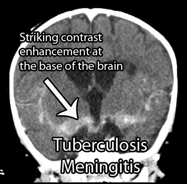
For unclear reasons, the inflammation seen in tuberculosis meningitis preferentially affects the basal cisterns and base of the brain. Autopsy specimens show a gelatinous material coating the undersurface of the brain. The inflammatory reaction is what causes the signs and symptoms of tuberculosis meningitis.

Signs and Symptoms

Tuberculosis meningitis can present in a number of different ways. Many patients present with days to months of non-specific symptoms such as headache, lethargy, nausea, and vomiting.

Cranial neuropathies are commonly seen, especially since tuberculosis meningitis affects the basal cisterns and base of the brain, which is where many of the cranial nerves run.

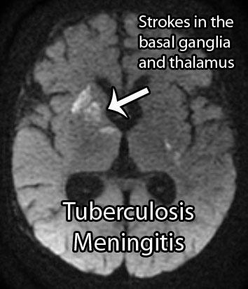
Additionally, up to 40% of patients may present with stroke. Strokes occur because the inflammation can "eat up" the linings of small blood vessels. The basal ganglia, internal capsule, and thalamus are the most common locations where strokes occur in tuberculosis meningitis.

Diagnosis

The clinical history is extremely important. Tuberculosis meningitis may affect both immunocompetent and immunocompromised (ie: think HIV/AIDs, diabetics, people on immunosuppressives, etc.) people. If the clinical history and physical exam findings are concerning for meningitis, than confirmatory studies should be performed.

TB Meningitis MRI
TB Meningitis CT
Cerebrospinal fluid (CSF) analysis shows elevated opening pressures, increased cellularity with inflammatory cells like neutrophils (earlier) and lymphocytes (later), an increased amount of protein, and a decreased amount of glucose. An acid-fast stain of the CSF to look for bacteria is sometimes positive. Culturing the CSF for mycobacterium is routinely done, but results can take weeks to months, and therefore is not useful in deciding whether or not to start treatment. Polymerase chain reactions (PCR) to look for mycobacterium DNA are also commonly used, and are much quicker than culturing.

Appropriate imaging studies include CT or MRI scans with contrast. The basal cisterns and base of the brain will "light up" with contrast because of inflammation. Imaging may also show strokes and hydrocephalus. Under the right clinical scenario imaging studies can help support the diagnosis, but are not specific.

Treatment

Treatment should consist of antibiotics that target mycobacterium tuberculosis. Commonly used antibiotics include rifampin, isoniazid, streptomycin, and pyrazinamide. Other antibiotics may be necessary if the strain of bacteria is resistant to these drugs.

Steroids are also frequently given to help reduce inflammation. Dexamethasone, a commonly used steroid, has been shown to improve survival rates (although it may not affect outcome in those that survive).

The intense inflammatory reaction seen in tuberculosis meningitis may gunk up the re-absorption of cerebrospinal fluid and cause a communicating hydrocephalus. Hydrocephalus occurs in 70% of cases and may require surgically inserted shunts to fix.

Overview

Tuberculosis meningitis is a devastating manifestation of a common infectious disease. It can cause cranial neuropathies, strokes, and hydrocephalus. Prompt diagnosis is mandatory and is made from the clinical history, CSF analysis, and imaging studies. Treatment is with anti-TB medications and steroids. Many survivors have long term disabilities despite appropriate treatment.

Related Articles

References and Resources

Giardia Lamblia (You Better Have a Toilet Nearby)

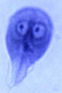
This little bastard to the left is called giardia lamblia and it is a single celled protozoan. It exits in two forms: trophozoite and cyst. The cyst form is highly resistant to destruction and can survive for weeks outside a host.

The life cycle of giardia is relatively simple. First, cysts are pooped out by a host (ie: animal or human) where they contaminate food and water supplies. They are then unknowingly ingested by a host. Once in the gut of the host, the cyst transforms into the trophozoite form. Trophozoites undergo asexual reproduction in the gut; as they reach the colon they re-encyst themselves. From there they are pooped out again to reinfect another unfortunate soul.

The trophozoite form has two nuclei (the two "eyes" in the photo to the right) and four pairs of flagella that allow it to move. The organism attaches itself to intestinal cells. How it causes symptoms is not entirely known. One possibility is that it causes inflammation of the intestinal cells resulting in a decreased ability to absorb nutrients from food.

Signs and Symptoms

Interestingly, most people infected by giardia lamblia are asymptomatic. However, in some individuals a foul smelling diarrhea results. The foul smell is due to steatorrhea (ie: undigested fat molecules). Many symptomatic people will also have belly pain, fatigue, flatulence, nausea, and possibly vomiting. All of these symptoms can give rise to weight loss.

Symptoms generally begin seven days after exposure. They generally last anywhere from two to four weeks with or without treatment. However, in a subset of patients who are untreated, symptoms may continue for months to years!

Diagnosis

Diagnosis is made most commonly by looking at the stool for proteins made by the organism (ie: giardia antigens). Also by looking at a stool sample under a microscope it is possible to directly see the trophozoites and cysts.

Treatment

Treatment is with an antibiotic known as metronidazole. This antibiotic is taken up by anaerobic bacteria and some protozoans where it is converted into toxic by products. These toxic metabolites damage DNA making the organism unable to divide; eventually, the damage causes cell death.

Overview

Giardia lamblia is a protozoan parasite that exists in two forms: trophozoite and cyst. The cyst is defecated and then ingested by another host. It turns into the trophozoite form and adheres to intestinal cells causing a foul smelling diarrhea in some individuals. Many people remain asymptomatic. Treatment is with an antibiotic known as metronidazole.

References and Resources

Ixodus, Burgdorferi, and Lyme Oh My!

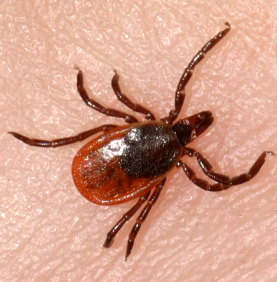
Borrelia burgdorferi is a gram negative spirochete bacterium. Its hosts are mice, deer, and humans. Spread from host to host is via a tick from the species Ixodes. Ixodes scapularis is the most common tick vector in the Northeastern United States, whereas Ixodes pacificus is the most common in the Western United States.

When an infected tick feeds from a human it can pass borrelia burgdorferi bacteria into the bloodstream. From there the bacterium is usually attacked by both the innate and adaptive immune systems. The innate immune system uses toll-like receptors on macrophages to bind to bacterial proteins. The adaptive immune response uses antibodies to bind to bacterial surface proteins, which results in complement activation and bacterial death.

Unfortunately, the immune response is often not enough to entirely eliminate this bacteria. This is because borrelia burgdorferi has the ability to evade the immune system by changing proteins on its cell surface.

Signs and Symptoms

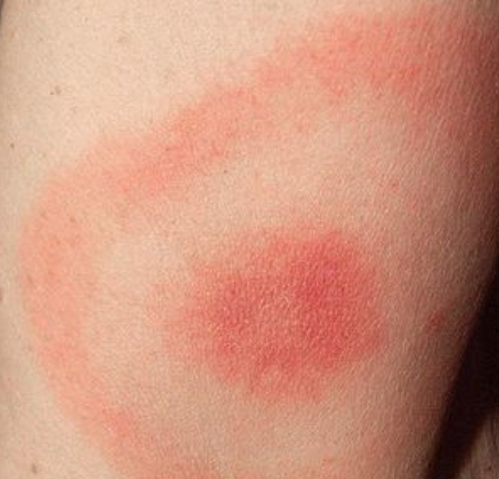
Borrelia burgdorferi causes Lyme disease. Lyme disease manifests in different ways depending on which stage of the disease the patient enters. The first stage of the disease presents with a rash known as erythema chronicum migrans. This rash appears as a red ring around a clear central area (ie: it resembles a bull’s eye). During this stage of the disease many patients also have flu-like symptoms.

If left untreated the disease can progress to stage two. During this stage neurological, joint, and cardiac symptoms can occur. The bacterium can cause cardiac arrhythmias, especially heart block. Facial drooping can be caused by damage to the facial nerve (cranial nerve seven). Meningitis, which ranges in severity, can occur. Finally, a migrating intermittent arthritis can also be seen during this stage of the disease.

If the disease continues to remain untreated, stage three ensues! This stage happens approximately a year or more after the initial tick bite. It is characterized by chronic arthritis, which can cause severe joint damage. Encephalitis (inflammation of the brain tissue) and myelitis (inflammation of the spinal cord) can also occur.

Diagnosis

The diagnosis of Lyme disease is a two step process. A blood sample is taken from the patient and a test known as an ELISA is performed. ELISA stands for “enzyme linked immunosorbent assay”. This test detects antibodies made by the patient against borrelia burgdorferi antigens (ie: it’s surface proteins). If the ELISA is positive, then a second confirmatory test called a "western blot" is performed. This test further separates the different antibodies to borrelia. If both the ELISA and western blot are "positive" the likelihood of Lyme disease is high.

Treatment

Treatment for early stage disease is with an antibiotic known as doxycycline. Children and pregnant women are often treated with amoxicillin. Other medications used include cefuroxime; erythromycin is sometimes used in patients who are allergic to pencillin. Later stages of the disease may need intravenous antibiotics.

If patients do not respond to initial antibiotic treatment it is important to think of co-existent infections with other tick-borne illnesses. Ticks may carry not only borrelia burgdorferi, but also other pathologic entities so treatment may need to be further tailored.

Overview

Lyme disease is caused by the bacterium borrelia burgdorferi. It is transmitted by the Ixodes tick and is common in the northeastern United States. Symptoms include a bull’s eye rash known as erythema migrans. Neurological, joint, and cardiac symptoms can also occur. Treatment for early stage disease is with oral antibiotics, usually doxycycline, although amoxicillin is sometimes used. Intravenous antibiotics may be necessary for later stages.

References and Resources