Colloid Cysts of the Third Ventricle

Colloid cysts of the third ventricle are slow-growing, benign cranial tumors. They are believed to be composed of an epithelial wall with either mucous or protein like material trapped inside a spherical structure. However, their exact etiology is still under debate.

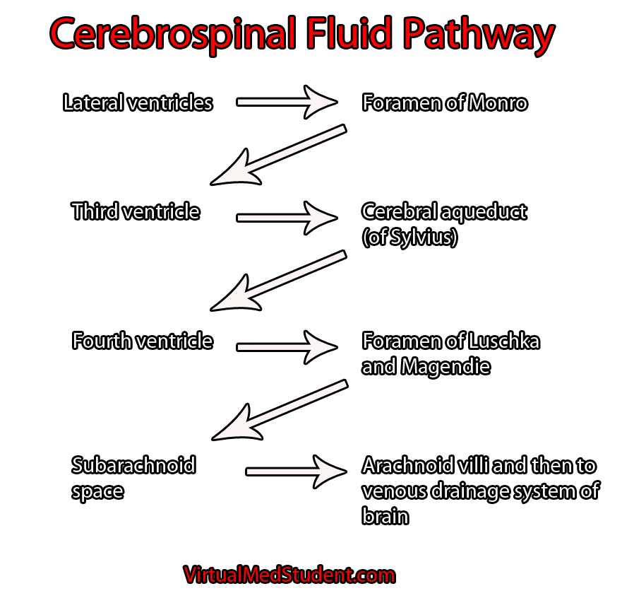
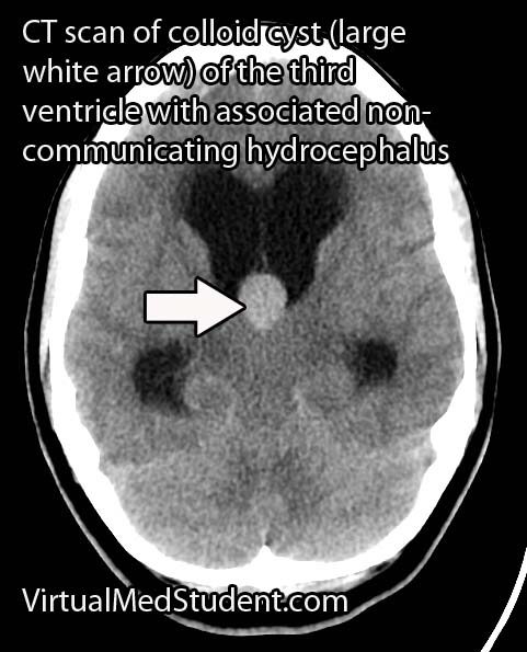
They are typically found in the anterior portion of the third ventricle near the foramen of Monroe (the channels that connect the lateral ventricles to the third ventricle). The third ventricle is one of the spinal fluid filled cavities of the brain.

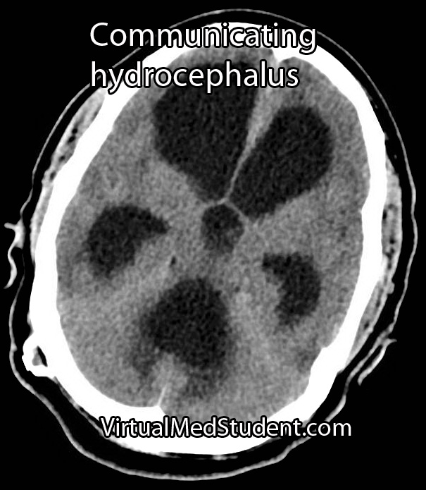
Colloid cysts are “benign” because they are not cancerous (ie: don’t invade other parts of the body); however, they have the potential to block the flow of cerebrospinal fluid, which can lead to acute hydrocephalus and brain herniation. Therefore, in this regards they are certainly not “benign” tumors!

Signs and Symptoms

The most common presenting symptom of a colloid cyst is headache and difficulty walking. Acute hydrocephalus (dilation of the ventricular system secondary to blocked cerebrospinal fluid) can occur if the cyst blocks the flow of cerebrospinal fluid; this can cause nausea, vomiting, headache, and lethargy. Changes in mental status may also be seen in patients with these lesions.

There are numerous reports of patients dying suddenly from colloid cysts of the third ventricle. This is believed to be due to rapid obstruction of cerebrospinal fluid at the foramen of Monroe. The fluid builds up behind the blockage which puts pressure on the brain. Too much pressure can cause the brain to herniate through the base of the skull (see the Monro-Kellie doctrine).
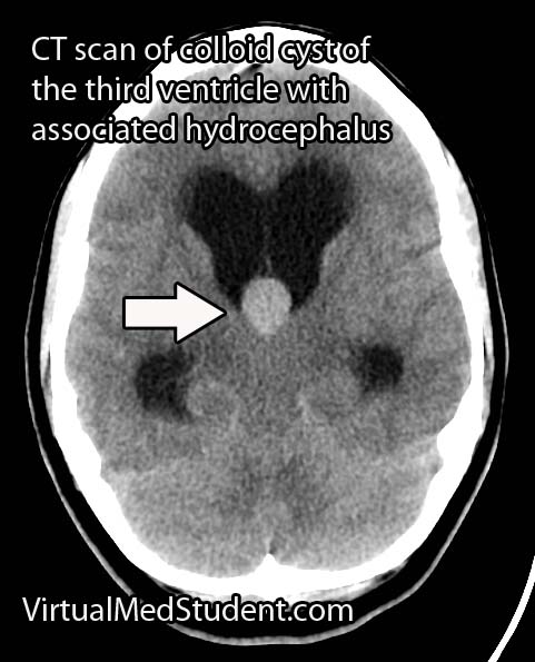
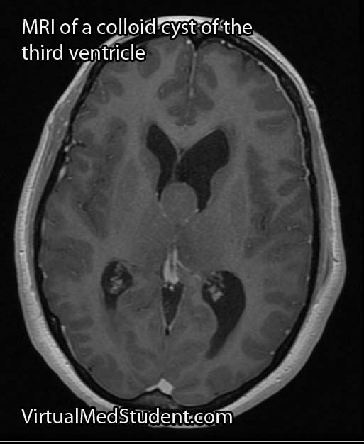
Diagnosis

Colloid Cyst of the 3rd Ventricle CT
MRI of colloid cyst of the third ventricle
Diagnosis can be made with MRI or CT scan. Head CT scans will reveal a hyperdense (ie: bright or white colored) lesion. MRI is beneficial because it provides a superior picture of the regional anatomy around the cyst. Lumbar puncture should never be performed in a patient with a colloid cyst due to the risk of brain herniation.

Treatment

Treatment of colloid cysts is surgical. There are numerous approaches including the use of an endoscope, or the use of stereotactic guidance systems. In patients with contraindications to surgery bilateral cerebrospinal fluid shunts can be placed to prevent acute hydrocephalus from developing.

Overview

Colloid cysts of the third ventricle are "benign" tumors. They have the potential to block the flow of cerebrospinal fluid leading to acute hydrocephalus. The most common symptom is headache followed by gait instability. Diagnosis is made with CT and MRI imaging. Treatment is surgical resection.

Related Articles

References and Resources

Hydrocephalus is Greek for “Water Head”

Hydrocephalus literally means "water head". It is a term used to describe a pathological increase in the amount of cerebrospinal fluid (CSF) within the ventricles (fluid filled cavities) of the brain.

In order to understand hydrocephalus we have to first appreciate the cerebrospinal fluid pathway and ventricular system of the brain. The brain has four ventricles: a pair of lateral ventricles, a third ventricle, and a fourth ventricle. They are connected to one another through narrow channels. The fourth ventricle drains into the subarachnoid space around the upper spinal cord. The spinal fluid travels down into the lumbar cistern and then back up again where it is absorbed by the arachnoid granulations overlying the cerebral hemispheres.

CSF pathway

Cerebrospinal fluid is created at a rate of roughly 500 mL per day. It is primarily secreted by specialized cells within the walls of the ventricles known as choroid plexus. As you can imagine, if it is secreted at 500 mL per day there must be an equal amount of re-absorption. This re-absorption occurs in the subarachnoid space by venous structures known as "arachnoid villi". Re-absorption does not occur in the ventricles themselves; this is an important point to keep in mind as we discuss the difference between communicating and non-communicating forms of the disease.

Hydrocephalus occurs when excess cerebrospinal fluid backs up. It is called "communicating" hydrocephalus if all of the ventricles are enlarged. Otherwise it is known as "non-communicating" hydrocephalus. There is another informal type of hydrocephalus that is known as "ex-vacuo"; it occurs when dilation of the ventricular system is a result of brain tissue loss, rather than a pathological increase in the amount of cerebrospinal fluid.

The term hydrocephalus usually implies an abnormally high pressure within the ventricular system. However, a different type of hydrocephalus known as "normal pressure" hydrocephalus defies this rule.

Communicating hydrocephalus usually develops when the arachnoid villi get "gunked up". Abnormal materials (such as blood in subarachnoid hemorrhage or proteins after meningitis) can block the arachnoid villi and prevent re-absorption. The cerebrospinal fluid then backs up and causes the entire ventricular system to enlarge.

Non-communicating hydrocephalus is slightly different because the re-absorption pathway is functioning properly, but CSF backs up behind a "road block" in one of the channels connecting the individual ventricles. "Road blocks" can be anything from tumors to developmental narrowing of the channel itself. For example, colloid cysts of the third ventricle can occlude the foramen of Monroe (the channel between the lateral and third ventricles); when this occurs CSF produced in the lateral ventricles is not able to flow into the third ventricle. CSF then backs up in the lateral ventricle(s) resulting in pathologic dilation.

Overall, there are crap tons of causes for both communicating and non-communicating hydrocephalus. Some patients have congenital forms secondary to improper development of the CSF pathways. Others may develop hydrocephalus later in life as a result of infection, tumor formation, or aneurysm rupture.

Signs and Symptoms

The signs and symptoms depend on the age of the patient and the rapidity of onset. In newborns hydrocephalus often presents as failure to thrive with an abnormally enlarging head.

If hydrocephalus develops rapidly, the increase in intracranial pressure (as a result of cerebrospinal fluid putting pressure on the brain) leads to nausea, vomiting, headache, and if severe enough, coma and potentially death!

Normal pressure hydrocephalus (NPH) is a unique type of hydrocephalus in which there is no elevation in pressure (hence the term "normal pressure"). The classic symptoms of NPH are difficulty walking, dementia, and urinary incontinence.

Diagnosis

Non-communicating hydrocephalus
Communicating Hydrocephalus

Diagnosis of hydrocephalus is made with CT scan coupled with clinical evidence of increased pressure within the ventricular system.

The scan will show an abnormally enlarged ventricular system. MRI scans with contrast are also routinely done to evaluate for tumors as a cause of hydrocephalus.

Treatment

Treatment consists of "shunting" the cerebrospinal fluid to another part of the body, usually the peritoneal cavity.

Other techniques such as third ventriculostomy, in which a surgically made "hole" is placed between the third ventricle and the subarachnoid space is also sometimes employed. This is very effective in cases of non-communicating hydrocephalus secondary to aqueductal stenosis (ie: the small passage between the third and fourth ventricles).

The ultimate treatment depends on the type of hydrocephalus and whether symptoms are severe enough to warrant surgical intervention.

Overview

Hydrocephalus is a pathological accumulation of cerebrospinal fluid. It can be communicating, non-communicating, or normal pressure. It can cause numerous signs and symptoms, but headache, nausea and vomiting are amongst the most common. Diagnosis is with CT scanning and clinical evidence of increased intracranial pressure. Treatment consists of shunting the CSF to another part of the body where it can be reabsorbed.

Related Articles

References and Resources

Tuberculous Meningitis: Basal Cisterns, Strokes, Hydrocephalus

Tuberculosis is one of the most common infectious diseases in the world. It is caused by a bacteria of the genus mycobacterium. Tuberculosis usually infects the lungs, but may also infect the lymph nodes, vertebral bodies, kidneys, gastrointestinal system, or central nervous system.

Central nervous system disease comes in two flavors: a focal abscess-like lesion known as a tuberculoma or tuberculosis meningitis. The remainder of this article will focus on tuberculosis meningitis, which is an uncommon (although not rare!) form of extra-pulmonary tuberculosis.

Let’s start by discussing how the bacteria get into the central nervous system. After being inhaled the bacteria infect cells known as macrophages. The infected macrophages move towards lymph nodes, and eventually end up in the blood stream. Once in the blood stream, the mycobacterium-infected macrophages can travel anywhere in the body!

One spot the bacterium hitch a ride to is the lining of the brain (aka: the meninges). Collections of mycobacterium-laden macrophages (“Rich foci”) can rupture into the subarachnoid space causing an inflammatory reaction (ie: a "meningitis", or inflammation of the meninges).

For unclear reasons, the inflammation seen in tuberculosis meningitis preferentially affects the basal cisterns and base of the brain. Autopsy specimens show a gelatinous material coating the undersurface of the brain. The inflammatory reaction is what causes the signs and symptoms of tuberculosis meningitis.

Signs and Symptoms

Tuberculosis meningitis can present in a number of different ways. Many patients present with days to months of non-specific symptoms such as headache, lethargy, nausea, and vomiting.

Cranial neuropathies are commonly seen, especially since tuberculosis meningitis affects the basal cisterns and base of the brain, which is where many of the cranial nerves run.

Additionally, up to 40% of patients may present with stroke. Strokes occur because the inflammation can "eat up" the linings of small blood vessels. The basal ganglia, internal capsule, and thalamus are the most common locations where strokes occur in tuberculosis meningitis.

Diagnosis

The clinical history is extremely important. Tuberculosis meningitis may affect both immunocompetent and immunocompromised (ie: think HIV/AIDs, diabetics, people on immunosuppressives, etc.) people. If the clinical history and physical exam findings are concerning for meningitis, than confirmatory studies should be performed.

TB Meningitis MRI
TB Meningitis CT
Cerebrospinal fluid (CSF) analysis shows elevated opening pressures, increased cellularity with inflammatory cells like neutrophils (earlier) and lymphocytes (later), an increased amount of protein, and a decreased amount of glucose. An acid-fast stain of the CSF to look for bacteria is sometimes positive. Culturing the CSF for mycobacterium is routinely done, but results can take weeks to months, and therefore is not useful in deciding whether or not to start treatment. Polymerase chain reactions (PCR) to look for mycobacterium DNA are also commonly used, and are much quicker than culturing.

Appropriate imaging studies include CT or MRI scans with contrast. The basal cisterns and base of the brain will "light up" with contrast because of inflammation. Imaging may also show strokes and hydrocephalus. Under the right clinical scenario imaging studies can help support the diagnosis, but are not specific.

Treatment

Treatment should consist of antibiotics that target mycobacterium tuberculosis. Commonly used antibiotics include rifampin, isoniazid, streptomycin, and pyrazinamide. Other antibiotics may be necessary if the strain of bacteria is resistant to these drugs.

Steroids are also frequently given to help reduce inflammation. Dexamethasone, a commonly used steroid, has been shown to improve survival rates (although it may not affect outcome in those that survive).

The intense inflammatory reaction seen in tuberculosis meningitis may gunk up the re-absorption of cerebrospinal fluid and cause a communicating hydrocephalus. Hydrocephalus occurs in 70% of cases and may require surgically inserted shunts to fix.

Overview

Tuberculosis meningitis is a devastating manifestation of a common infectious disease. It can cause cranial neuropathies, strokes, and hydrocephalus. Prompt diagnosis is mandatory and is made from the clinical history, CSF analysis, and imaging studies. Treatment is with anti-TB medications and steroids. Many survivors have long term disabilities despite appropriate treatment.

Related Articles

References and Resources

Monro-Kellie and Their Doctrine: Blood, Brain, Spinal Fluid

The Monro-Kellie doctrine states that three things exist within the fixed dimensions of the skull: blood, cerebrospinal fluid, and brain. An increase in any one component must necessarily lead to a decrease in one (or both) of the other components, otherwise intracranial pressure will increase.

Increases in one of the three components can take many different shapes and sizes. For example, abnormal bleeding within the cranium such as in epidural and subdural hematomas are common examples, which typically occur after traumatic events. Bleeding within the brain tissue itself – known as an intraparenchymal or intracerebral hematoma – can also occur, especially in patients with untreated high blood pressure. Brain tumors of any type effectively increase the amount of brain tissue. And last, but not least, the cerebrospinal fluid can back up in a condition known as hydrocephalus.

Regardless of the cause, the end result is an abnormal increase in either blood, brain, or cerebrospinal fluid within the confines of the skull.

So what’s the big deal? If the abnormality becomes large enough, the pressure within the skull can increase rapidly. Eventually the pressure can become so great that the brain gets squished, and will pop over rigid boundaries and out the small holes within the skull.

This is known as “herniating” the brain tissue. It can occur in numerous places depending on where the pressure is greatest. However, the most important herniation clinically occurs at the base of the skull where a hole known as the foramen magnum exists.

When the brain herniates here it really pisses off a vital structure known as the brainstem. The brainstem is responsible for all the stuff we don’t consciously think about (heart rate, breathing, etc.), which ultimately keeps us alive. When herniation of the brainstem through the foramen magnum occurs it stretches all the “wires” that allow our brainstem to function properly. If severe enough, all those autopilot functions (ie: breathing, beating of the heart, etc.) stop working and brain death occurs.

Overview

The skull contains three components within it: blood, brain, and cerebrospinal fluid. An abnormal increase in any one of these components causes an increase in pressure, which if severe enough, can cause herniation of brain tissue out of the skull. This can lead to coma and brain death.

References and Resources

How To Systematically Interpret a Head CT: Blood Can Be Bad

Head CTs are a common, inexpensive, and fast way of evaluating intracranial pathology. Although they do not give the anatomical detail of an MRI, they are still extremely important in diagnosing “gross” pathology that needs emergent intervention.

CT scans are based on the Hounsfield unit (HU), which is an indirect way to measure density. Interestingly, Sir Godfrey Newbold Hounsfield won a Nobel prize for his work on developing the CT scanner, but I digress…

The importance of the Hounsfield unit is that things that are hyper-dense (very dense) appear bright; those things that are hypo-dense (not very dense) appear dark. The different tissues and fluids within the confines of the skull have varying densities. The most dense materials, like bone, have very high Hounsfield units; less dense materials such as air and cerebrospinal fluid have very low Hounsfield units.

It is important to approach head CTs in a systematic fashion so that subtle (and not so subtle) pathology is not missed. The easiest way I have found to read a head CT is to remember the following mnemonic:

Blood Can Be Very Bad

The first “B” in the mnemonic stands for, you guessed it, blood. There are five different pathological locations that blood can be located: epidural, subdural, subarachnoid, intraventricular, and intraparenchymal. Depending on the age of the blood, it may be hyper-dense (acute/active bleeding), isodense (roughly 3 to 7 days old), or hypo-dense (older than 7 days).

CT scans of Intracerebral Hemorrhages
The "C" in the mnemonic stands for "cisterns". Cisterns are enlarged subarachnoid spaces where cerebrospinal fluid pools. The most important cisterns are around the brainstem. They include the interpeduncular, suprasellar, ambient, quadrigeminal and pre-pontine cisterns. A healthy amount of cerebrospinal fluid should “bathe” the brainstem; if there is increased intracranial pressure cerebrospinal fluid will get pushed out of these cisterns as brain tissue starts to herniate into them. And that as they say is “no bueno”.

The second "B" stands for "brain". Although blatant pathology such as blood clots are usually readily apparent, more subtle pathology can also be obtained from a CT. For example, blurring of the gray-white junction may indicate evolving stroke. Any areas of hypodensity (ie: dark areas) within the brain may indicate edema associated with a tumor.

The "V" represents the ventricular system. The ventricular system consists of a pair of lateral ventricles, a third ventricle, and a fourth ventricle (don’t ask me what happened to the first and second ventricle!). The ventricles are in communication with one another via holes known as foramen. The paired foramen of Monroe connect the lateral ventricles to the third ventricle; the cerebral aqueduct of Sylvius connects the third ventricle to the fourth ventricle. The fourth ventricle drains into the subarachnoid space surrounding the spinal cord via the foramen of Magendie and Lushka.

The ventricular system is quite symmetric. Any obvious asymmetries may indicate a pathologic process "pushing" on a ventricle causing it to become distorted. In addition, if the ventricles are larger than normal it may indicate the presence of hydrocephalus, a condition in which cerebrospinal fluid is not reabsorbed appropriately.

The final "B" in the mnemonic stands for "bone". The skull should be assessed for fractures, especially in trauma patients. A common place for fractures is at the skull base. Time should be spent assessing this area to rule out fractures that extend across the canals and foramen that house the carotid arteries, jugular veins, and cranial nerves.

Reading a head CT is the first step in determining what additional imaging studies are necessary, or what treatment should be given. By using the above mnemonic it allows the interpreter of the scan to quickly and effectively assess if there is underlying pathology that needs further evaluation.

Overview

The mnemonic – blood can be very bad – can be used to systematically interpret a head CT. The first "B" stands for blood. The "C" stands for cisterns. The second "B" stands for brain. The "V" represents the ventricular system. And the last "B" stands for bone. By looking at these five components it is possible to assess all the important pathology that may require further imaging and/or treatment.

References and Resources