Pyogenic Liver Abscesses: Pus, Needle Drainage, and Antibiotics

Pyogenic liver abscesses are localized collections of pus and bacteria. The initial infection occurs when bacteria travel through the portal vein. This most commonly occurs after bowel contents leak into the peritoneal cavity from the gut. Other causes of liver abscesses include direct spread of infection from the bile duct system, or from other bacterial infections of the blood. Rarely, penetrating injuries (gunshots, stab wounds, surgery) may directly introduce infection. There are numerous “bugs” that can cause pyogenic liver abscesses. They include, but are not limited to, streptococcus species, klebsiella pneumoniae, and staphylococcus species.

Signs and Symptoms

Similar to other infections, liver abscesses can cause fevers, chills, decreased appetite, abdominal pain, and a generalized sense of not feeling well (ie: malaise). Interestingly, hiccups may also be present if the abscesses are causing adjacent inflammation/irritation of the diaphragm.

Diagnosis

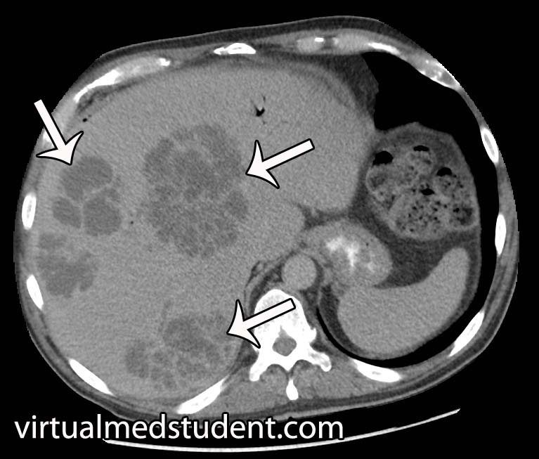
Pyogenic Liver Abscesses
The diagnosis of pyogenic liver abscesses is made with imaging. The most commonly used method is a CT scan of the abdomen with and without contrast. If present, a liver abscess will look like a collection of fluid, with or without septated dividers.

Another commonly employed imaging modality is the use of ultrasound to detect the fluid filled pockets within the liver.

However, it is important to note that imaging studies alone cannot distinguish between the different types of liver abscesses. Imaging can also not tell you what bacteria is responsible for the abscess.

Treatment

Pyogenic abscesses must be drained and treated with antibiotics. Drainage is usually done under image guidance using a needle placed through the skin into the abscess. Although occasionally direct surgical evacuation of the abscess is necessary.

Draining the abscess is important for two reasons. First, it decreases the size of the abscess allowing antibiotic therapy to work more effectively. And secondly, it provides abscess fluid that can be sent to the lab for bacterial culture and gram stain.

The results of the culture help guide subsequent antibiotic therapy. Commonly used antibiotics include piperacillin-tazobactam (Zosyn®), vancomycin, metronidazole (Flagyl®), and ceftriaxone; once cultures confirm the causative bug antibiotic therapy can be narrowed.

Overview

Pyogenic liver abscesses are collections of pus and bacteria. They occur most commonly after the spilling of gut bacteria into the peritoneal space (ie: peritonitis). There are numerous causative bacteria. Symptoms include fever, chills, decreased appetite, and abdominal discomfort. Treatment is with drainage and antibiotics.

References and Resources

  • Hasper D, Schefold JC, Baumgart DC. Management of severe abdominal infections. Recent Pat Antiinfect Drug Discov. 2009 Jan;4(1):57-65.
  • Benedetti NJ, Desser TS, Jeffrey RB. Imaging of hepatic infections. Ultrasound Q. 2008 Dec;24(4):267-78.
  • Mortelé KJ, Segatto E, Ros PR. The infected liver: radiologic-pathologic correlation. Radiographics. 2004 Jul-Aug;24(4):937-55.
  • Kurland JE, Brann OS. Pyogenic and amebic liver abscesses. Curr Gastroenterol Rep. 2004 Aug;6(4):273-9.

Pelvic Inflammatory Disease: Gonococcus, Chlamydia, and the Chandelier Sign

Pelvic inflammatory disease (PID) refers to infection of the uterus, fallopian tubes, and/or ovaries. The most common infections that cause pelvic inflammatory disease are neisseria gonorrhoeae and chlamydia trachomatis. In fact, both bacteria may be present in the same patient! However, it is not uncommon for other pathogenic bacteria to be present as well. The exact role of most of these other pathogenic bacteria is not well understood.

In order for these organisms to gain access to the uterus, fallopian tubes, and ovaries there must be some breakdown of the normal barriers that separate the vagina from the upper genital tract. Interestingly, a significant number of cases of pelvic inflammatory disease occur just after menstruation when the possibility of pathogenic bacteria gaining access to the upper genital structures is at its greatest. In addition, normal vaginal bacteria can act as a barrier to invading pathogenic strains. Therefore, in some patients with PID the normal vaginal bacterial landscape has been altered in some way, which allows the overgrowth of pathogenic bacteria.

Patients who are at risk for pelvic inflammatory disease are generally those women who are at increased risk of sexually transmitted diseases. Therefore, women with multiple sexual partners, younger age (most cases occur between 15 and 25 years of age), previous history of sexually transmitted disease, and those that do not use barrier contraception (ie: condoms) are all at increased risk.

Signs and Symptoms

Bilateral lower quadrant abdominal pain is a common presenting symptom. Infection and the resulting inflammation that occurs can make having sex painful (ie: "dyspareunia") and can make urinating painful (ie: "dysuria"). In addition, abnormal vaginal odors and discharges are also possible. Like most other serious infections, systemic signs such as fever, nausea, vomiting, and lethargy may also occur.

On pelvic examination patients are extremely tender to palpation of the uterus and ovaries. This is often referred to as the “Chandelier sign” because women will “reach for the chandeliers” because of the discomfort during the exam.

Complications

Complications can be severe if left untreated. Pelvic inflammatory disease can lead to scarring of the fallopian tubes and possible infertility. The risk of ectopic pregnancies is increased in women with a history of PID. Abscesses can form in the ovaries and tubes, as well as in the peritoneal cavity (ie: abdominal cavity). In addition, inflammation of the liver’s capsule can occur in a complication known as Fitz-Hugh-Curtis syndrome. Pelvic inflammatory disease can also lead to infection of the peritoneal cavity (aka: peritonitis).

Diagnosis

Diagnosis of pelvic inflammatory disease is based on clinical signs and symptoms. If lower abdominal discomfort with pain during pelvic examination (ie: cervical motion, uterine, and/or ovarian tenderness) is present, patients should be treated as though they have pelvic inflammatory disease.

There are numerous other criteria that can “support” the diagnosis, but are not necessary to have. They include other signs of infection like abnormal vaginal discharge, fever, elevated white blood cell count, and evidence of gonorrhea or chlamydial infection from laboratory tests.

Definitive diagnosis is made either by biopsy of the upper genital tract, imaging (ie: CT or MRI) that shows edematous fallopian tubes, or disease seen laparoscopically.

Treatment

Treatment of pelvic inflammatory disease is based on the severity of the clinical signs and symptoms, as well as the likely underlying organisms. Patients who are stable can be given oral antibiotics.

Generally a single dose of a third generation cephalosporin like ceftriaxone is given to cover gonorrhea; another antibiotic called doxycycline is given to cover the possibility of co-existent chlamydial infection. Metronidazole may be added to these medications if anaerobic bacteria are suspected.

If the patient appears very sick, is pregnant, unlikely to continue treatment as an outpatient (ie: homeless, destitute, etc), or unable to take oral antibiotics they should be hospitalized and treated with intravenous (IV) antibiotics. Usually a second generation cephalosporin like cefoxitin plus doxycycline (either IV or oral) is given. Clindamycin and gentamicin, as well as ampicillin-sulbactam (Unasyn®) can also be used in certain circumstances. In general, there are numerous antibiotic regimens that can be considered.

Overview

Pelvic inflammatory disease is an infection of the upper genital tract in females. This includes the uterus, fallopian tubes, and/or ovaries. Gonorrhea and chlamydia are the two most commonly isolated bacteria, but many infections are caused by other pathogenic bacteria. Diagnosis is made on clinical signs and symptoms. Treatment is with at least two antibiotics: one to cover gonorrhea and one to cover chlamydia.

Related Articles

References and Resources

Appendicitis: A Vestigial Remnant to Belly Pain

The appendix is a small out-pouching off a part of the large intestine known as the cecum. It functions similar to normal large intestine by secreting mucous and absorbing water. Its overall importance, however, is not well understood, and it is likely a vestigial remnant from a distant ancestor. Unfortunately for some unlucky folk it can become inflamed; when this occurs it is called “appendicitis”.

Appendicitis occurs when something blocks the opening of the appendix into the cecum. There are numerous causes. The most common in younger individuals is a mass of inflammatory cells known as lymphoid hyperplasia, which can occur after a viral or bacterial infection of the gut. In older individuals, the most common cause is a small, hard piece of poop known as a “fecalith”.

When lymphoid hyperplasia or a fecalith (or any other obstructing thing) blocks the opening of the appendix, any mucous secreted by it gets trapped. When the appendix becomes distended enough, it literally chokes off its own blood supply and starts to die.

The dying appendix sets off a cascade of inflammation. Bacteria within the intestine are able to move in and wreak further havoc! The end result is a nasty inflammatory process, that if left unchecked, can lead to a very serious surgical illness.

Signs and Symptoms

Appendicitis initially presents with periumbilical pain (ie: pain around the belly button) that quickly migrates to involve the right lower quadrant of the abdomen.

The reason pain occurs in this sequence is because the initial discomfort of appendicitis is due to inflammation of the visceral peritoneum and appendix itself. The visceral peritoneum is a layer of tissue that envelopes the gut tube. This type of pain is carried back to the spinal cord by autonomic nerves, and due to their embryological origin, that pain gets referred to the midline of the abdomen (the belly button).

Over the course of the disease, the parietal peritoneum eventually becomes inflamed. This pain is carried by somatosensory nerves with a very specific dermatomal distribution, thus the pain gets localized directly to the anatomical location of the appendix – the right lower quadrant. This pain is often very well localized at an area known as McBurney’s point.

Patients often have nausea, vomiting, and a decreased appetite. In fact, if patients are hungry and want to eat, appendicitis becomes a highly unlikely diagnosis for abdominal pain. This is referred to informally as the "cheeseburger sign".

There are also several physical exam findings. Pain with palpation of the right lower quadrant associated with rebound tenderness (ie: pain that occurs when you release pressure during palpation) is frequently seen. Sometimes palpating the left lower quadrant (ie: the area on the other side of the abdomen from the appendix) will cause discomfort in the right lower quadrant. This is known as "Rovsing’s sign".

Any maneuvers that irritate the peritoneum will also cause discomfort. The first of these signs occurs when you flex the hip. This causes the iliopsoas muscle to rub up against an inflamed peritoneum. This is known as the "psoas sign". Another way to irritate the peritoneum is to internally rotate the leg while the patient’s knee and hip are flexed. This is known as the "obturator sign".

Diagnosis

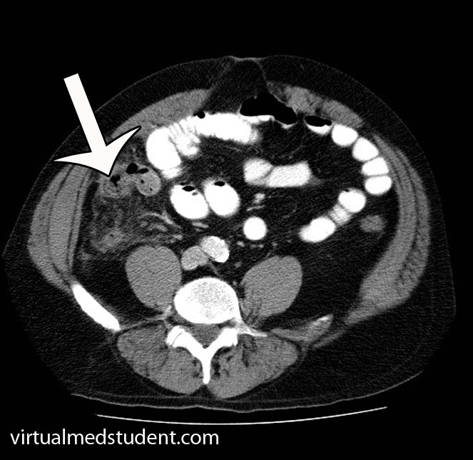
Appendicitis CT Scan
Appendicitis is first and foremost a clinical diagnosis. Therefore, in patients with a history of periumbilical pain that migrates to the right lower quadrant appendicitis is the most likely diagnosis.

However, in a world with advanced imaging technologies we can quickly get pictures that support the diagnosis. Both ultrasound (frequently used in children, pregnant patients, and younger adults) or CT scans can be obtained quickly and inexpensively. The CT scan will show fat stranding and fluid around an enlarged appendix (see image to the left).

Blood tests such as a complete blood count (CBC) will show an elevated white blood cell count (ie: the cells that fight off infection and are responsible for inflammation).

Treatment

Treatment is surgical (ie: appendectomy)! Get that nasty inflamed appendix out of there STAT! In addition, patients are started on intravenous fluids and antibiotics that cover anaerobic organisms.

Commonly used antibiotics for a non-ruptured appendix are metronidazole, ampicillin/sulbactam, and ciprofloxacin. If the appendix has ruptured, broad spectrum coverage with piperacillin/tazobactam or a combination of ampicillin, ciprofloxacin, and clindamycin is started and continued for at least 5 days.

Occasionally a ruptured appendix will wall itself off into an abscess. If this is the case, the abscess must be drained with a needle. Antibiotics are started, and the patient is taken to surgery roughly six weeks later to remove the appendix after the inflammation has "calmed down".

Overview

Appendicitis occurs when something blocks the opening of the appendix into the cecum. Progressive enlargement of the appendix occurs eventually chocking off its own blood supply. Pain in the right lower quadrant of the abdomen is a common symptom of appendicitis. Diagnosis is clinical, but ultrasound and CT scanning can be helpful in elucidating unclear cases. Treatment is appendectomy (removal of the appendix), IV fluids, and antibiotics.

References and Resources

  • Merlin MA, Shah CN, Shiroff AM. Evidence-based appendicitis: the initial work-up. Postgrad Med. 2010 May;122(3):189-95.
  • Kim JK, Ryoo S, Oh HK, et al. Management of appendicitis presenting with abscess or mass. J Korean Soc Coloproctology. 2010 Dec;26(6):413-9. Epub 2010 Dec 31.
  • Lee SL, Islam S, Cassidy LD. Antibiotics and appendicitis in the pediatric population: an American Pediatric Surgical Association Outcomes and Clinical Trials Committee systematic review. J Pediatr Surg. 2010 Nov;45(11):2181-5.
  • Markides G, Subar D, Riyad K. Laparoscopic versus open appendectomy in adults with complicated appendicitis: systematic review and meta-analysis. World J Surg. 2010 Sep;34(9):2026-40.
  • Grundmann RT, Petersen M, Lippert H, et al. The acute (surgical) abdomen – epidemiology, diagnosis and general principles of management. Z Gastroenterol. 2010 Jun;48(6):696-706. Epub 2010 Jun 1.