Hypertrophic Cardiomyopathy: Athletes and Genetic Mutations

Hypertrophic cardiomyopathy occurs when the size of heart muscle cells increase (aka: hypertrophy). Genetic mutations in DNA that codes for heart muscle cell proteins are responsible for the development of hypertrophic cardiomyopathy. Most of these mutations are in DNA that code for sarcomere proteins (ie: myosin, actin, troponin, etc.). The mutated proteins cause decreased contractile function. As a result, the muscle cell hypertrophies (enlarges) in an attempt to overcome the decreased contractility. The result is a disorganized pattern of muscle cell fibers with intervening fibrosis (ie: scar tissue).

Signs and Symptoms

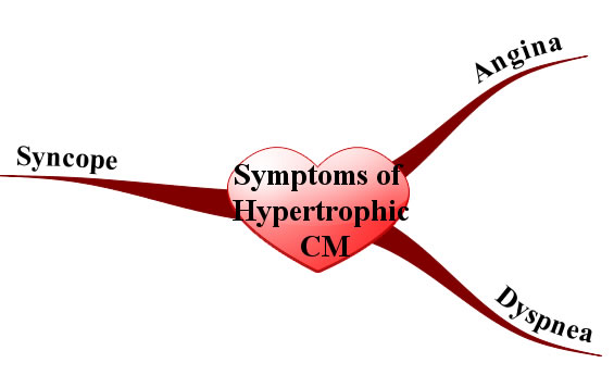
Since the myocardium is hypertrophied there is less ventricular compliance (ie: the heart becomes stiff). This stiffness decreases the filling capacity of the ventricle. The result is diastolic dysfunction, or a decreased ability of the heart to fill during its relaxation phase. High diastolic pressures occur leading to the back-up of blood into the left atrium, pulmonary veins, and pulmonary capillaries. Excess fluid in the pulmonary capillaries causes pulmonary edema with resultant shortness of breath and exercise intolerance.

In addition, angina (chest pain) can occur even without co-existing coronary artery disease because the increased muscle mass of the ventricle results in a higher oxygen demand. Under strenuous conditions the hypertrophied muscle cannot get enough oxygen, which causes chest pain.

Symptoms and Signs of Hypertrophic Cardiomyopathy
Syncope (ie: fainting) is another common symptom that is usually due to arrhythmias caused by the abnormal myocyte architecture.

Physical exam can reveal an S4 gallop (aka: atrial gallop), which is caused by the atrium forcing blood into a stiff left ventricle during the "atrial kick" at the end of diastole.

Murmurs can also be heard, usually mitral regurgitation and a systolic outflow obstruction murmur. Mitral regurgitation occurs because the hypertrophied ventricular septum acts as a barrier to blood flow into the aorta. As a result, during systole blood will flow backwards through the mitral valve into the left atrium. Blood flowing across the septal barrier into the aorta will create an obstruction murmur. The obstruction murmur worsens with valsalva, which distinguishes it from the murmur of aortic stenosis.

Diagnosis

The work-up is very similar to dilated cardiomyopathy, except ancillary studies are usually not helpful. Echocardiography (ie: ultrasound of the heart) is the gold standard and will show the hypertrophic myocardium. ECG will often reveal left ventricular hypertrophy and left atrial hypertrophy. Arrhythmias may sometimes be observed on ECG as well. Prominent Q-waves can be seen in the lateral leads (ie: V4-V6) and inferior leads (II, III, aVF); this is the result of greater depolarization of the hypertrophied septum (remember depolarization of the septum starts on the left side and moves rightward creating a downward deflection in leads on the opposite side of the body, before the left ventricle "overpowers" the ECG findings).

Treatment

β-blockers are the mainstay of treatment. They decrease the heart rate and allow increased diastolic filling times, which leads to decreased outflow obstruction; they also decrease myocardial oxygen demand leading to decreased anginal symptoms.

Prevention of fatal arrhythmias is important in hypertrophic cardiomyopathy. Medical management of arrhythmias is accomplished with amiodarone and/or disopyramide. In some patients, strong consideration should be given to an implantable cardiac defibrillator, especially those at high risk of sudden death. Surgery with partial myomectomy to remove some of the hypertrophied muscle can also be done if the patient is unresponsive to medical management.

Since hypertrophic cardiomyopathy is caused by genetic mutations, genetic counseling should be offered to children of affected parents. First degree relatives should undergo screening with echocardiography as well.

Unlike dilated cardiomyopathy, diuretics should be used sparingly because they can worsen outflow obstruction by causing decreased venous return to the left ventricle. Digoxin is also contraindicated because it can worsen outflow obstruction. When considering treatment options it is important to remember that the problem in hypertrophic cardiomyopathy is diastolic, not systolic dysfunction.

Prognosis depends on the type and severity of the genetic mutation involved. Some mutations result in minimal morbidity and a normal life span, whereas others can cause significant heart failure symptoms. Overall mortality is roughly 5% per year secondary to ventricular fibrillation; therefore, even minimally symptomatic patients must be monitored closely.

Overview

The cause of hypertrophic cardiomyopathy is genetic. Diagnosis is made with echocardiography (ie: ultrasound of the heart). Treatment is generally with beta blockers, amiodarone, implantable cardiac defibrillators (ICD), and myomectomy in select patients. All 1st degree relatives should be offered genetic counseling and undergo screening echocardiography. Prognosis is variable and depends on the mutation type.

Related Articles

References and Resources

  • Bos JM, Ommen SR, Ackerman MJ. Genetics of hypertrophic cardiomyopathy: one, two, or more diseases? Curr Opin Cardiol. 2007 May;22(3):193-9.
  • Bo CY, López B, Coelho-Filho OR, et al. Myocardial fibrosis as an early manifestation of hypertrophic cardiomyopathy. N Engl J Med. 2010 Aug 5;363(6):552-63.
  • Kumar V, Abbas AK, Fausto N. Robbins and Cotran Pathologic Basis of Disease. Seventh Edition. Philadelphia: Elsevier Saunders, 2004.
  • Lilly LS, et al. Pathophysiology of Heart Disease: An Introduction to Cardiovascular Disease. Seventh Edition. Lippincott Williams and Wilkins, 2006.
  • Flynn JA. Oxford American Handbook of Clinical Medicine (Oxford American Handbooks of Medicine). First Edition. Oxford University Press, 2007.

Restrictive Cardiomyopathy: Amyloid, Diastolic Dysfunction, and Kussmaul’s Sign

Restrictive cardiomyopathy is the least common type of cardiomyopathy. The restriction refers primarily to diastolic dysfunction (ie: problems with relaxation of the heart); systolic function is generally well preserved. From a clinical perspective the restrictive cardiomyopathies present similarly to the hypertrophic cardiomyopathies.

There are two main causes of restriction: infiltration of the heart muscle with abnormal substances or scarring of the heart muscle. Each of these causes has many underlying etiologies, a few of which are listed below.

   (1) Infiltration
        (a) Amyloidosis
        (b) Sarcoidosis
        (c) Storage diseases
            (i) Hemochromatosis
            (ii) Glycogen storage diseases
   (2) Fibrosis and scarring
        (a) Post-radiation
        (b) Endomyocardial fibrosis

Regardless of the cause, the heart muscle gets "gunked up" with stuff that shouldn’t be there. The result is decreased contractile ability.

Signs and Symptoms

Patient’s with restrictive cardiomyopathy often present with signs and symptoms of diastolic dysfunction. Pulmonary edema (ie: fluid in the lungs) results in dyspnea (difficulty breathing), paroxysmal nocturnal dyspnea (waking up gasping for air), and orthopnea (inability to lay flat secondary to shortness of breath). Reduced cardiac output results in fatigue, dizziness, and weakness.

On physical exam, Kussmaul’s sign can sometimes be appreciated. Kussmaul’s sign occurs when you can see the jugular vein distend when a patient inspires (breathes in). Normally jugular venous distension decreases with inspiration as venous return to the heart increases due to decreased intrathoracic pressure. However, in restrictive cardiomyopathy the ventricle cannot accommodate the increased blood flow and it backs up into the jugular veins during inspiration, hence the paradoxical worsening.

In addition, signs of systemic volume overload can occur including hepatomegaly (ie: an enlarged liver), ascites (ie: fluid in the abdomen), and bilateral lower extremity edema (ie: leg swelling). Arrhythmias are also common because amyloid deposits can wreak havoc on the conduction system of the heart.

Diagnosis and Work-Up

It is important to distinguish restrictive cardiomypathy from restrictive pericarditis, which is treatable. The gold standard test is a myocardial biopsy, which will reveal infiltrating substances in restrictive cardiomyopathy. MRI and CT can show a thickened pericardium, which is more consistent with restrictive pericarditis.

Treatment and Prognosis

Treatment is aimed at the underlying cause. For example, chemotherapy for multiple myeloma or phlebotomy for hemochromatosis can slow disease progression. Since diastolic function is most affected, treatment is aimed at ensuring adequate filling time for the left ventricle. Diuretics should be used cautiously because patient’s are dependent on higher blood volumes to fill as much of the restricted ventricle as possible. Digoxin and vasodilator therapies are usually not helpful since systolic function is usually well-preserved.

Prognosis is generally poor unless treatment of the underlying condition is curative.

Overview

Restrictive cardiomyopathy is caused by infiltrative processes (most commonly amyloidosis). Physical exam reveals signs of diastolic dysfunction resulting in blood backing up in the lungs and body. The gold standard for diagnosis is myocardial biopsy, although this is not routinely performed in clinical practice. Treatment is aimed at the underlying cause, if identifiable. In addition, since diastolic dysfunction is present, treatments should be used that allow the heart adequate time to fill.

Related Articles

References and Resources

Dilated Cardiomyopathy: Poor Pump, An S3, and Crackles

Cardiomyopathy = cardio (heart) + myo (muscle) + pathy (pathology). In other words, cardiomyopathies are pathologic processes that affect the heart muscle. Dilated cardiomyopathy, which this article is about, is the most common form of cardiomyopathy. It has some known causes, but interestingly the majority of cases have no known cause and may in fact be inherited. In order to be diagnosed with dilated cardiomyopathy you must have left ventricular dilation and a low ejection fraction on echocardiography.

   (1) Idiopathic (ie: no known cause) and/or genetic
   (2) Alcoholism (chronic)
   (3) Inflammatory
       (a) Infectious
           (i) Viral
                 1. Adenovirus
                 2. Coxsackie virus
                 3. Parvovirus
                 4. HIV
           (ii) Protozoan
                 1. Trypanosomiasis (Chagas’ disease)
       (b) Non-infectious
           (i) Collagen vascular disorders
           (ii) Sarcoidosis
   (4) Drug/medicine related
       (a) Chemotherapeutics (daunorubicin/doxorubicin)
       (b) Cocaine
       (c) Methamphetamines
       (d) Heavy metals
   (5) Metabolic
       (a) Hypothyroidism
       (b) Hypocalcemia (chronic)
       (c) Hypophosphatemia (chronic)
   (6) Neuromuscular diseases

Regardless of the cause, the left ventricle of the heart dilates, which decreases its ability to pump effectively.

Signs and Symptoms

The symptoms of dilated cardiomyopathy are directly related to the decreased pumping ability of the heart (ie: systolic dysfunction). Blood backs up into the rest of the body starting with the lungs. This causes pulmonary edema, which can manifest as orthopnea (ie: inability to sleep flat due to shortness of breath), dyspnea (ie: shortness of breath with exertion), and paroxysmal nocturnal dyspnea (ie: waking up in the middle of the night short of breath). Patients also complain of exercise intolerance, dizziness, and fatigue.

The physical exam for someone with dilated cardiomyopathy will often reveal an S3 gallop (aka: ventricular gallop). An S3 gallop is caused by excess blood in the left ventricle after systole; during diastole the blood from the left atrium rushes into a relatively full left ventricle creating the S3 gallop, which can be heard with a stethoscope.

In addition, “crackles” may be heard in the lung fields secondary to pulmonary edema. If right sided heart failure has also occurred (usually after many years of left sided cardiomyopathy) there may be signs of systemic volume overload. These signs include hepatomegaly (a larger than normal liver), bilateral lower extremity edema (ie: pitting edema), and jugular vein distension.

Diagnosis

Work Up for Dilated Cardiomyopathy
Echocardiography (ie: an ultrasound of the heart) is the gold standard test and will traditionally show a dilated ventricle(s) with a depressed ejection fraction (EF < 55%).

Additional studies can be ordered depending on the clinical scenario. It is especially important to not miss alcoholism or hypothyroidism as these can be easily treated. Cardiac catheterization is often performed to determine if there is co-existing (or causative) coronary artery disease.

Treatment

Treatment for patients with dilated cardiomyopathy consists of the similar treatments used for other heart failure patients. Many patients will be on an angiotensin converting enzyme inhibitor (ACEI, lisinopril is a commonly used one), or angiotensin receptor blocker (ARB) and a beta blocker (carvedilol is commonly used due to its beneficial lipid profile compared to other beta blockers). Other considerations include spironolactone (an aldosterone receptor antagonist).

ACEI/ARBs, beta blockers, and spironolactone improve survival rates in patients with dilated cardiomyopathy. In addition, an implantable cardiac defibrillator (ICD) should be considered in all patients with an ejection fraction of less than 35% because it has been shown to reduce death from abnormal heart rhythms.

Blood thinning medications like warfarin are indicated if the patient has a thrombus (ie: a “blood clot”) seen on echocardiogram, atrial fibrillation, or previous embolic event, although some physicians may recommend thinning the blood prophylactically if ventricular function is severely impaired (EF < 30%).

Symptomatic management consists of diuretics for volume overload (ie: pitting edema, shortness of breath secondary to pulmonary edema, etc.) and digoxin to increase cardiac contractility and improve forward blood flow.

Curative treatment is a heart transplant. Overall prognosis without a transplantation is poor. Over 50% of non-transplant patients are deceased at 5 years compared to 25% of transplanted patients.

Overview

There are many causes of dilated cardiomyopathy some of which are reversible. An S3 gallop and symptoms of volume overload are often seen on physical exam. Echocardiography is the gold standard for diagnosis. It is important to treat with at least a beta blocker and ACEI; spironolactone is another option. Symptomatic management includes diuretics and digoxin. Prognosis is poor without transplant.

Related Articles

References and Resources

  • Wexler RK, Elton T, Pleister A, et al. Cardiomyopathy: an overview. Am Fam Physician. 2009 May 1;79(9):778-84.
  • Abdo AS, Kemp R, Barham J, et al. Dilated cardiomyopathy and role of antithrombotic therapy. Am J Med Sci. 2010 Jun;339(6):557-60.
  • Fatkin D, Otway R, Richmond Z. Genetics of dilated cardiomyopathy. Heart Fail Clin. 2010 Apr;6(2):129-40.
  • Kumar V, Abbas AK, Fausto N. Robbins and Cotran Pathologic Basis of Disease. Tenth Edition. Philadelphia: Elsevier Saunders, 2004.
  • Lilly LS, et al. Pathophysiology of Heart Disease: A Collaborative Project of Medical Students and Faculty. Seventh Edition. Lippincott Williams and Wilkins, 2006.
  • Flynn JA. Oxford American Handbook of Clinical Medicine (Oxford American Handbooks of Medicine). First Edition. Oxford University Press, 2007.