Ventricular Fibrillation: Shock Me Baby!

Cardiac muscle contracts in a predictable, regular pattern, which allows it to generate enough force to eject blood to the body. However, in ventricular fibrillation the heart muscle “quivers” in a rapid, irregular, and unsynchronized manner. These feeble contractions are not strong, nor coordinated enough to eject blood from the heart. As a result, cardiac output drops and the body quickly goes into cardiogenic shock.

There are numerous causes of ventricular fibrillation. The most common cause of abnormal electrical activity occurs in diseased heart tissue that has lost its normal architecture. For example, muscle damage from heart attacks or disorganized heart structures seen in cardiomyopathies can serve as abnormal areas of electrical impulse formation; these irritated areas can predispose patients to develop ventricular fibrillation.

Other causes of ventricular fibrillation include electrolyte abnormalities. For example, hyperkalemia (ie: an elevated blood potassium level) can depolarize heart muscle cells and make them more likely to "fire" an action potential. In general, any electrolyte disturbance that makes the resting potential of the cardiac muscle fiber more positive (ie: more depolarized) can cause abnormal electrical impulse formation; these abnormal impulses can degenerate into ventricular fibrillation.

Signs and Symptoms

Ventricular fibrillation is a highly fatal rhythm because the heart fails to pump blood, and more specifically oxygen, to the bodies’ organs. As a result, every organ system in the body, including the heart becomes ischemic and dies.

The rapid decline in blood flow to the brain causes people to lose consciousness. If treatment is not sought quickly the patient will have anoxic brain injury (ie: a massive global stroke), which will lead to brain death.

Diagnosis

Ventricular fibrillation is diagnosed by looking at an electrocardiogram (ECG). The ECG will show disorganized and chaotic electrical activity.

Ventricular fibrillation ECG
ECG of ventricular fibrillation

Treatment

Treatment of ventricular fibrillation is with immediate un-synchronized electrical cardioversion (ie: the paddle "thingies" they use to shock someone’s heart). The goal of shocking the heart with electricity is to reset (ie: repolarize) all the cardiac muscle fibers at the same time. From there the sinus node should theoretically take over, and reset the heart back into a normal rhythm.

If a patient survives their first episode of ventricular fibrillation they often have a cardiac defibrillator implanted. Implantable cardiac defibrillators shock the heart when they detect an abnormal rhythm.

Overview

Ventricular fibrillation is a rapidly fatal, disorganized, and inefficient “quivering” of heart muscle. It causes cardiogenic shock and organ death if left untreated. It is most commonly due to underlying heart disease seen in people with coronary artery disease, previous heart attacks, and cardiomyopathies, although other causes exist. Treatment is with immediate electrical cardioversion (ie: “shocking” the heart).

Related Articles

References and Resources

  • Marcus GM, Scheinman MM, Keung E. The year in clinical cardiac electrophysiology. J Am Coll Cardiol. 2010 Aug 17;56(8):667-76.
  • Dosdall DJ, Fast VG, Ideker RE. Mechanisms of defibrillation. Annu Rev Biomed Eng. 2010 Aug 15;12:233-58.
  • Braunwald E. Hypertrophic cardiomyopathy: the early years. J Cardiovasc Transl Res. 2009 Dec;2(4):341-8. Epub 2009 Oct 7.
  • Rea TD, Page RL. Community approaches to improve resuscitation after out-of-hospital sudden cardiac arrest. Circulation. 2010 Mar 9;121(9):1134-40.
  • Schaer B, Kühne M, Koller MT, et al. Therapy with an implantable cardioverter defibrillator (ICD) in patients with coronary artery disease and dilated cardiomyopathy: benefits and disadvantages. Swiss Med Wkly. 2009 Nov 14;139(45-46):647-53.
  • Callans DJ. Out-of-hospital cardiac arrest–the solution is shocking. N Engl J Med. 2004 Aug 12;351(7):632-4.
  • Lilly LS, et al. Pathophysiology of Heart Disease: An Introduction to Cardiovascular Medicine. Seventh Edition. Lippincott Williams and Wilkins, 2020.

Hypertrophic Cardiomyopathy: Athletes and Genetic Mutations

Hypertrophic cardiomyopathy occurs when the size of heart muscle cells increase (aka: hypertrophy). Genetic mutations in DNA that codes for heart muscle cell proteins are responsible for the development of hypertrophic cardiomyopathy. Most of these mutations are in DNA that code for sarcomere proteins (ie: myosin, actin, troponin, etc.). The mutated proteins cause decreased contractile function. As a result, the muscle cell hypertrophies (enlarges) in an attempt to overcome the decreased contractility. The result is a disorganized pattern of muscle cell fibers with intervening fibrosis (ie: scar tissue).

Signs and Symptoms

Since the myocardium is hypertrophied there is less ventricular compliance (ie: the heart becomes stiff). This stiffness decreases the filling capacity of the ventricle. The result is diastolic dysfunction, or a decreased ability of the heart to fill during its relaxation phase. High diastolic pressures occur leading to the back-up of blood into the left atrium, pulmonary veins, and pulmonary capillaries. Excess fluid in the pulmonary capillaries causes pulmonary edema with resultant shortness of breath and exercise intolerance.

In addition, angina (chest pain) can occur even without co-existing coronary artery disease because the increased muscle mass of the ventricle results in a higher oxygen demand. Under strenuous conditions the hypertrophied muscle cannot get enough oxygen, which causes chest pain.

Symptoms and Signs of Hypertrophic Cardiomyopathy
Syncope (ie: fainting) is another common symptom that is usually due to arrhythmias caused by the abnormal myocyte architecture.

Physical exam can reveal an S4 gallop (aka: atrial gallop), which is caused by the atrium forcing blood into a stiff left ventricle during the "atrial kick" at the end of diastole.

Murmurs can also be heard, usually mitral regurgitation and a systolic outflow obstruction murmur. Mitral regurgitation occurs because the hypertrophied ventricular septum acts as a barrier to blood flow into the aorta. As a result, during systole blood will flow backwards through the mitral valve into the left atrium. Blood flowing across the septal barrier into the aorta will create an obstruction murmur. The obstruction murmur worsens with valsalva, which distinguishes it from the murmur of aortic stenosis.

Diagnosis

The work-up is very similar to dilated cardiomyopathy, except ancillary studies are usually not helpful. Echocardiography (ie: ultrasound of the heart) is the gold standard and will show the hypertrophic myocardium. ECG will often reveal left ventricular hypertrophy and left atrial hypertrophy. Arrhythmias may sometimes be observed on ECG as well. Prominent Q-waves can be seen in the lateral leads (ie: V4-V6) and inferior leads (II, III, aVF); this is the result of greater depolarization of the hypertrophied septum (remember depolarization of the septum starts on the left side and moves rightward creating a downward deflection in leads on the opposite side of the body, before the left ventricle "overpowers" the ECG findings).

Treatment

β-blockers are the mainstay of treatment. They decrease the heart rate and allow increased diastolic filling times, which leads to decreased outflow obstruction; they also decrease myocardial oxygen demand leading to decreased anginal symptoms.

Prevention of fatal arrhythmias is important in hypertrophic cardiomyopathy. Medical management of arrhythmias is accomplished with amiodarone and/or disopyramide. In some patients, strong consideration should be given to an implantable cardiac defibrillator, especially those at high risk of sudden death. Surgery with partial myomectomy to remove some of the hypertrophied muscle can also be done if the patient is unresponsive to medical management.

Since hypertrophic cardiomyopathy is caused by genetic mutations, genetic counseling should be offered to children of affected parents. First degree relatives should undergo screening with echocardiography as well.

Unlike dilated cardiomyopathy, diuretics should be used sparingly because they can worsen outflow obstruction by causing decreased venous return to the left ventricle. Digoxin is also contraindicated because it can worsen outflow obstruction. When considering treatment options it is important to remember that the problem in hypertrophic cardiomyopathy is diastolic, not systolic dysfunction.

Prognosis depends on the type and severity of the genetic mutation involved. Some mutations result in minimal morbidity and a normal life span, whereas others can cause significant heart failure symptoms. Overall mortality is roughly 5% per year secondary to ventricular fibrillation; therefore, even minimally symptomatic patients must be monitored closely.

Overview

The cause of hypertrophic cardiomyopathy is genetic. Diagnosis is made with echocardiography (ie: ultrasound of the heart). Treatment is generally with beta blockers, amiodarone, implantable cardiac defibrillators (ICD), and myomectomy in select patients. All 1st degree relatives should be offered genetic counseling and undergo screening echocardiography. Prognosis is variable and depends on the mutation type.

Related Articles

References and Resources

  • Bos JM, Ommen SR, Ackerman MJ. Genetics of hypertrophic cardiomyopathy: one, two, or more diseases? Curr Opin Cardiol. 2007 May;22(3):193-9.
  • Bo CY, López B, Coelho-Filho OR, et al. Myocardial fibrosis as an early manifestation of hypertrophic cardiomyopathy. N Engl J Med. 2010 Aug 5;363(6):552-63.
  • Kumar V, Abbas AK, Fausto N. Robbins and Cotran Pathologic Basis of Disease. Seventh Edition. Philadelphia: Elsevier Saunders, 2004.
  • Lilly LS, et al. Pathophysiology of Heart Disease: An Introduction to Cardiovascular Disease. Seventh Edition. Lippincott Williams and Wilkins, 2006.
  • Flynn JA. Oxford American Handbook of Clinical Medicine (Oxford American Handbooks of Medicine). First Edition. Oxford University Press, 2007.

Restrictive Cardiomyopathy: Amyloid, Diastolic Dysfunction, and Kussmaul’s Sign

Restrictive cardiomyopathy is the least common type of cardiomyopathy. The restriction refers primarily to diastolic dysfunction (ie: problems with relaxation of the heart); systolic function is generally well preserved. From a clinical perspective the restrictive cardiomyopathies present similarly to the hypertrophic cardiomyopathies.

There are two main causes of restriction: infiltration of the heart muscle with abnormal substances or scarring of the heart muscle. Each of these causes has many underlying etiologies, a few of which are listed below.

   (1) Infiltration
        (a) Amyloidosis
        (b) Sarcoidosis
        (c) Storage diseases
            (i) Hemochromatosis
            (ii) Glycogen storage diseases
   (2) Fibrosis and scarring
        (a) Post-radiation
        (b) Endomyocardial fibrosis

Regardless of the cause, the heart muscle gets "gunked up" with stuff that shouldn’t be there. The result is decreased contractile ability.

Signs and Symptoms

Patient’s with restrictive cardiomyopathy often present with signs and symptoms of diastolic dysfunction. Pulmonary edema (ie: fluid in the lungs) results in dyspnea (difficulty breathing), paroxysmal nocturnal dyspnea (waking up gasping for air), and orthopnea (inability to lay flat secondary to shortness of breath). Reduced cardiac output results in fatigue, dizziness, and weakness.

On physical exam, Kussmaul’s sign can sometimes be appreciated. Kussmaul’s sign occurs when you can see the jugular vein distend when a patient inspires (breathes in). Normally jugular venous distension decreases with inspiration as venous return to the heart increases due to decreased intrathoracic pressure. However, in restrictive cardiomyopathy the ventricle cannot accommodate the increased blood flow and it backs up into the jugular veins during inspiration, hence the paradoxical worsening.

In addition, signs of systemic volume overload can occur including hepatomegaly (ie: an enlarged liver), ascites (ie: fluid in the abdomen), and bilateral lower extremity edema (ie: leg swelling). Arrhythmias are also common because amyloid deposits can wreak havoc on the conduction system of the heart.

Diagnosis and Work-Up

It is important to distinguish restrictive cardiomypathy from restrictive pericarditis, which is treatable. The gold standard test is a myocardial biopsy, which will reveal infiltrating substances in restrictive cardiomyopathy. MRI and CT can show a thickened pericardium, which is more consistent with restrictive pericarditis.

Treatment and Prognosis

Treatment is aimed at the underlying cause. For example, chemotherapy for multiple myeloma or phlebotomy for hemochromatosis can slow disease progression. Since diastolic function is most affected, treatment is aimed at ensuring adequate filling time for the left ventricle. Diuretics should be used cautiously because patient’s are dependent on higher blood volumes to fill as much of the restricted ventricle as possible. Digoxin and vasodilator therapies are usually not helpful since systolic function is usually well-preserved.

Prognosis is generally poor unless treatment of the underlying condition is curative.

Overview

Restrictive cardiomyopathy is caused by infiltrative processes (most commonly amyloidosis). Physical exam reveals signs of diastolic dysfunction resulting in blood backing up in the lungs and body. The gold standard for diagnosis is myocardial biopsy, although this is not routinely performed in clinical practice. Treatment is aimed at the underlying cause, if identifiable. In addition, since diastolic dysfunction is present, treatments should be used that allow the heart adequate time to fill.

Related Articles

References and Resources

Dilated Cardiomyopathy: Poor Pump, An S3, and Crackles

Cardiomyopathy = cardio (heart) + myo (muscle) + pathy (pathology). In other words, cardiomyopathies are pathologic processes that affect the heart muscle. Dilated cardiomyopathy, which this article is about, is the most common form of cardiomyopathy. It has some known causes, but interestingly the majority of cases have no known cause and may in fact be inherited. In order to be diagnosed with dilated cardiomyopathy you must have left ventricular dilation and a low ejection fraction on echocardiography.

   (1) Idiopathic (ie: no known cause) and/or genetic
   (2) Alcoholism (chronic)
   (3) Inflammatory
       (a) Infectious
           (i) Viral
                 1. Adenovirus
                 2. Coxsackie virus
                 3. Parvovirus
                 4. HIV
           (ii) Protozoan
                 1. Trypanosomiasis (Chagas’ disease)
       (b) Non-infectious
           (i) Collagen vascular disorders
           (ii) Sarcoidosis
   (4) Drug/medicine related
       (a) Chemotherapeutics (daunorubicin/doxorubicin)
       (b) Cocaine
       (c) Methamphetamines
       (d) Heavy metals
   (5) Metabolic
       (a) Hypothyroidism
       (b) Hypocalcemia (chronic)
       (c) Hypophosphatemia (chronic)
   (6) Neuromuscular diseases

Regardless of the cause, the left ventricle of the heart dilates, which decreases its ability to pump effectively.

Signs and Symptoms

The symptoms of dilated cardiomyopathy are directly related to the decreased pumping ability of the heart (ie: systolic dysfunction). Blood backs up into the rest of the body starting with the lungs. This causes pulmonary edema, which can manifest as orthopnea (ie: inability to sleep flat due to shortness of breath), dyspnea (ie: shortness of breath with exertion), and paroxysmal nocturnal dyspnea (ie: waking up in the middle of the night short of breath). Patients also complain of exercise intolerance, dizziness, and fatigue.

The physical exam for someone with dilated cardiomyopathy will often reveal an S3 gallop (aka: ventricular gallop). An S3 gallop is caused by excess blood in the left ventricle after systole; during diastole the blood from the left atrium rushes into a relatively full left ventricle creating the S3 gallop, which can be heard with a stethoscope.

In addition, “crackles” may be heard in the lung fields secondary to pulmonary edema. If right sided heart failure has also occurred (usually after many years of left sided cardiomyopathy) there may be signs of systemic volume overload. These signs include hepatomegaly (a larger than normal liver), bilateral lower extremity edema (ie: pitting edema), and jugular vein distension.

Diagnosis

Work Up for Dilated Cardiomyopathy
Echocardiography (ie: an ultrasound of the heart) is the gold standard test and will traditionally show a dilated ventricle(s) with a depressed ejection fraction (EF < 55%).

Additional studies can be ordered depending on the clinical scenario. It is especially important to not miss alcoholism or hypothyroidism as these can be easily treated. Cardiac catheterization is often performed to determine if there is co-existing (or causative) coronary artery disease.

Treatment

Treatment for patients with dilated cardiomyopathy consists of the similar treatments used for other heart failure patients. Many patients will be on an angiotensin converting enzyme inhibitor (ACEI, lisinopril is a commonly used one), or angiotensin receptor blocker (ARB) and a beta blocker (carvedilol is commonly used due to its beneficial lipid profile compared to other beta blockers). Other considerations include spironolactone (an aldosterone receptor antagonist).

ACEI/ARBs, beta blockers, and spironolactone improve survival rates in patients with dilated cardiomyopathy. In addition, an implantable cardiac defibrillator (ICD) should be considered in all patients with an ejection fraction of less than 35% because it has been shown to reduce death from abnormal heart rhythms.

Blood thinning medications like warfarin are indicated if the patient has a thrombus (ie: a “blood clot”) seen on echocardiogram, atrial fibrillation, or previous embolic event, although some physicians may recommend thinning the blood prophylactically if ventricular function is severely impaired (EF < 30%).

Symptomatic management consists of diuretics for volume overload (ie: pitting edema, shortness of breath secondary to pulmonary edema, etc.) and digoxin to increase cardiac contractility and improve forward blood flow.

Curative treatment is a heart transplant. Overall prognosis without a transplantation is poor. Over 50% of non-transplant patients are deceased at 5 years compared to 25% of transplanted patients.

Overview

There are many causes of dilated cardiomyopathy some of which are reversible. An S3 gallop and symptoms of volume overload are often seen on physical exam. Echocardiography is the gold standard for diagnosis. It is important to treat with at least a beta blocker and ACEI; spironolactone is another option. Symptomatic management includes diuretics and digoxin. Prognosis is poor without transplant.

Related Articles

References and Resources

  • Wexler RK, Elton T, Pleister A, et al. Cardiomyopathy: an overview. Am Fam Physician. 2009 May 1;79(9):778-84.
  • Abdo AS, Kemp R, Barham J, et al. Dilated cardiomyopathy and role of antithrombotic therapy. Am J Med Sci. 2010 Jun;339(6):557-60.
  • Fatkin D, Otway R, Richmond Z. Genetics of dilated cardiomyopathy. Heart Fail Clin. 2010 Apr;6(2):129-40.
  • Kumar V, Abbas AK, Fausto N. Robbins and Cotran Pathologic Basis of Disease. Tenth Edition. Philadelphia: Elsevier Saunders, 2004.
  • Lilly LS, et al. Pathophysiology of Heart Disease: A Collaborative Project of Medical Students and Faculty. Seventh Edition. Lippincott Williams and Wilkins, 2006.
  • Flynn JA. Oxford American Handbook of Clinical Medicine (Oxford American Handbooks of Medicine). First Edition. Oxford University Press, 2007.

Carotid Stenosis: TIAs, Strokes, and Amaurosis Fugax

Carotid stenosis is narrowing of the carotid arteries. This is usually due to atherosclerotic disease present in the internal carotid arteries.

The internal carotid arteries supply a significant portion of the brain with blood, nutrients, and oxygen. If carotid stenosis is present it can result in transient ischemic attacks and possibly stroke.

Signs and Symptoms

Carotid stenosis may be either symptomatic or asymptomatic. Symptomatic carotid stenosis presents in one of three ways: transient ischemic attacks, stroke, or amaurosis fugax.

Transient ischemic attacks (TIA) occur when blood to the brain is blocked temporarily. TIAs cause the same symptoms as a stroke, but the symptoms resolve over 24 hours and there is no permanent brain injury.

A sub-category of TIA is a symptom known as amaurosis fugax, which is a temporary loss in vision, usually in one eye. It is described by patients as a curtain being drawn over the affected eye. It is caused by a small blood clot that breaks off from an atherosclerotic plaque present in the internal carotid artery. This small clot enters the retinal artery and occludes blood flow causing temporary blindness.

Stroke is by far the scariest problem associated with carotid stenosis. When a stroke occurs it can cause severe and irreversible brain injury. Strokes can cause life changing symptoms such as paralysis, aphasia (inability to speak or understand language), and even death!

Carotid Stenosis

Diagnosis

Diagnosis of carotid stenosis is based on imaging studies. The most commonly employed studies are carotid ultrasound, CT angiogram, MR angiogram, and formal angiography.

Treatment

Treatment is dependent on the location and degree of stenosis. Lesions that are near the origin of the internal carotid artery are frequently fixed with surgery in a procedure known as an endarterectomy. In a carotid endarterectomy the artery is surgically opened and the atherosclerotic material is "scooped" out.

The benefit of carotid endarterectomy on stroke risk for symptomatic patients is dependent on the degree of narrowing in the vessel. A landmark study in 1991 showed that carotid endarterectomy was very beneficial in preventing stroke in already symptomatic patients when the degree of narrowing was high (defined as 70 to 99%). A different study looked at lesser degrees of stenosis (50 to 69%) and found a benefit, albeit less robust. There is no clear benefit to having surgery in patients who have narrowing that is 50% or less. Surgery in asymptomatic patients with higher degrees of stenosis follows a more complicated algorithm.

If the diseased segment is higher up on the internal carotid, and surgical access is anatomically difficult, then procedures like carotid stenting or angioplasty can be performed. Stenting is a procedure in which a tiny metal tube-like device is threaded up through the femoral artery in the groin and then opened to “re-expand” the diseased arterial segment.

Patients who may not tolerate a procedure are often managed medically with antiplatelet medications such as aspirin, aspirin/dipyridamole combo (Aggrenox®), ticlopidine (Ticlid®) or clopidogrel (Plavix®). Currently there is no "best" medication to prescribe; the choice is highly dependent on the individual patient and physician.

Overview

Carotid stenosis refers to narrowing of the internal carotid artery. It is usually due to atherosclerotic disease. It can cause transient ischemic attacks, stroke, and amaurosis fugax. Diagnosis is with CT angiogram, carotid ultrasound, and/or formal invasive angiography. Treatment is highly variable, but usually involves a combination of surgery (endarterectomy), stenting, angioplasty, and antiplatelet medications like aspirin.

References and Resources

  • Barnett HJ, Taylor DW, Eliasziw M, et al. Benefit of carotid endarterectomy in patients with symptomatic moderate or severe stenosis. North American Symptomatic Carotid Endarterectomy Trial Collaborators. N Engl J Med. 1998 Nov 12;339(20):1415-25.
  • Chambers BR, Donnan GA. Carotid endarterectomy for asymptomatic carotid stenosis. Cochrane Database Syst Rev. 2005 Oct 19;(4):CD001923.
  • [No authors listed]. Beneficial effect of carotid endarterectomy in symptomatic patients wth high-grade carotid stenosis. North American symptomatic carotid endarterectomy trial collaborators. N Eng J Med. 1991 Aug 15;325(7):445-53.
  • Biller J, Feinberg WM, Castaldo JE, et al. Guidelines for carotid endarterectomy: a statement for healthcare professionals from a Special Writing Group of the Stroke Council, American Heart Association. Circulation. 1998 Feb 10;97(5):501-9.

Aortic Dissection: Intima, Media, DeBakey

Pathology

Aortic dissection occurs when blood flows into a “false” pathway created by damage to a layer of the aorta. In order to understand dissections, we have to first appreciate the layers of the aorta. Most blood vessels have three layers: the intima, media, and adventitia (see image below). You can think of these layers as insulation around a pipe. The adventitia is the outermost portion of the “pipe”; it helps connect the blood vessel to adjacent structures in the body. The media is the middle most layer, and is composed of smooth muscle; it helps control the diameter, and therefore pressure within the vessel. The intima is the layer that is immediately adjacent to blood flow.

Blood Vessel Layers
It is this intimal layer where all the “action” in aortic dissection occurs. Damage to the intima is the initiating event in a dissection, and can be caused by many different things. The most common cause is years and years of untreated high blood pressure (ie: hypertension). The hypertension “beats” down the intimal layer making it more likely to tear. In addition, other less common causes such as Marfans Disease or Ehlers-Danlos Syndrome can also cause aortic dissection. These genetic disorders result in weak connective tissue that can predispose the intimal layer to tearing.

And this is precisely what happens in aortic dissection. The intimal layer of the vessel tears. As a result blood can now take one of two possible pathways:

(1) Down into the aorta (ie: its normal path).

(2) Into the “false” space between the intimal and medial layer (aka: a "false lumen").

If blood takes the later pathway it ultimately “dissects” the intimal and medial layer away from one another, hence the name aortic “dissection”. Ultimately, the classification and treatment of aortic dissections depends on where along the aorta the dissection occurs.

Classification Systems

Aorta Schematic
There are two different systems for classifying aortic dissections based on their location along the aorta. The first system is the DeBakey system (named after a world renowned heart surgeon, Michael DeBakey). It is divided into 3 subtypes:

– DeBakey type 1 -> involves the ascending and descending aorta.
– DeBakey type 2 -> involves the ascending aorta only.
– DeBakey type 3 -> involves the descending aorta only.

The second classification system is more simple. It is called the Stanford classification, and is divided into 2 subtypes:

– Stanford type A -> Involves the ascending aorta (may or may not involve the descending aorta)

– Stanford type B -> involves the descending aorta (does not involve the ascending aorta)

The reason these classification systems exist is because the location of the dissection dictates treatment. Ascending dissections are treated much differently than descending dissections.

Signs and Symptoms

The symptoms of aortic dissection depend on its location. If the dissection affects the ascending aorta the classical presentation is a tearing or ripping chest pain. If the descending aorta is involved the pain is often referred to the upper back between the scapulae. In addition, patients usually come in with elevated blood pressures (which is usually the precipitating cause in most cases). However, the blood pressure readings can be different between arms. Pulses from blood vessels that originate before the dissection are often stronger. This can lead to pulse asymmetry on physical exam.

New heart murmurs can also occur. The most common one is the murmur of aortic regurgitation. New murmurs occur when the aortic root is involved in the dissection. This can cause mechanical damage to the aortic valve leading to abnormal function. Blood is then able to regurgitate (ie: flow backwards) into the heart due to the deficient valve.

Since the aortic arch (the portion between the ascending and descending sections) contains vessels that eventually go to the brain, patients sometimes have neurological symptoms as well, although this is a relatively uncommon finding in clinical practice.

Diagnosis

The diagnosis of aortic dissection is based on clinical suspicion combined with imaging studies. Chest x-ray will sometimes show a “widened mediastinum”. This occurs because the enlarged aorta casts a larger shadow on the x-ray detector. If this is seen, and there is a high clinical suspicion of a dissection, a CT scan of the chest with intravenous contrast is ordered (see image).

Aortic Dissection

The CT scan will show the true and false lumens associated with dissection. Transesophageal echocardiography (TEE) is another way of visualizing the aorta and is highly sensitive and specific for detecting dissections.

Treatment

Treatment is dependent on the location of the dissection along the aorta. Dissections of the ascending aorta (DeBakey type 1 and 2, and Stanford class A) are surgical emergencies and require operative repair.

Descending aortic dissections (DeBakey type 3, and Stanford class B) are often managed medically through blood pressure control.

With the advent of endovascular techniques, many dissections can be treated endovascularly (ie: "through the groin"), which may be superior to open operative techniques for certain types of dissections.

Overview

Aortic dissections occur after damage to the intimal layer of the blood vessel. There are different classification systems depending on what part of the aorta is involved. Symptoms are usually a severe ripping chest pain that can radiate to the back. Pulses and blood pressures may vary between the right and left arms. In addition, neurological symptoms may occur if the dissections includes the vessels leading to the head. If the ascending aorta is involved surgery is indicated; descending aortic dissections are usually managed with aggressive blood pressure control.

References and Resources

  • Kumar V, Abbas AK, Fausto N. Robbins and Cotran Pathologic Basis of Disease. Seventh Edition. Philadelphia: Elsevier Saunders, 2004.
  • Lilly LS, et al. Pathophysiology of Heart Disease: A Collaborative Project of Medical Students and Faculty. Fourth Edition. Lippincott Williams and Wilkins, 2006.
  • Flynn JA. Oxford American Handbook of Clinical Medicine (Oxford American Handbooks of Medicine). First Edition. Oxford University Press, 2007.
  • Blackbourne LH. Surgical Recall, Fifth North American Edition (Recall Series). Fifth Edition. Philadelphia: Lippincott Williams and Wilkins, 2009.
  • Hata M, Sezai A, Yoshitake I, et al. Clinical trends in optimal treatment strategy for type a acute aortic dissection. Ann Thorac Cardiovasc Surg. 2010 Aug;16(4):228-35.
  • Nordon IM, Hinchliffe RJ, Loftus IM, et al. Management of Acute Aortic Syndrome and Chronic Aortic Dissection. Cardiovasc Intervent Radiol. 2010 Nov 12. [Epub ahead of print].

Atherosclerosis: Gruel and Hardening

Pathology

What does the term atherosclerosis mean? If we break the term down into its components, "athero" is Greek for a gruel or paste, and sclerosis means hardening. This is precisely what is happening in the blood vessels of people with this disease; a paste-like material hardens to form a plaque. The specifics of how that paste-like material forms are much more complicated.

The paste-like material is composed of several different elements. The first, and perhaps most important element, is low density lipoprotein (ie: LDL). LDL, or the “bad cholesterol” as it is commonly referred, is a mixture of lipid (ie: fat and cholesterol) and protein. These molecules are highly atherogenic, which means they accelerate the plaque forming process.

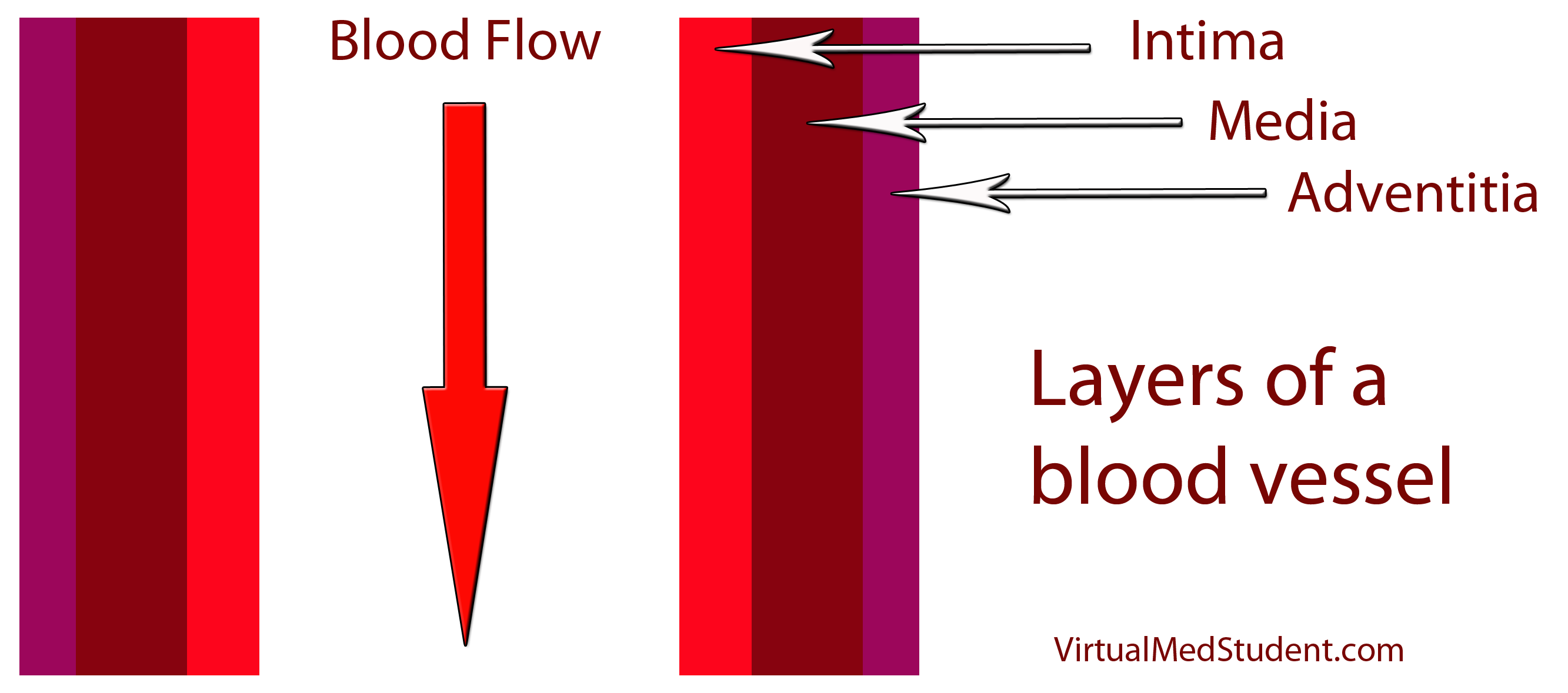
How does LDL lead to a plaque? The first step involves disruption of part of the blood vessel known as the intima. Blood vessels have three layers: intima, media, and adventitia. The intimal layer is the portion of the vessel directly in contact with blood flow. It is composed of endothelial cells, which have numerous important functions. The medial layer is smooth muscle that controls the diameter of the blood vessel; the adventitial layer is a connective tissue coating (ie: similar to the plastic coating surrounding an electrical wire) that anchors the vessel to adjacent structures in the body.

Blood Vessel with Plaque

When the intimal layer is disrupted, LDL particles floating in the blood get trapped in the vessel wall. Once trapped, they start an inflammatory reaction. White blood cells (a key component of inflammation) known as macrophages are recruited to the vessel’s walland gobble up the intruding LDL particles. At this point the macrophages are known as “foam cells”. Foam cells get enmeshed in a web of scar and smooth muscle. All of this together becomes a "plaque".

Symptoms

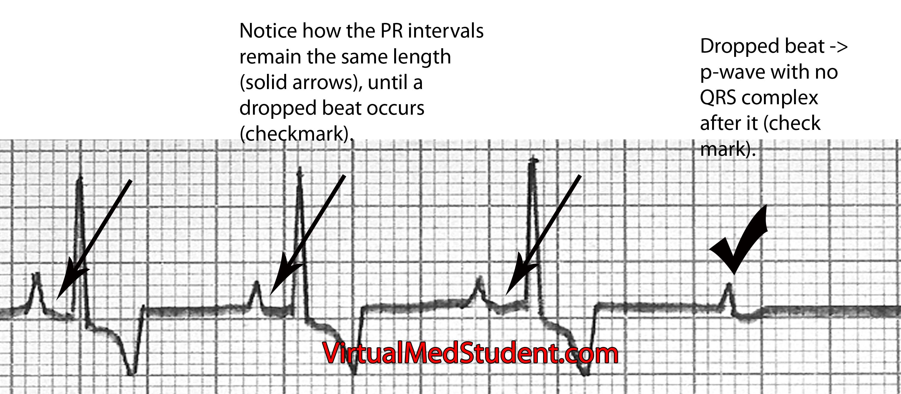
The symptoms of atherosclerosis depend on how large the plaques are, and where they are located. The three common symptoms associated with atherosclerosis:

(1) Angina (chest pain) – read about acute coronary syndromes
(2) Transient ischemic attacks (TIA)
(3) Vascular claudication

Angina refers to chest pain that occurs with exercise (by exercise we refer to any activity above normal daily activity). The pain is a result of atherosclerotic plaque blocking the increased blood flow needed to supply the heart during strenuous activities. In essence, what is happening is that the heart is "starving" for oxygen, which results in chest pain. When the patient stops exercising the heart no longer needs as much oxygen; the amount of blood flow is again adequate, and the chest pain ceases. Angina is a harbinger of a potential heart attack.

Transient ischemic attacks are due to atherosclerosis in the blood vessels leading to the brain. The temporary decrease in blood flow to the brain caused by the blockages can result in stroke-like symptoms that resolve within twenty-four hours. TIAs are harbingers of potential strokes in the future.

If atherosclerosis develops in the vessels going to the lower extremities, vascular claudication occurs. Claudication refers to pain in the legs that worsens while walking (or running). Similar to angina, the pain is due to blocked blood flow to the legs secondary to the plaques. A condition known as "Leriche’s syndrome" occurs when there is decreased blood flow to not only the legs, but also the vessels leading to the penis. This causes not only lower extremity claudication, but also impotence.

The complications of atherosclerosis stem from the symptoms. They include myocardial infarction (eg: heart attack), of which angina is a warning sign; as well as cerebrovascular accidents (eg: strokes), of which transient ischemic attacks serve as a warning. Worsening disease in the lower extremities can lead to gangrenous limbs and the need for potential amputation.

(1) Myocardial infarction (eg: heart attack)
(2) Cerebrovascular accidents (eg: stroke)
(3) Worsening lower extremity disease -> amputations

Diagnosis

Diagnosis of atherosclerotic disease depends on the location of symptoms. If anginal chest pain is the main symptom several different studies can be done. One such study is the “stress test”. There are several different permutations of the stress test.

All of them include two components. The first is a method of "visualizing" the heart. This can be done by ECG, echocardiography (ie: ultrasound of the heart), or nuclear/radioactive scans. The second component involves "stressing" the heart. This is most commonly done by exercising on a treadmill, but medications like dobutamine can also be used if the patient cannot exercise for whatever reason.

Stress Testing
Methods for visualizing the heart’s function – Electrocardiogram (ECG)
– Echocardiogram (echo)
– Nuclear studies
   
Methods for stressing the heart – Exercise (ie: treadmill)
– Medications (ie: dobutamine, persantine, etc.)

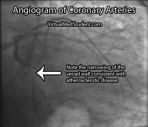
If the stress test is positive, or there is a high index of suspicion for atherosclerosis involving the coronary arteries, the next test performed is an angiogram. This involves injecting radio-opaque contrast material into the coronary arteries through a tiny catheter. The coronary arteries can then be visualized using x-rays. Areas where there is less contrast in the vessel indicate coronary atherosclerotic disease. The image below is an example of narrowing in the circumflex artery (indicated by the arrow).

Angiogram Circumflex Atherosclerosis

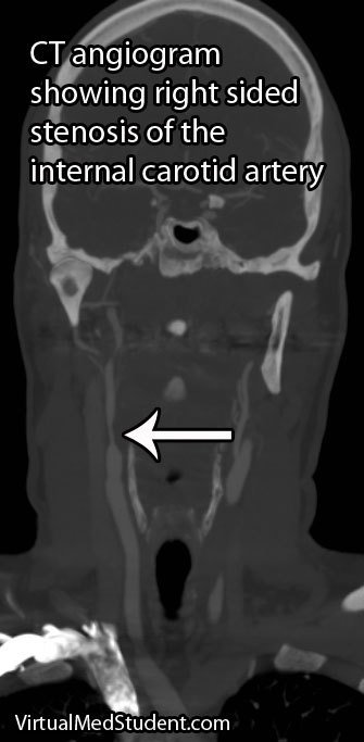
If the involved areas are the carotid arteries, a test known as "carotid artery duplex scanning" can be done. This involves using ultrasound to detect the amount of blood flow present in the vessel(s). Decreased rates are often attributable to atherosclerotic disease.

Finally, if disease is thought to be present in the lower extremities, a test known as the arterial brachial-ankle index (ABI) is obtained. This test involves taking the blood pressure in the arm and the leg and comparing the results. Blood pressure should be about equal in the upper and lower extremities resulting in a brachial to ankle ratio of 1.0. However, if there is atherosclerotic diseases in the lower extremities the ABI decreases to less than 1.0.

Location of suspected atherosclerotic disease: Initial test performed:
– Coronary arteries – Stress test
– Carotid arteries – Duplex scanning
– Arteries of lower extremities – Ankle-Brachial index (ABI)

Occasionally cardiologists or vascular surgeons will recommend a more invasive procedure known as a "catheterization". In this procedure a small catheter is threaded from a blood vessel in the groin to the area of interest (ie: the heart, carotids, vessels of the lower extremities). From there radio-opaque dye is injected. A fancy X-ray machine can then pick up locations of significant plaque formation.

Treatment

Treatment is designed to prevent further plaque formation. Since LDL plays a crucial role in this process, it is a target of medical therapy, but in order to understand treatment we have to first understand how cholesterol gets into the body. Cholesterol is either (a) absorbed by the gut from your diet, or (b) produced naturally by your body through a series of biosynthetic reactions.

The first interventions for decreasing cholesterol in the body are, you guessed it, dietary modifications. By decreasing the amount of cholesterol in the diet it is possible to decrease the amount of cholesterol, either free or in the form of LDL, that is in the blood stream. There is also some evidence that replacing saturated fats with polyunsaturated fats (ie: α-linolenic acid, an ω-3 fatty acid) can decrease atherosclerotic plaque formation.

Unfortunately, diet and exercise are not always enough to decrease blood cholesterol levels to acceptable limits. Several medications have been developed, and as one could imagine, they target either cholesterol in the gut, or the biosynthetic machinery that produces cholesterol naturally.

There are several types of drugs that bind cholesterol in the gut to keep it from being absorbed. They are referred to as "bile acid binding medications" or "bile acid sequestrants". It is important to realize that bile acids, which are secreted by the liver and gallbladder, are rich in cholesterol.

Under normal circumstances they are reabsorbed by the gut and recycled back into the bile pathway. By inhibiting their re-absorption, cholesterol from the blood stream is recruited by the liver to replace the bile acids that were previously being recycled. The ultimate effect is that these drugs decrease the amount of cholesterol in the blood stream. There are three common medications in this category: colesevelam, cholestyramine, and colestipol.

Another medication, ezetimibe, directly binds to free cholesterol in the gut. As a result, less cholesterol is delivered to the liver. To compensate, the liver increases its ability to absorb LDL and other forms of cholesterol from the blood resulting in decreased plasma levels.

Atherosclerosis
treatments:

(1) diet and
      exercise
(2) bile acid
      sequestrants
(3) statins
(4) niacin
(5) fibrates
Perhaps the most widely used class of medications used to treat atherosclerosis are known as “statins.” Statins such as atorvastatin and pravastatin inhibit an enzyme (ie: HMG-CoA reductase) that normally produces cholesterol in the body. These medications dramatically reduce the risk of stroke and heart attack in patients with atherosclerotic disease.

A vitamin, niacin (ie: vitamin B3), can also lead to favorable effects on the lipid profile of the blood. It increases "good" cholesterol (ie: HDL). It is believed to perform these feats by increasing the activity of an enzyme, lipoprotein lipase, which normally breaks down VLDL particles (a precursor of LDL).

A final category of medications known as the "fibrates" decrease fat content in the blood. They do not necessarily affect LDL levels significantly, but they do decrease another atherosclerotic forming fat type known as triglycerides. The two common fibrates in use today are gemfibrozil and fenofibrate.

Overview

Atherosclerosis is due to LDL trapping in blood vessel walls. This trapping results in inflammation and the formation of a fibromuscular plaque. Symptoms include angina (ie: chest pain), lower extremity claudication, and transient ischemic attacks. Diagnosis is made by stress testing, carotid ultrasound, and ankle-brachial index; more invasive testing with groin catheters can be performed as well. Treatment is based on decreasing cholesterol absorption from the gut, or decreasing the bodies natural mechanism for creating cholesterol.

References and Resources

  • Negi S, Nambi V. Coronary heart disease risk stratification: pitfalls and possibilities. Methodist Debakey Cardiovasc J. 2010 Dec;6(4):26-32.
  • Amarenco P, Lavallée PC, Labreuche J, et al. Prevalence of Coronary Atherosclerosis in Patients With Cerebral Infarction. Stroke. 2010 Nov 18. [Epub ahead of print].
  • Kumar V, Abbas AK, Fausto N. Robbins and Cotran Pathologic Basis of Disease. Seventh Edition. Philadelphia: Elsevier Saunders, 2004.
  • Lilly LS, et al. Pathophysiology of Heart Disease: A Collaborative Project of Medical Students and Faculty. Fourth Edition. Lippincott Williams and Wilkins, 2006.
  • Flynn JA. Oxford American Handbook of Clinical Medicine (Oxford American Handbooks of Medicine). First Edition. Oxford University Press, 2007.

Atrioventricular Heart Block: PR, QRS, Mobitz

Heart block, also known as atrioventricular (AV) conduction system block, refers to decreased conduction of electrical impulses through the heart. This decreased conduction occurs at the atrioventricular node of the heart’s conduction system. Heart block occurs in three flavors: type 1, type 2, and type 3. Each type has different causes and different treatments.

Pathology and Types

In type 1 AV block the PR interval on an ECG is longer than normal (> 200 msec), but every QRS complex has a preceding P-wave indicating that the electrical impulses from the atria (ie: the top chambers in the heart) are "making" it through the AV node to the ventricles. In type 1 block, the AV nodal conduction is slowed compared to normal "healthy" individuals, but the impulses are still able to get all the way through the conduction system of the heart.

Type 1 AV node block = PR interval > 200 msec, no missed beats.

Type 2 heart block occurs in 2 flavors: Mobitz type 1 and type 2. In Mobitz type 1 the amount of delay between the P-wave and QRS complex (ie: the PR interval) gradually increases until a beat is dropped. It is caused by impaired conduction in the AV node. Mobitz type 2 occurs when the PR interval remains stable, and then out of the blue a missed beat occurs; this is caused most commonly by slowed conduction through the bundle of His or bundle branches of the heart’s conduction system.

Mobitz type 1 = increasing PR interval until dropped beat occurs (image below). Mobitz type 1 is also referred to as "Wenckeback" block.

Mobitz Type 1

Mobitz type 2 = PR interval remains the same until a dropped beat occurs (image below). Mobitz type 2 is also referred to as "Hay" block.

Mobitz Type 2

Finally, type 3 AV nodal block occurs when the atria and ventricles of the heart beat independently of one other. The most common causes of 3rd degree block are severe diseases of the AV nodal system due to age, previous heart attack, drug and medication toxicity (ie: digitalis), and untreated Lyme disease. The atria generally beat at a faster rate than the ventricles. In other words, the ECG will show more P-waves than QRS complexes. The QRS complexes may be narrow or wide depending on where in the conduction system the escape rhythm originates.

Type 3 heart block = atria (p-waves) and ventricles (QRS) beat independently of each other; no impulses from the atria make it to the ventricles (image below).

AV nodal block type 3

Symptoms

The symptoms of heart block depend on how slow the rate becomes. Patients with type 1 block rarely become symptomatic because the atria are still setting the pace of the entire hearts rhythm. However, in Mobitz type 2 and type 3 block the heart rate can become very slow. When this occurs symptoms such as syncope (ie: fainting), extreme fatigue, shortness of breath, and dizziness can occur. These symptoms occur because the heart is not pumping blood fast enough to keep up with the body’s demand; the result is a decrease in cardiac output.

Diagnosis

Diagnosis of heart block can be made by looking at the characteristic findings on ECG.

Treatment

Treatment of AV nodal dysfunction is dependent on the type. Most patients with type 1 heart block do not need treatment. Type 1 heart block can be considered a "healthy" variant of AV nodal conduction, which is slower than most people in the population. The only instance where type 1 block may need further work up is in elderly patients who have other signs or symptoms of coronary artery disease.

Type Treatment
Type 1

Usually none, look for causes such as coronary artery disease in elderly patients

Type 2 – Mobitz type 1

Usually none, but pacemaker if symptomatic. Also discontinue any drugs that slow AV node.
Type 2 – Mobitz type 2 Pacemaker
Type 3 Pacemaker

Mobitz type 1 (ie: one of the 2 subgroups of type 2 heart block) also generally requires no treatment. If patients are symptomatic pacemaker insertion may be considered. Mobitz type 2 is more worrisome because it often progresses to 3rd degree heart block. In these patients, pacemakers are often inserted to ensure that the heart beats at a pre-defined and safe rate.

Type 3 heart block is almost always controlled with pacemaker insertion. The reason is that the ventricles often beat at a rate that is too slow for normal daily activity. A pacemaker will keep the heart beating at a pre-defined rate to ensure that symptoms do not develop.

Overview

Heart block (aka: atrioventricular block) occurs when impulses travel too slowly, or not at all through the AV node. There are different types depending on how severe the block is. In type 1 AV block impulses are slowed, but cause no conduction block. In type 2 missed beats can occur, and in type 3 the atria beat independently of the ventricles indicating complete conduction block. Symptoms depend on the severity of block but can include fainting, shortness of breath, dizziness, and fatigue. Diagnosis is made by ECG. Treatment is dependent on the type of block, but can include placement of a pacemaker.

References and Resources

Hypertension: Understanding and Managing High Blood Pressure

The pathology of essential hypertension is not well understood. However, there are numerous theories, each with supporting evidence.

Genetic causes are supported by the fact that children of hypertensive parents have an increased risk of developing the disease. However, specific known genetic mutations are not common. When mutations are responsible for essential hypertension they often involve the sodium and chloride channels in kidney cells, or mutations in the genes responsible for producing the proteins of the renin-angiotensin-aldosterone system. Sodium, renin, angiotensin, and other molecules play a vital role in blood pressure. In some hypertensive patients these systems are abnormal and can lead to elevated blood pressure.

Some patients with essential hypertension appear to be “salt sensitive”. Salt, or more specifically sodium, appears to play a role in the development of hypertension. Some patients likely have a genetic predisposition to retain sodium. Excess sodium enters the blood stream where it exerts an osmotic pull on water in adjacent tissues. Fluid from body tissues is "sucked" into the blood stream resulting in increased blood volumes. The expanded blood volume increases the pressure within the blood vessel causing hypertension. Although salt plays a role in hypertension, new research has shown its importance may not be as clear as once thought.

There are other known causes of hypertension, but they constitute a relatively small proportion of cases. They are discussed in separate articles.

Signs and Symptoms

Essential hypertension in its earliest stages does not cause any signs or symptoms. Many people do not know they have high blood pressure. However, if left untreated hypertension can cause serious long term consequences.

One such consequence is an increase in the risk of cardiovascular disease. The risk of cardiovascular disease doubles with each 20/10 mmHg increase in blood pressure beyond a "baseline" of 115/75 mmHg! After years of pumping at elevated pressures the heart undergoes physical changes. Like any good muscle, it becomes larger because it is having to pump harder than normal. The result is a process known as concentric hypertrophy. The added muscle mass of the heart results in increased oxygen demand and the potential for heart attacks and heart failure.

Overall, nearly a third of heart attacks are attributable to high blood pressure. In addition, the risk of stroke is also dramatically higher in untreated hypertensive patients. The small blood vessels in the kidney and retina can also be damaged by years of high blood pressure resulting in kidney failure (hypertensive nephropathy) and blindness (hypertensive retinopathy).

Diagnosis

The diagnostic parameters of hypertension are constantly shifting. Currently the diagnosis can be made if a patient presents with a severely elevated blood pressure (systolic blood pressure > 200 and/or a diastolic blood pressure > 120) and/or signs or symptoms referable to the elevated blood pressure.

Severity Classification:
(1) Pre-hypertensive:
       130-140/80-90
(2) Grade 1:
       140-160/90-100
(3) Grade 2:
        >160/>100
In patients with less severe elevations, it is generally recommended that the blood pressure be measured several times over a period of multiple weeks. If the average of these readings is a systolic blood pressure of 140 or greater, or a diastolic blood pressure of 90 or greater than the diagnosis can be made. If the blood pressue stays between 120 and 130 systolic, and 80 to 90 diastolic the diagnosis of "pre-hypertension" is made meaning the patient is at risk of becoming hypertensive.

Some patients may have "white coat" hypertension simply by being in a doctor’s office (and the anxiety that this can provoke! Gosh, I hate going to the doctor’s office!). If this is the case the patient should be instructed to take their blood pressure at home and keep a log of the results.

Treatment

Treatment of hypertension consists of lifestyle modifications and/or using medications from several different pharmacologic categories. These medications may be used alone, or in combination, depending on each individual patient’s needs.

Patients who are pre-hypertensive or have stage 1 hypertension should be started on “lifestyle” modification therapy for at least 6 months prior to starting medicines (assuming there are no other health issues like diabetes or coronary artery disease). Lifestyle therapy consists of increasing aerobic exercise, as well as adhering to the “DASH” diet. DASH stands for "dietary approaches to stop hypertension" and consists of eating fruits, vegetables, low-fat dairy, whole grains, poultry and fish while reducing (or eliminating) red meat and sugars. This intervention alone can decrease systolic pressures over 10 mmHg and diastolic pressures over 5 mmHg.

Sometimes diet and exercise aren’t enough so we may have to resort to medications to control blood pressure. The first category of medications are known as diuretics, most commonly of the thiazide class. Thiazide diuretics work by inhibiting the re-absorption of sodium at the distal convoluted tubule of the kidney. Less sodium means less circulating blood volume, and therefore decreased blood pressure. Hydrochlorothiazide is a commonly used thiazide diuretic.

The second category of medications is known as β-blockers. β-blockers lower blood pressure by slowing the heart rate and indirectly lowering the amount of angiotensin II (angiotensin II is a potent natural blood vessel constrictor produced by the body). β-blockers reduce renin synthesis by specialized cells in the kidney.

A third category of medications known as angiotensin converting enzyme inhibitors (ACEIs) interfere with the formation of angiotensin II. ACEIs inhibit an enzyme present in the lung, which converts angiotensin I into the more potent vasoconstrictor angiotensin II. Interestingly, ACEIs also increase the level of bradykinin; this molecule causes blood vessels to dilate, which helps to further lower pressure.

There are also medications called direct angiotensin receptor blockers (ARBs), which inhibit the actions of angiotensin II. They do this by blocking angiotensin IIs ability to bind to its normal receptor sites.

Some medications, the calcium channel blockers (CCBs), lower blood pressure by inhibiting the contraction of smooth muscle cells that line the blood vessel walls. A specific subcategory of calcium channel blockers known as dihydropyridines block calcium from flowing into vascular smooth muscle cells. This results in decreased contraction of the muscle surrounding the vessel. As a result the vessel remains dilated, which lowers pressure.

Overview

The pathology of essential hypertension is not well understood. Several theories have been postulated, but it is likely a combination of genetic predispositions that result in this type of high blood pressure. Years of elevated blood pressure lead to cardiovascular disease including heart failure, heart attacks, and stroke, as well as damage to the kidneys and retinas. Treatment is with lifestyle modifications, diuretics, β-blockers, calcium channel blockers, angiotensin converting enzyme inhibitors, and angiotensin receptor blockers.

References and Resources

Systemic Vascular Resistance: Radius, Length, Viscosity

The systemic vascular resistance is the resistance that blood “sees” as it travels throughout the circulatory system of the body.

It is controlled by three different factors: length of the blood vessel (l), radius of the blood vessel (r), and the viscosity of the blood (η). The equation that relates these three factors to resistance is known as Poiseuilles’ equation:

R ≈ (η x l) / r4

Let’s discuss how each of the factors above influences resistance. The longer the vessel, the more likely blood will sludge and stick up against the walls; this tendency intuitively causes an increase in overall resistance. Blood vessel length does not typically change during adulthood, therefore its contribution to vascular resistance in humans is relatively constant.

When blood is more viscous than usual the resistance also increases. For example, molasses flows very slowly because it is highly viscous; blood is no different. There are a few uncommon instances in human physiology where blood viscosity increases; therefore, like vessel length, its contribution to vascular resistance is also relatively constant.

The most important parameter is the radius of the vessel. When the radius of a vessel shrinks, the resistance increases; when the radius of a vessel dilates, the resistance drops. Imagine trying to pump a set amount of fluid through a small opening versus a larger opening. There is much less resistance encountered through the larger opening. The same is true for large and small blood vessels. Even more importantly, as indicated by Poiseuille’s equation above, when the radius of a vessel is halved, the resistance increases by 16 fold! Therefore, even small changes in blood vessel diameter can have dramatic consequences on the bodies’ vascular resistance.

How the Body Controls Resistance

How do you think the body controls resistance most effectively? You guessed it! By controlling the radius of blood vessels. When there is an abnormal drop in vascular resistance, the body compensates by pumping out hormones (ie: epinephrine and norepinephrine); these hormones cause muscle cells surrounding blood vessels to constrict. Constriction leads to a decreased radius of the vessel, which leads to increased resistance.

Clinical Measurements

How do you measure systemic vascular resistance? Well, first off, systemic vascular resistance is not measured, but is calculated from other cardiovascular vital signs such as the mean arterial pressure (MAP), cardiac output (CO), and central venous pressures (CVP). The equation that relates these three values to resistance is:

SVR = [(MAP – CVP) / CO] x 80

Mean arterial pressure is a sort-of average between the systolic and diastolic blood pressure readings. It can be calculated using blood pressures obtained from an arterial catheter or a blood pressure cuff.

Central venous pressures are normally low, and do not contribute much to vascular resistance, but when very accurate measurements are needed they can be obtained from a central venous and/or a pulmonary artery catheter.

Cardiac output can be obtained from a pulmonary artery catheter if accurate values are needed; it can also be roughly estimated from a cardiac echocardiogram (ie: an ultrasound of the heart).

Role in Disease

So what’s the big deal? Why all this talk about vascular resistance? Vascular resistance is important because it is one determinant of blood pressure, and therefore organ perfusion.

The mean arterial pressure is calculated by multiplying the cardiac output by the systemic vascular resistance, and then adding the result to the central venous pressure (MAP = CO x SVR + CVP, a reorganization of the equation above).

Since the mean arterial pressure is the driving force behind the delivery of blood and oxygen to the organs, it is of vital importance that it stays above a certain threshold. Otherwise organs will not receive enough oxygen and will eventually die. One way of ensuring that the mean arterial pressure stays within a set range is for the body to constrict and/or dilate the vessels as needed.

On the flip side, if systemic vascular resistance is too "clamped" down then hypertension occurs. If severe enough, the heart may not be able to generate enough force to eject an adequate amount of blood into the tightened arterial system, which over time could result in systolic heart failure.

Overview

Systemic vascular resistance is the resistance blood sees as it travels throughout the bodies blood vessels. It is influenced by the length and radius of the blood vessel(s), as well as the viscosity of blood. It is calculated from the mean arterial pressure, central venous pressure, and cardiac output. Systemic vascular resistance plays a vital role in maintaining blood pressures within set ranges so that organ perfusion is maximized.

References and Resources