Hypertrophic Cardiomyopathy: Athletes and Genetic Mutations

Hypertrophic cardiomyopathy occurs when the size of heart muscle cells increase (aka: hypertrophy). Genetic mutations in DNA that codes for heart muscle cell proteins are responsible for the development of hypertrophic cardiomyopathy. Most of these mutations are in DNA that code for sarcomere proteins (ie: myosin, actin, troponin, etc.). The mutated proteins cause decreased contractile function. As a result, the muscle cell hypertrophies (enlarges) in an attempt to overcome the decreased contractility. The result is a disorganized pattern of muscle cell fibers with intervening fibrosis (ie: scar tissue).

Signs and Symptoms

Since the myocardium is hypertrophied there is less ventricular compliance (ie: the heart becomes stiff). This stiffness decreases the filling capacity of the ventricle. The result is diastolic dysfunction, or a decreased ability of the heart to fill during its relaxation phase. High diastolic pressures occur leading to the back-up of blood into the left atrium, pulmonary veins, and pulmonary capillaries. Excess fluid in the pulmonary capillaries causes pulmonary edema with resultant shortness of breath and exercise intolerance.

In addition, angina (chest pain) can occur even without co-existing coronary artery disease because the increased muscle mass of the ventricle results in a higher oxygen demand. Under strenuous conditions the hypertrophied muscle cannot get enough oxygen, which causes chest pain.
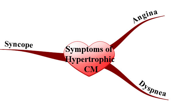
Symptoms and Signs of Hypertrophic Cardiomyopathy
Syncope (ie: fainting) is another common symptom that is usually due to arrhythmias caused by the abnormal myocyte architecture.

Physical exam can reveal an S4 gallop (aka: atrial gallop), which is caused by the atrium forcing blood into a stiff left ventricle during the "atrial kick" at the end of diastole.

Murmurs can also be heard, usually mitral regurgitation and a systolic outflow obstruction murmur. Mitral regurgitation occurs because the hypertrophied ventricular septum acts as a barrier to blood flow into the aorta. As a result, during systole blood will flow backwards through the mitral valve into the left atrium. Blood flowing across the septal barrier into the aorta will create an obstruction murmur. The obstruction murmur worsens with valsalva, which distinguishes it from the murmur of aortic stenosis.

Diagnosis

The work-up is very similar to dilated cardiomyopathy, except ancillary studies are usually not helpful. Echocardiography (ie: ultrasound of the heart) is the gold standard and will show the hypertrophic myocardium. ECG will often reveal left ventricular hypertrophy and left atrial hypertrophy. Arrhythmias may sometimes be observed on ECG as well. Prominent Q-waves can be seen in the lateral leads (ie: V4-V6) and inferior leads (II, III, aVF); this is the result of greater depolarization of the hypertrophied septum (remember depolarization of the septum starts on the left side and moves rightward creating a downward deflection in leads on the opposite side of the body, before the left ventricle "overpowers" the ECG findings).

Treatment

β-blockers are the mainstay of treatment. They decrease the heart rate and allow increased diastolic filling times, which leads to decreased outflow obstruction; they also decrease myocardial oxygen demand leading to decreased anginal symptoms.

Prevention of fatal arrhythmias is important in hypertrophic cardiomyopathy. Medical management of arrhythmias is accomplished with amiodarone and/or disopyramide. In some patients, strong consideration should be given to an implantable cardiac defibrillator, especially those at high risk of sudden death. Surgery with partial myomectomy to remove some of the hypertrophied muscle can also be done if the patient is unresponsive to medical management.

Since hypertrophic cardiomyopathy is caused by genetic mutations, genetic counseling should be offered to children of affected parents. First degree relatives should undergo screening with echocardiography as well.

Unlike dilated cardiomyopathy, diuretics should be used sparingly because they can worsen outflow obstruction by causing decreased venous return to the left ventricle. Digoxin is also contraindicated because it can worsen outflow obstruction. When considering treatment options it is important to remember that the problem in hypertrophic cardiomyopathy is diastolic, not systolic dysfunction.

Prognosis depends on the type and severity of the genetic mutation involved. Some mutations result in minimal morbidity and a normal life span, whereas others can cause significant heart failure symptoms. Overall mortality is roughly 5% per year secondary to ventricular fibrillation; therefore, even minimally symptomatic patients must be monitored closely.

Overview

The cause of hypertrophic cardiomyopathy is genetic. Diagnosis is made with echocardiography (ie: ultrasound of the heart). Treatment is generally with beta blockers, amiodarone, implantable cardiac defibrillators (ICD), and myomectomy in select patients. All 1st degree relatives should be offered genetic counseling and undergo screening echocardiography. Prognosis is variable and depends on the mutation type.

Related Articles

References and Resources

  • Bos JM, Ommen SR, Ackerman MJ. Genetics of hypertrophic cardiomyopathy: one, two, or more diseases? Curr Opin Cardiol. 2007 May;22(3):193-9.
  • Bo CY, López B, Coelho-Filho OR, et al. Myocardial fibrosis as an early manifestation of hypertrophic cardiomyopathy. N Engl J Med. 2010 Aug 5;363(6):552-63.
  • Kumar V, Abbas AK, Fausto N. Robbins and Cotran Pathologic Basis of Disease. Seventh Edition. Philadelphia: Elsevier Saunders, 2004.
  • Lilly LS, et al. Pathophysiology of Heart Disease: An Introduction to Cardiovascular Disease. Seventh Edition. Lippincott Williams and Wilkins, 2006.
  • Flynn JA. Oxford American Handbook of Clinical Medicine (Oxford American Handbooks of Medicine). First Edition. Oxford University Press, 2007.

Hypertension: Understanding and Managing High Blood Pressure

The pathology of essential hypertension is not well understood. However, there are numerous theories, each with supporting evidence.

Genetic causes are supported by the fact that children of hypertensive parents have an increased risk of developing the disease. However, specific known genetic mutations are not common. When mutations are responsible for essential hypertension they often involve the sodium and chloride channels in kidney cells, or mutations in the genes responsible for producing the proteins of the renin-angiotensin-aldosterone system. Sodium, renin, angiotensin, and other molecules play a vital role in blood pressure. In some hypertensive patients these systems are abnormal and can lead to elevated blood pressure.

Some patients with essential hypertension appear to be “salt sensitive”. Salt, or more specifically sodium, appears to play a role in the development of hypertension. Some patients likely have a genetic predisposition to retain sodium. Excess sodium enters the blood stream where it exerts an osmotic pull on water in adjacent tissues. Fluid from body tissues is "sucked" into the blood stream resulting in increased blood volumes. The expanded blood volume increases the pressure within the blood vessel causing hypertension. Although salt plays a role in hypertension, new research has shown its importance may not be as clear as once thought.

There are other known causes of hypertension, but they constitute a relatively small proportion of cases. They are discussed in separate articles.

Signs and Symptoms

Essential hypertension in its earliest stages does not cause any signs or symptoms. Many people do not know they have high blood pressure. However, if left untreated hypertension can cause serious long term consequences.

One such consequence is an increase in the risk of cardiovascular disease. The risk of cardiovascular disease doubles with each 20/10 mmHg increase in blood pressure beyond a "baseline" of 115/75 mmHg! After years of pumping at elevated pressures the heart undergoes physical changes. Like any good muscle, it becomes larger because it is having to pump harder than normal. The result is a process known as concentric hypertrophy. The added muscle mass of the heart results in increased oxygen demand and the potential for heart attacks and heart failure.

Overall, nearly a third of heart attacks are attributable to high blood pressure. In addition, the risk of stroke is also dramatically higher in untreated hypertensive patients. The small blood vessels in the kidney and retina can also be damaged by years of high blood pressure resulting in kidney failure (hypertensive nephropathy) and blindness (hypertensive retinopathy).

Diagnosis

The diagnostic parameters of hypertension are constantly shifting. Currently the diagnosis can be made if a patient presents with a severely elevated blood pressure (systolic blood pressure > 200 and/or a diastolic blood pressure > 120) and/or signs or symptoms referable to the elevated blood pressure.

Severity Classification:
(1) Pre-hypertensive:
       130-140/80-90
(2) Grade 1:
       140-160/90-100
(3) Grade 2:
        >160/>100
In patients with less severe elevations, it is generally recommended that the blood pressure be measured several times over a period of multiple weeks. If the average of these readings is a systolic blood pressure of 140 or greater, or a diastolic blood pressure of 90 or greater than the diagnosis can be made. If the blood pressue stays between 120 and 130 systolic, and 80 to 90 diastolic the diagnosis of "pre-hypertension" is made meaning the patient is at risk of becoming hypertensive.

Some patients may have "white coat" hypertension simply by being in a doctor’s office (and the anxiety that this can provoke! Gosh, I hate going to the doctor’s office!). If this is the case the patient should be instructed to take their blood pressure at home and keep a log of the results.

Treatment

Treatment of hypertension consists of lifestyle modifications and/or using medications from several different pharmacologic categories. These medications may be used alone, or in combination, depending on each individual patient’s needs.

Patients who are pre-hypertensive or have stage 1 hypertension should be started on “lifestyle” modification therapy for at least 6 months prior to starting medicines (assuming there are no other health issues like diabetes or coronary artery disease). Lifestyle therapy consists of increasing aerobic exercise, as well as adhering to the “DASH” diet. DASH stands for "dietary approaches to stop hypertension" and consists of eating fruits, vegetables, low-fat dairy, whole grains, poultry and fish while reducing (or eliminating) red meat and sugars. This intervention alone can decrease systolic pressures over 10 mmHg and diastolic pressures over 5 mmHg.

Sometimes diet and exercise aren’t enough so we may have to resort to medications to control blood pressure. The first category of medications are known as diuretics, most commonly of the thiazide class. Thiazide diuretics work by inhibiting the re-absorption of sodium at the distal convoluted tubule of the kidney. Less sodium means less circulating blood volume, and therefore decreased blood pressure. Hydrochlorothiazide is a commonly used thiazide diuretic.

The second category of medications is known as β-blockers. β-blockers lower blood pressure by slowing the heart rate and indirectly lowering the amount of angiotensin II (angiotensin II is a potent natural blood vessel constrictor produced by the body). β-blockers reduce renin synthesis by specialized cells in the kidney.

A third category of medications known as angiotensin converting enzyme inhibitors (ACEIs) interfere with the formation of angiotensin II. ACEIs inhibit an enzyme present in the lung, which converts angiotensin I into the more potent vasoconstrictor angiotensin II. Interestingly, ACEIs also increase the level of bradykinin; this molecule causes blood vessels to dilate, which helps to further lower pressure.

There are also medications called direct angiotensin receptor blockers (ARBs), which inhibit the actions of angiotensin II. They do this by blocking angiotensin IIs ability to bind to its normal receptor sites.

Some medications, the calcium channel blockers (CCBs), lower blood pressure by inhibiting the contraction of smooth muscle cells that line the blood vessel walls. A specific subcategory of calcium channel blockers known as dihydropyridines block calcium from flowing into vascular smooth muscle cells. This results in decreased contraction of the muscle surrounding the vessel. As a result the vessel remains dilated, which lowers pressure.

Overview

The pathology of essential hypertension is not well understood. Several theories have been postulated, but it is likely a combination of genetic predispositions that result in this type of high blood pressure. Years of elevated blood pressure lead to cardiovascular disease including heart failure, heart attacks, and stroke, as well as damage to the kidneys and retinas. Treatment is with lifestyle modifications, diuretics, β-blockers, calcium channel blockers, angiotensin converting enzyme inhibitors, and angiotensin receptor blockers.

References and Resources