Hemangioblastoma of the Central Nervous System

Hemangioblastomas are benign tumors that occur in the central nervous system, most commonly in the cerebellum. The exact cell from which they arise is unknown; however, they are believed to be meningeal (ie: the cells that make up the covering of the brain) in origin.

Hemangioblastomas can be solid or cystic in appearance. However, all hemangioblastomas have capillary networks that are lined by endothelial cells. Interspersed between the capillaries are pericytes and stromal cells with a polygonal appearance.

The arrangement of these various cell types can occur in three different architectures: juvenile, clear cell, or transitional. Each architecture has a specific ratio of capillaries to stroma (connective tissue), with the juvenile type having the greatest amount of capillary tissue, and the clear cell type having the greatest amount of stromal tissue.

About a quarter of cases are related to a genetic disorder known as von Hippel-Lindau syndrome. von Hippel-Lindau syndrome is the result of a genetic mutation in the VHL gene on chromosome 3. The protein product of this gene is a tumor suppressor; when mutated it is unable to suppress the abnormal growth of tumor cells. As a result, patients with von Hippel-Lindau syndrome develop hemangioblastomas of the brain and retina, renal cell carcinoma, and other tumor types.

Hemangioblastomas are most commonly found in the cerebellum, but on occassion will affect the cervical spinal cord and brainstem. They are most commonly seen in males starting at around 20 years of age.

Signs and Symptoms

Patients with cerebellar hemangioblastomas present with numerous signs and symptoms. Many patients will complain of headache, nausea, and vomiting. This is often due to the tumor compressing the cerebral aqueduct, which causes cerebrospinal fluid to “back up” in the brain leading to hydrocephalus and increased pressure inside the head.

In addition, many people with hemangioblastomas will have evidence of cerebellar dysfunction on physical exam. These signs include ataxia (ie: wobbly gait), dysmetria (ie: uncoordinated movements of the limbs), and dysdiadochokinesia (ie: difficulty repeating rapid alternating movements).

If the tumor is present in the spinal cord, symptoms may include weakness, spasticity, numbness, or other sensory changes.

Interestingly, hemangioblastomas can secrete an analogue of the hormone erythropoietin. This hormone causes bone marrow to pump out more red blood cells. As a result, some patients may have an increased number of red blood cells; this is known as polycythemia.

Diagnosis
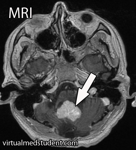
Hemangioblastoma MRI
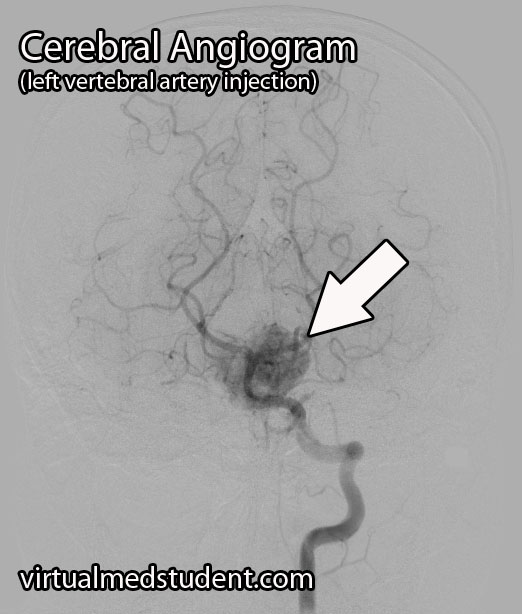
Hemangioblastoma Angiogram
A presumptive diagnosis can be made using epidemiology and imaging studies.

A tumor located in the cerebellum of an adult with certain characteristics on CT, MRI, and cerebral angiogram can make the diagnosis of hemangioblastoma likely.

However, the final diagnosis can only be made by looking at a sample of the tumor under the pathology microscope.

Treating These Bastards

Definitive treatment is surgical resection. However, hemangioblastomas can be highly vascular, which means they tend to bleed like stink! Therefore, preoperative embolization by an interventional neuro-radiologist can decrease the amount of bleeding that occurs during surgery.

Radiation therapy is also sometimes used as an adjunctive treatment. It may help slow the growth of the tumor, but will not cure it. Radiation therapy can be used in patients who are unable to undergo surgical resection, or if the tumor is inaccessible via traditional surgical means.

Recap It All…

Hemangioblastomas are highly vascular, but benign central nervous system tumors of undetermined origin. They are associated with von Hippel-Lindau syndrome. Signs and symptoms include headache, nausea, and vomiting, as well as cerebellar dysfunction. Diagnosis is based on imaging and pathological findings at the time of surgical resection. Surgery is the treatment of choice, although embolization and radiation therapy may also be used as an adjunct.

More Interesting Neurological Problems…

Some More Expert Ideas Below

Chiari Malformation: Type1, Tonsils and Syrinx

The Chiari malformations are a group of disorders characterized, at least in part, by herniation of hindbrain structures through the foramen magnum at the base of the skull. They are categorized as type 1, type 2, and type 3 depending on clinical and radiographic findings.

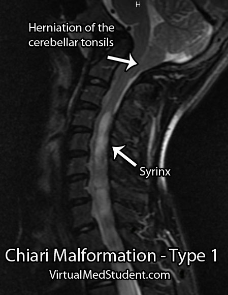
This article will focus on type 1 Chiari malformations. The definition of this malformation has been debated, but most agree that the combination of herniated cerebellar tonsils (usually defined as greater than 5mm below the foramen magnum), with or without a syrinx, in the setting of referable symptoms is sufficient to make the diagnosis.

So why do the cerebellar tonsils herniate? Nobody knows for sure! We do know that type 1 malformations can be congenital or acquired. One theory is that tonsillar herniation is a result of an abnormally small posterior fossa (ie: the bones that compose the base of the skull). A small posterior fossa may be caused by under-development of the occipital somites in-utero (the fetal precursors that form bone and connective tissues), premature fusion of the cranial bones (ie: craniosynostosis), or medical conditions that promote abnormal bony growth.

Other experts advocate that abnormal cerebrospinal fluid pressures between the brain and spine may cause the tonsils to herniate downwards.

Regardless of how you slice it, we can say with certainty that there are multiple potential etiologies for type 1 Chiari malformations.

Type 1 Chiari malformations are commonly associated with a finding known as a “syrinx”. A syrinx is an abnormal fluid filled cavity that is seen in the cervical and/or thoracic spinal cord. It may represent an enlargement and extension of the central canal of the cord, in which case it is termed hydromyelia; it may also represent a complex glial lined cavity, which is referred to as syringomyelia. Regardless, syrinxes are found in 30% to 70% of type 1 Chiari malformations.

For unclear reasons, type 1 Chiari malformations with a syrinx are associated with scoliotic curves of the spine (especially left sided curves). It is believed that the syrinx puts pressure on the motor pathways of the spinal cord. This results in weakness of the paraspinal muscles causing the vertebral column to curve.

Signs and Symptoms

The most common presenting symptom of a type 1 Chiari malformation is pain. The pain is usually located at the back of the head and upper neck. Additionally, a cape-like sensation loss, as well as problems with vision and/or hearing may also be present.

Myelopathic signs or symptoms may be present if there is a syrinx. Myelopathic patients present with a combination of gait dysfunction, hand clumsiness, weakness, abnormally brisk reflexes, spasticity, and Lhermitte’s sign.

Diagnosis

Type 1 Chiari Malformation
Diagnosis of a type 1 Chiari malformation is made when an MRI shows abnormal herniation of the cerebellar tonsils, with or without an associated syrinx, in the context of appropriate signs and/or symptoms.

Treatment

The treatment of Chiari malformation is with surgical decompression. Most commonly this involves "shaving" off part of the occipital bone and removing the C1 lamina. This effectively decompresses the spinal cord and cerebellar tonsils.

If an associated syrinx is present, many neurosurgeons will open the dura (ie: the lining of the spinal cord) and perform a "duraplasty"; during the duraplasty a patch is sewn into place to give the spinal cord and cerebellar tonsils more room. Duraplasty generally improves the size and severity of the syrinx over time, but adds risk and complications to the procedure.

Some neurosurgeons will surgically shrink the cerebellar tonsils after opening the dura. This is done with bipolar electrocautery and serves to further decompress the area.

Overview

Type 1 Chiari malformations are hindbrain abnormalities characterized by herniation of the cerebellar tonsils below the foramen magnum. They are associated with cervicothoracic syrinxes as well as neuromuscular scoliosis. Symptoms can range from pain to neurological deficits. Treatment is with surgical decompression, although the exact type of decompression has been the subject of intense research.

References and Resources

Cerebellum, Purkinje, Mossy, and All That Jazz!

Cellular Anatomy

The cerebellum is a large outgrowth on the backside of the brainstem that looks like a piece of cauliflower. It is the great "modulator" of movement. It compares the movement that the brain wants to do with what the body is actually doing. It then makes fine adjustments to ensure that the intended movement is smooth and fluid. It works in conjunction with the basal ganglia and motor cortex to modulate movement.

As can be expected, the cerebellum is highly complex with inputs and outputs going to various regions of the brain and spinal cord. Before we delve into the circuitry of the cerebellum, we need to first understand its cellular architecture…

Unlike most of the cerebral cortex, the cerebellar cortex has only three layers. They include – from most superficial to deep – the molecular layer, the Purkinje cell layer, and the granular layer.

The molecular layer is composed of connections between the dendrites (ie: information receiving processes) of Purkinje cells and the axons (ie: information sending processes) of granule cells. The molecular layer also contains stellate and basket cells, which help modulate the connections between Purkinje and granule neurons.

The Purkinje layer is, you guessed it, composed of Purkinje neurons. These cells send dendrites into the molecular layer where they receive information from granule cells. Purkinje cells also directly receive signals from other areas of the brain and spinal cord. Purkinje cells then send information along to the deep cerebellar nuclei, which are discrete collections of neurons within the cerebellum.

Finally, the granular layer is populated with granule and Golgi cells. They send axons into the molecular layer, which serve to pass on information to the dendrites of Purkinje cells in the molecular layer.

Confused yet???

Input Circuitry

In order for the cerebellum to modulate movement it must receive input from multiple sources. At this point, we have to introduce three new terms: climbing fibers, mossy fibers, and aminergic fibers.

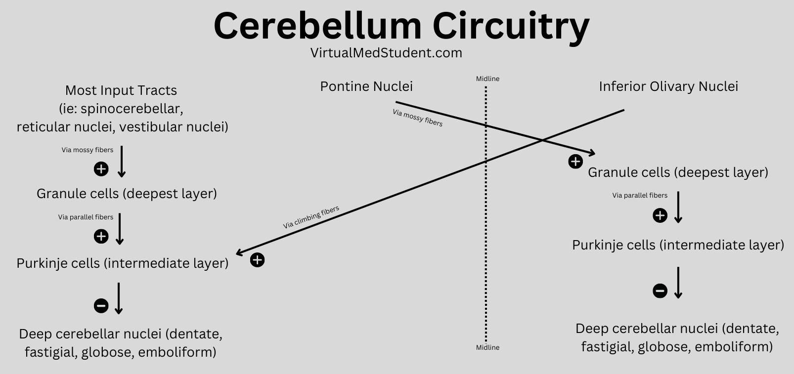
Neurons in an area of the brainstem known as the "olive" (a part of the medulla oblongata) send climbing fibers into the cerebellum. Each climbing fiber forms direct and powerful excitatory connections with multiple Purkinje cells (interestingly, each Purkinje cell only receives input from one climbing fiber).

The second input, mossy fibers, originate from several different areas outside the cerebellum (see table below). These axons form connections with the granule cells. The granule cells, if you remember from our discussion above, send axons into the molecular layer where they form connections to Purkinje cell dendrites. Therefore, mossy fibers indirectly influence Purkinje cells through the "intermediary" granule cells.

Input Connections (ie: Afferent Fibers) to the Cerebellum

Source of Input Type of Fiber Target of Input
Olivary nuclei (brainstem) Climbing fibers Contralateral cerebellum
Pontine nuclei (brainstem) Mossy fibers Contralateral cerebellum
Reticular nuclei (brainstem) Mossy fibers Ipsilateral cerebellum
Ventral spinocerebellar tract Mossy fibers Ipsilateral cerebellum
Dorsal spinocerebellar tract Mossy fibers Ipsilateral cerebellum
Vestibular nuclei Mossy fibers Ipsilateral cerebellum
Locus ceruleus Norepinephrine Bilateral projections
Raphe nucleus Serotonin Bilateral projections

Aminergic fibers originate from the locus ceruleus in the pons, and the raphe nuclei of the midbrain, pons, and medulla. The locus ceruleus fibers "spit" norepinephrine and the raphe nuclei "spit" serotonin onto multiple areas within the cerebellum.

All of these inputs (as well as basket, stellate and Golgi cells, which are intrinsic to the cerebellum itself) are trying to influence the output of the Purkinje neurons. The Purkinje cells ultimately synthesize and pass along all of this competing information via an inhibitory message to the deep cerebellar nuclei.

And that folks brings us to our next section: the output circuitry…

Output from the cerebellum passes exclusively from the deep cerebellar nuclei. The deep nuclei are four discrete collections of neurons, which are given specific (and funky) names; they include the fastigial, globose, emboliform, and dentate nuclei.

Remember that the Purkinje cells inhibit the output of the deep cerebellar nuclei. Therefore, the more active the incoming messages (via mossy and climbing fibers) –> the more active the Purkinje cells –> the less active the output of the deep cerebellar nuclei.

The output of the deep nuclei goes to four major structures outside the cerebellum: the red nucleus, the vestibular nucleus, the reticular formation, and the thalamus. From these structures the information is passed along to the cerebral cortex and/or the spinal cord for additional processing.

Output Connections (ie: Efferent Fibers) from the Cerebellar Nuclei

Source of Output Target of Output Function
Globose nucleus Contralateral red nucleus
Contralateral thalamus
Influences tone of flexor
muscles
Emboliform nucleus Contralateral red nucleus
Contralateral thalamus
Influences tone of flexor
muscles
Dentate nucleus Contralateral thalamus Influences motor cortex
and coordination
Fastigial nucleus Bilateral vestibular nucleus
Bilateral reticular formation
Influences motor neurons in
spinal cord and helps
control tone of extensor
muscles

Ultimately, the output of the cerebellum influences not only coordination, but also the tone of flexor and extensor muscles. This allows movement to be smooth and coordinated (unless of course, you are me on the dance floor… in that case all bets are off!).

Overview

The cerebellum is an extremely complex part of the brain. It receives information about an intended movement from the cerebral cortex and compares that to sensory information coming back from the spinal cord. If the intended movement doesn’t match the actual movement the output of the cerebellum attempts to restore balance.

References and Resources

  • Ikai Y, Takada M, Shinonaga N, et al. Dopaminergic and non-dopaminergic neurons in the ventral tegmental area of the rat project, respectively, to the cerebellar cortex and deep cerebellar nuclei. Neuroscience, V51:3, p 719-28.
  • Asanuma C, Thach WT, Jones EG. Brainstem and spinal projections of the deep cerebellar nuclei in the monkey, with observations on the brainstem projections of the dorsal column nuclei. Brain Research Reviews. V5:3, May 1983. pp 299-322.
  • Huang CC, Sugino K, Shima Y, et al. Convergence of pontine and proprioceptive streams onto multimodal cerebellar granule cells. Elife. 2013;2:e00400. Epub 2013 Feb 26.
  • Baehr M, Frotscher M. Duus’ Topical Diagnosis in Neurology: Anatomy, Physiology, Signs, Symptoms. Fourth Edition. Stuttgart: Thieme, 2005.
  • Purves D, Augustine GJ, Fitzpatrick D, et al. Neuroscience. Fourth Edition. Sinauer Associates, Inc., 2007.