Hemangioblastoma of the Central Nervous System

Hemangioblastomas are benign tumors that occur in the central nervous system, most commonly in the cerebellum. The exact cell from which they arise is unknown; however, they are believed to be meningeal (ie: the cells that make up the covering of the brain) in origin.

Hemangioblastomas can be solid or cystic in appearance. However, all hemangioblastomas have capillary networks that are lined by endothelial cells. Interspersed between the capillaries are pericytes and stromal cells with a polygonal appearance.

The arrangement of these various cell types can occur in three different architectures: juvenile, clear cell, or transitional. Each architecture has a specific ratio of capillaries to stroma (connective tissue), with the juvenile type having the greatest amount of capillary tissue, and the clear cell type having the greatest amount of stromal tissue.

About a quarter of cases are related to a genetic disorder known as von Hippel-Lindau syndrome. von Hippel-Lindau syndrome is the result of a genetic mutation in the VHL gene on chromosome 3. The protein product of this gene is a tumor suppressor; when mutated it is unable to suppress the abnormal growth of tumor cells. As a result, patients with von Hippel-Lindau syndrome develop hemangioblastomas of the brain and retina, renal cell carcinoma, and other tumor types.

Hemangioblastomas are most commonly found in the cerebellum, but on occassion will affect the cervical spinal cord and brainstem. They are most commonly seen in males starting at around 20 years of age.

Signs and Symptoms

Patients with cerebellar hemangioblastomas present with numerous signs and symptoms. Many patients will complain of headache, nausea, and vomiting. This is often due to the tumor compressing the cerebral aqueduct, which causes cerebrospinal fluid to “back up” in the brain leading to hydrocephalus and increased pressure inside the head.

In addition, many people with hemangioblastomas will have evidence of cerebellar dysfunction on physical exam. These signs include ataxia (ie: wobbly gait), dysmetria (ie: uncoordinated movements of the limbs), and dysdiadochokinesia (ie: difficulty repeating rapid alternating movements).

If the tumor is present in the spinal cord, symptoms may include weakness, spasticity, numbness, or other sensory changes.

Interestingly, hemangioblastomas can secrete an analogue of the hormone erythropoietin. This hormone causes bone marrow to pump out more red blood cells. As a result, some patients may have an increased number of red blood cells; this is known as polycythemia.

Diagnosis

Hemangioblastoma MRI
Hemangioblastoma Angiogram
A presumptive diagnosis can be made using epidemiology and imaging studies.

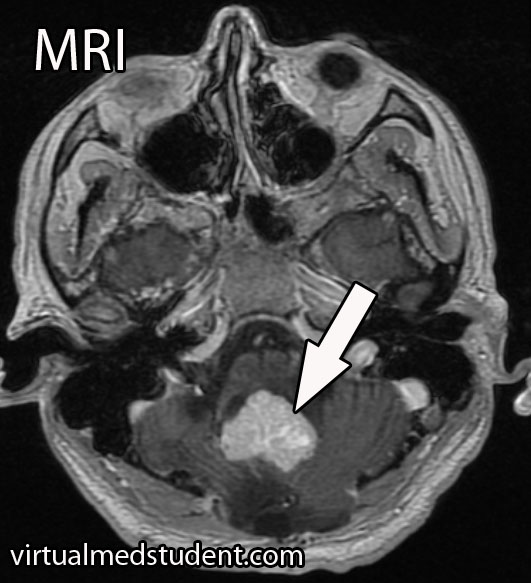
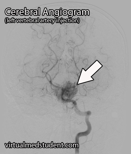
A tumor located in the cerebellum of an adult with certain characteristics on CT, MRI, and cerebral angiogram can make the diagnosis of hemangioblastoma likely.

However, the final diagnosis can only be made by looking at a sample of the tumor under the pathology microscope.

Treating These Bastards

Definitive treatment is surgical resection. However, hemangioblastomas can be highly vascular, which means they tend to bleed like stink! Therefore, preoperative embolization by an interventional neuro-radiologist can decrease the amount of bleeding that occurs during surgery.

Radiation therapy is also sometimes used as an adjunctive treatment. It may help slow the growth of the tumor, but will not cure it. Radiation therapy can be used in patients who are unable to undergo surgical resection, or if the tumor is inaccessible via traditional surgical means.

Recap It All…

Hemangioblastomas are highly vascular, but benign central nervous system tumors of undetermined origin. They are associated with von Hippel-Lindau syndrome. Signs and symptoms include headache, nausea, and vomiting, as well as cerebellar dysfunction. Diagnosis is based on imaging and pathological findings at the time of surgical resection. Surgery is the treatment of choice, although embolization and radiation therapy may also be used as an adjunct.

More Interesting Neurological Problems…

Some More Expert Ideas Below

Meningioma and the Arachnoid Cap Cell

A meningioma is a tumor that arises from the lining of the brain or spinal cord (ie: the “meninges”). They arise from cells known as “arachnoid cap cells”. Meningiomas are usually slow growing, “benign” tumors, which means that they are not usually considered cancerous in the strictest sense of the term.

When viewed under the light microscope, meningiomas can have many different appearances. The most common type is a dense sheet-like formation of cells interspersed with closely packed blood vessels. Sometimes the sheets of cells can be separated by connective tissue.

Frequently, meningiomas will have calcium deposits in them known as “psammoma bodies”. Uncommonly, meningioma cells may take on a malignant appearance characterized by increased cellular division (ie: “mitotic figures”) and invasion of the tumor cells into surrounding brain or bone.

Meningiomas test positive for epithelial membrane antigen (EMA) and vimentin (a marker of connective tissue). Ki-67 (a marker of proliferation) can be elevated in more aggressively behaving tumors.

The most common genetic abnormality seen in patients with meningiomas is found on chromosome 22. If present, a mutation in the NF2 gene on this chromosome causes type 2 neurofibromatosis. This disease predisposes individuals to developing multiple tumor types including meningiomas. Other less common genetic abnormalities can be seen on other chromosomes as well.

Signs and Symptoms

Due to their slow growing and benign nature, many meningiomas cause no symptoms. However, if they become too large or start to compress adjacent brain tissue they can cause headache, seizures, confusion, or visual problems. Spinal cord compression can result in myelopathy.

The most common location for a meningioma is in between the two hemispheres of the brain – the so called “parasagittal” location. Parasagittal meningiomas near the portion of the brain responsible for muscle movements may cause weakness of the opposite leg.

Diagnosis

Diagnosis of meningioma can reliably be made on characteristic findings seen on CT or MRI scans. Interestingly, many meningiomas are found incidentally when a CT or MRI is done for other reasons.

Meningioma MRIs

However, like any other tumor, meningiomas can only be truly diagnosed once a specimen is sent to the pathology lab for analysis. Pathologists can reliably make the diagnosis based on typical histological features.

Meningiomas must be distinguished from a more malignant tumor known as a hemangiopericytoma. Hemangiopericytomas can look similar to meningiomas on imaging studies.

Dish Me Up Some Treatment Sir

Many meningiomas can be watched over time with repeat imaging studies; this is especially true if they are small and not causing neurological signs or symptoms.

On the other hand, large or symptomatic meningiomas require surgical resection. Many meningiomas can be removed completely. However, some meningiomas may be near vital structures such as the carotid artery, cranial nerves, or venous draining systems of the brain where complete surgical removal may be very difficult without causing significant neurologic impairment. In these cases the tumor is debulked as much as possible. The residual tumor can be followed or irradiated depending on the grade of meningioma.

Residual tumor after incomplete surgical resection, or meningiomas in difficult to access locations are candidates for radiation therapy. Many studies have shown long term growth control rates.

Overview

Meningiomas are considered “benign” tumors of the brain. They arise from arachnoid cap cells, which are located in a layer of the meninges (ie: the covering of the brain) known as the arachnoid. Symptoms include headache, weakness, vision problems, paresthesias (ie: abnormal sensations), amongst many other possible symptoms. Diagnosis can be made reliably from imaging studies such as CT or MRI. If symptomatic, or large, treatment is surgical resection. Small asymptomatic meningiomas can be managed with repeat imaging to assess for growth over time.

Related Articles

Curated References for Your Pleasure…

Chordoma and that Pesky Notochord

In order to understand chordomas we have to first learn a little bit about how the nervous system develops. Enter the notochord.

The notochord is a midline structure in the developing fetus that sends out various molecules (the most well known of which is “sonic hedgehog”). These molecules influence the development of the layers of embryonic cells that surround the notochord. One of these layers, the ectoderm, which is immediately behind (ie: posterior) the notochord eventually forms the brain and spinal cord. The mesoderm, which is immediately adjacent to the notochord forms the vertebral column and axial skeleton (amongst other things).

The purpose of the notochord is to ensure that each layer of tissue forms what it is supposed to. Once this occurs, the notochord ultimately becomes the nucleus pulposus of the intervertebral discs.

In some people, nests of cells that composed the fetal notochord remain (unnaturally) after birth. These collections of cells are known formally as ecchordoses physaliphora (try saying that 5 times fast). These cells can divide and turn into a slow growing tumor… What is that tumor called? You guessed it! A chordoma!

Chordomas are slow growing tumors that are most commonly located at the ends of the vertebral column. The most common place to see them is in the sacrococcygeal region, followed in frequency by a bone known as the clivus at the base of the skull. However, chordomas can occur anywhere along the vertebral column. The reason they occur most frequently at the "ends" of the vertebral column (ie: skull base and sacrum) is because these are the last areas to fuse in-utero.

Additionally, since the notochord is a midline structure in the fetus, chordomas are almost always midline in location.

Microscopically chordomas contain large polygonal shaped cells embedded in a mesh of long repetitive sugar and nitrogen containing molecules known as mucopolysaccharides.

Chordoma Highlights:
– Arise from notochord cells
– S-100 positive
– Cytokeratin positive
– Polygonal cells
– Slow growing
– Midline location
– Sacrum and clivus most
   common locations
– Worse prognosis than
   chondrosarcomas
Less than a third of chordomas will show cartilage like features. These chordomas are classically called “chondroid” chordomas because of the chondrocyte-like (ie: cartilage-like) cells and extracellular material within them. Chondroid chordomas must be distinguished from a similar looking tumor known as a chondrosarcoma.

Distinguishing chondrosarcomas from chordomas is possible with immunocytologic staining techniques. Chondromas and chondrosarcomas stain positive for a protein known as S-100. S-100 proteins have numerous intracellular functions and are commonly present in cells such as adipocytes (fat cells), chondrocytes (cartilage forming cells), melanocytes (pigment producing cells), and Schwann cells, amongst others.

So if chondrosarcomas and chordomas can look alike, and both stain positive for S-100, how the heck do we distinguish between the two? Using another molecule known as cytokeratin! Cytokeratin is a molecule that forms part of the intracellular frame for many cells. It is present in chordomas, but not in chondrosarcomas.

On to the Clinic I Say…

Chordomas are slow growing tumors and will usually start to cause symptoms in mid-adulthood. Symptoms are based on the location of the tumor.

If the tumor is located in the sacrum or coccyx then pain is the most frequent presenting symptom. If undiscovered this may progress to bowel and bladder problems as the tumor slowly envelopes the sacral nerves that go to the bowel and bladder. Additionally, patients may present with radicular symptoms such as numbness, tingling, or sharp pain in the distribution of the sacral nerves.

Clivus Chordoma
Chordomas of the clivus, the second most common location, can present with headache and if large enough symptoms of brainstem or upper cervical spinal cord compression. These symptoms may include vertigo, difficulty moving the tongue, double vision, hearing problems, spastic gait, increased reflexes, clumsiness, amongst other symptoms.

Diagnose Me McCoy

Diagnosis of chordoma can only be officially made by looking at the specimen under a microscope. However, imaging studies with x-rays, CT scans, and MRI imaging can support the diagnosis. Imaging studies will typically show a midline lytic lesion centered in the bone. Invasion of adjacent anatomical structures can occur, but is a late manifestation of the disease course.

Treating These Ugly Tumors

The gold-standard treatment for chordomas is en-bloc surgical resection with wide margins followed by radiation therapy. Complete resection is difficult, if not impossible to achieve in the skull base, but may be possible in the spine and/or sacrum with very skilled surgical teams.

Without an en-bloc surgical resection the risk of tumor re-growth and recurrence is very high. If you are planning to biopsy of a sacral lesion you should mark the biopsy tract with methylene blue so that the tract can also be resected during surgery as tumor cells can seed the tract as the needle is being pulled out.

Let’s Remix This Overview

Chordomas are slow growing, but malignant tumors that arise from notochord cells that fail to regress after birth. They are most frequently found in the sacrum and clivus (one of the bones constituing . Diagnosis is made with CT, MRI, and x-rays, as well as via tissue diagnosis at the time of surgical removal. Chordomas stain positive for S-100 and cytokeratin proteins, which helps distinguish them from chondrosarcomas that only stain positive for S-100. Symptoms are based on where the lesion is located (skull base or spine). Treatment is surgery followed by radiation therapy.

Just Warming Up…

Want to Research More About Chordomas?

Hemangiopericytoma: A Tumor of Pericytes

In order to understand hemangiopericytomas we have to define a few terms. The first term is mesenchyma. Mesenchyma is a word used to describe the different tissues that provide structure to the bodies’ organ systems. A type of mesenchymal cell known as a “pericyte” provides structural support to blood vessels. When pericytes go haywire they form hemangiopericytomas.

Hemangiopericytomas can occur anywhere blood vessels are located, but are most commonly located in the lower extremities, pelvis, head, and neck.

Intracranial hemangiopericytomas are uncommon. They represent less than 1% of tumors within the confines of the skull. They typically arise from blood vessels adjacent to the dura (ie: lining of the brain) and often form dural attachments. They are therefore commonly lumped into the category of “dural-based tumors”, but should be distinguished from their more benign meningeal cousins (ie: meningiomas).

Since hemangiopericytomas are mesenchymal in origin, they typically have lots of reticulin (a collagen fiber) that envelopes individual cells (see pathology slide). They are highly cellular tumors, and vascular channels in the shape of "staghorns", may be seen under the microscope. Actively dividing cells (aka: "mitotic" figures) are commonly seen and are a testament to their more malignant nature. Unlike meningiomas, calcifications are absent.

Hemangiopericytomas test positive for vimentin (a marker of connective tissue), Ki-67 (a marker of proliferation), vascular endothelial growth factor (VEGF, a marker of blood vessel proliferation), CD34 (a marker of blood and vascular cell lineage), and reticulin (a collagen fiber). These tumors do not stain positive for epithelial membrane antigen. Genetic mutations have been found on different chromosomes , but the importance of these abnormalities is not well understood.

Intracranial hemangiopericytomas are considered malignant tumors. This means that they can spread to other areas of the body. In addition, hemangiopericytomas that have been removed surgically have a high recurrence rate.

Hemangiopericytoma

Signs and Symptoms

Hemangiopericytomas are relatively slow growing and often do not cause symptoms until they become quite large. However, once they start to compress adjacent brain tissue they may cause headaches, seizures, confusion, weakness, or visual problems.

Diagnosis

MRIs and CT scans of the brain typically reveal a contrast enhancing dural-based lesion. Cerebral angiograms show a highly vascular tumor with blood supply coming from the dura, as well as the underlying brain tissue.

Based on imaging alone, hemangiopericytomas are often mistaken for meningiomas. Subtle characteristics such as a lack of calcification seen on CT scans may help distinguish one from the other, but this is not reliable.

The only reliable way to diagnose hemangiopericytoma is to look at a specimen of the tumor under a microscope. Special stains and features of the tumor can help delineate it from a meningioma (see pathology section above).

Did I Hear Someone Say “Treatment”?

Intracranial hemangiopericytomas should be surgically resected when feasible. Unfortunately, even after complete resection, they frequently recur and/or spread to other areas of the body.

Because of their aggressive nature, patients with hemangiopericytomas should also have adjuvant radiation therapy. Radiation treatment after surgical removal of the tumor has been shown to lengthen survival and slows (but doesn’t appear to prevent) the time to recurrence.

The role of chemotherapy is less clear and is still being investigated. At this point, chemotherapy is typically used in patients where radiation and surgery have failed to control the disease.

Let’s Recap It…

Intracranial hemangiopericytomas are malignant dural-based tumors that arise from pericytes. They are highly vascular tumors that enhance on MRI and CT scans. Symptoms are variable and depend on the size and location of the tumor. Treatment is with surgical removal followed by radiation therapy. Recurrence rates are high despite optimal treatment.

You Should Learn About This Stuff Too…

Want Some More?

Acoustic Neuroma, and Really We Mean Vestibular Schwannoma

Both the term “acoustic” and “neuroma” are incorrect ways of describing a tumor that arises from the 8th cranial nerve (vestibulocochlear nerve). An "acoustic neuroma" is a tumor that arises from Schwann cells that myelinate the peripheral portion of the nerve; this technically makes them “schwannomas”.

In addition, the tumor does not arise from the acoustic division of the 8th cranial nerve (ie: the portion of the nerve responsible for hearing), but instead arises most commonly from the vestibular division (ie: the balance portion of the nerve). Therefore, the appropriate medical term given to these tumors is a “vestibular schwannoma”.

These tumors are frequently caused by mutations in genes responsible for controlling cell cycle, cell morphogenesis, cell development, cell death, and cell adhesion. A well known cause of vestibular schwannomas occurs in patients with neurofibromatosis (NF) type II.

In this condition, which is responsible for about 5% of acoustic neuromas, a mutation in the NF gene on chromosome 22 causes an absent or dysfunctional protein product. This protein normally serves as a tumor suppressor; once mutated, it is no longer able to suppress tumor growth. The growth of various cells, including Schwann cells, becomes unchecked. The end result? A vestibular schwannoma.

When viewed under a pathology microscope, vestibular schwannomas are composed of different patterns of tissue. The first pattern is referred to as Antoni A; it consists of densely packed, elongated cells with nuclear free areas of cytoplasmic extensions referred to as "Verocay bodies". The second pattern is, you guessed it – Antoni B. This pattern has fewer cells and appears "looser" than the type A pattern.

These tumors are considered "benign", which means that they do not spread (metastasize) to other areas of the body. Overall, acoustic neuromas increase in size at the rate of roughly 1mm per year, but about 50% of tumors show no growth at all! Although they are not malignant tumors they can still cause symptoms.

Signs and Symptoms

Vestibular schwannomas cause local signs and symptoms. Since they arise from the 8th cranial nerve (vestibulocochlear nerve), which is responsible for hearing and balance, almost all patients present with some degree of hearing loss. In type II neurofibromatosis acoustic neuromas arising from both vestibulocochlear nerves may cause deafness. Some patients have tinnitus (ie: ear ringing), as well as a sense of vertigo.

Symptoms that are less common are a result of the tumor pressing on adjacent cranial nerves. Dysfunction of the 7th cranial nerve (facial nerve) can cause weakness of the facial muscles. If the tumor presses on the 5th cranial nerve (trigeminal nerve) it can cause face numbness; if it touches the 6th nerve (abducens nerve) diploplia (ie: double vision) may occur.

Finally, if the tumor continues to grow, it can cause compression of the brainstem. This can block the flow of cerebrospinal fluid (CSF) leading to a condition called hydrocephalus. These patients often have headaches, nausea, and vomiting secondary to increased pressure within the skull.

Diagnosis and Classification

Vestibular Schwannoma
MRI is the imaging study of choice. It will show a well encapsulated tumor that sits in the cerebellopontine angle and/or involves the internal acoustic meatus.

Audiometric analysis is important in order to document hearing loss and for monitoring treatment outcomes. The most useful test is a pure tone audiogram. Differences in hearing ability between the two ears is suspicious for an acoustic neuroma, but not specific.

Although these tumors are commonly diagnosed from characteristic MRI findings, the definitive diagnosis is made when a pathologist looks at the tumor under a microscope.

A common classification system known as the Koos grading scale is frequently used. Grade 1 tumors involve only the internal auditory canal. Grade 2 tumors extend into the cerebellopontine angle, but do not encroach on the brainstem. A grade 3 tumor fills the entire cerebellopontine angle and a grade 4 tumor displaces the brainstem and adjacent cranial nerves.

Treatment

Treatment of these tumors depends on several factors, such as how large the tumor is, and whether or not the patient has symptoms from it (ie: hearing loss, face weakness, etc). If the tumor is small it can be followed with repeat MRI to monitor for enlargement. If the tumor grows, or begins to cause symptoms, then definitive treatment should be provided.

The two most commonly used treatment modalities are surgical resection and radiation. Surgery is most useful for very large tumors or when the patient is clinically deaf. Radiation comes in two flavors: single session stereotactic radiosurgery and fractionated radiotherapy.

Stereotactic radiosurgery is a single dose of radiation delivered directly to the tumor, typically with a dose of 12 to 13 Gy. The ability to preserve useful hearing with radiosurgery ranges from 32 to 71%. For tumors less than 3 cm in diameter, the ability of radiosurgery to halt the growth of the tumor has been shown to be between 92 and 100%.

Radiation can be harmful, especially when large doses are used in one session. Inadvertent injury to the facial nerve, acoustic nerve, trigeminal nerve, and brainstem are all possible adverse events. The use of fractionated radiotherapy has been tried to decrease these risks while still delivering large doses of radiation to the tumor.

Fractionated radiotherapy spreads the total radiation dose over multiple distinct sessions. For example, a total of 40 to 58 Gy can be delivered to the tumor in 2 Gy sessions over the course of several weeks. This is more radiation delivered to the tumor compared to single session radiosurgery (13 Gy), but it is delivered over a longer time frame, which helps mitigate the risk of damaging the adjacent cranial nerves and brainstem. Hearing preservation with fractionated radiotherapy has been shown to be superior to single-session radiosurgery.

Overview

A vestibular schwannoma is a benign tumor that arises from the vestibular portion of the 8th cranial nerve. It cause hearing loss and may cause compression of adjacent cranial nerves. It is diagnosed by clinical history, audiometric studies, and MRI. Treatment consists of surgical excision, radiation therapy, or both depending on the clinical situation.

More Brain Tumors…

References and Resources

  • Ferrer M, Schulze A, Gonzalez S, et al. Neurofibromatosis type 2: molecular and clinical analyses in Argentine sporadic and familial cases. Neurosci Lett. 2010 Aug 9;480(1):49-54. Epub 2010 Jun 8.
  • Cayé-Thomasen P, Borup R, Stangerup SE, et al. Deregulated genes in sporadic vestibular schwannomas. Otol Neurotol. 2010 Feb;31(2):256-66.
  • Harner SG, Laws ER Jr. Clinical findings in patients with acoustic neurinoma. Mayo Clin Proc. 1983 Nov;58(11):721-8.
  • Bederson JB, von Ammon K, Wichmann WW, et al. Conservative treatment of patients with acoustic tumors. Neurosurgery. 1991 May;28(5):646-50; discussion 650-1.
  • Kumar V, Abbas AK, Fausto N. Robbins and Cotran Pathologic Basis of Disease. Seventh Edition. Philadelphia: Elsevier Saunders, 2004.
  • Koos WT, Day JD, Matula C, et al. Neurotopographic considerations in the microsurgical treatment of small acoustic neurinomas. J Neurosurg. 1998 Mar;88(3):506-12.

Ependymoma: Myxopapillary, Anaplastic, and Perivascular Pseudorosettes

Ependymomas are tumors that develop from cells known as ependymal cells (duh!). Ependymal cells are a type of glial cell that line the ventricles (ie: fluid filled cavities) of the brain and central canal of the spinal cord.

Normal ependyma have cilia and microvilli on the side of the cell that faces cerebrospinal fluid (ie: the "apical" side). Cilia are hair like extensions that are believed to "beat" cerebrospinal fluid around the ventricles. Microvilli are folds in the cellular membrane that are thought to aid in the reabsorption of cerebrospinal fluid.

Unlike other epithelial cells in the body, of which ependyma are considered a subgroup, they do not rest on a basement membrane. Instead their basal surfaces (the surface not in contact with cerebrospinal fluid) intertwine with the overlying brain tissue.

Like any other cell in the body, ependymal cells can decide to turn naughty and form a tumor. Ependymomas can occur anywhere there are ependymal cells, and therefore develop in both the brain and spinal cord. Intracranial ependymomas are more common in younger age groups, whereas spinal forms are more common in older individuals. Of those that form within the confines of the skull, the most common location is in the fourth ventricle near the brainstem.

There are three "grades" of ependymoma. There are two subsets of grade one: myxopapillary and subependymomas. The second grade of ependymoma has four distinct variants. They are cellular, papillary, clear cell, and tanycytic. The third grade is also referred to as "anaplastic" ependymoma. Regardless of the grade, each type has its own distinct characteristics when viewed under the pathology microscope.

Surgical specimens of ependymomas are often "stained" by pathologists to help aid in diagnosis, and more importantly, distinguish them from other tumor types. Ependymomas stain positive for the glial fibrillary acidic protein (GFAP), as well as phosphotungstic acid hematoxylin (PTAH).

Ependymomas may have perivascular pseudorosettes, which helps support the diagnosis. Pseudorosettes may not be apparent in tumors with dense cellularity such as anaplastic ependymomas.

In addition, ependymomas can spread throughout the cerebrospinal fluid space. For example, a tumor that arises in the fourth ventricle may "drop" tumor cells down into the spinal cord forming a secondary tumor. These secondary tumors are referred to as "drop mets".

Signs and Symptoms

The signs and symptoms depend on the location of the ependymoma.

The most common symptom of intracranial ependymoma is headache associated with nausea and/or vomiting. These symptoms occur when the ependymoma blocks the flow of cerebrospinal fluid, which causes a condition known as non-communicative hydrocephalus.

You can think of non-communicative hydrocephalus as a clog in a pipe. Everything upstream of the clog starts to back up, which eventually leads to increasing pressures. When this increased pressure occurs in the ventricular system of the brain it causes worsening headaches, nausea, and vomiting. This is especially true if the ependymoma is in the fourth ventricle of the brain, which even without tumor, is an anatomically narrow "pipe" to begin with.

Additionally, if the tumor pushes on brainstem structures a patient may present with dysfunction of the nerves that go to the various muscles of the head and face. The most commonly involved nerves are the facial nerve, which can cause weakness of the face, as well as the abducens nerve, which can cause weakness of the eye.

Tumors located in the spinal cord cause weakness and sensory disturbances.

Diagnosis

Ependymoma

MRI scans can be very useful and can support (but not prove) the diagnosis of ependymoma, especially when the tumor is in a common anatomical location.

If there is a high index of suspicion for ependymoma then the entire neuro-axis, meaning the brain and entire spinal column, should be imaged using MRI. This will detect “drop” mets, which, if present, further support the diagnosis.

Diagnosis can only be officially made when a sample of tumor (either surgical or at autopsy) is seen under the pathology microscope.

Treatment

Treatment of ependymoma is with surgical resection followed by radiation therapy. Patient outcome is most effective if the entire tumor can be removed during surgery. This is known as "gross total resection". However, the extent of surgical resection should always be weighed against the risk of harming the patient, especially if the tumor has invaded vital structures like the brainstem.

Fortunately, ependymomas are very radio-sensitive, which means that they respond well to getting zapped with radiation. Chemotherapy is not typically helpful except in very young children where the effects of radiation can be devastating.

Overview

Ependymomas arise from the cells that line the ventricular system of the brain and spinal cord. There are different subtypes depending on what it looks like under the pathology microscope. Diagnosis is based on pathological analysis and characteristic MRI findings. Treatment is with surgery and radiation.

Other Diseases You Should Know About…

References and Resources

Hodgkin’s Lymphoma: B-Cells, Pel-Ebstein, and EBV

Lymphoma is a cancer that develops from cells in the body known as lymphocytes. Lymphocytes are a subcategory of white blood cells, which are the cells that help ward off infection. There are two different types of lymphocytes: B-cells and T-cells. The majority of lymphomas, including Hodgkin’s disease, stem from B-cells.

In Hodgkin’s disease a B-cell, for unknown reasons, becomes cancerous. The cell then makes many clones of itself. These cells bundle together to form a solid tumor known as a lymphoma.

Why B-cells in Hodgkin’s lymphoma become cancerous is not entirely known. One belief is that infection with Epstein-Barr virus (the same virus that causes infectious mononucleosis) can cause the cells to turn cancerous in genetically susceptible people. Other theories are that certain genetic translocations may be the underlying factor. As of yet, no particular theory has significant supporting data to call it the true cause. It is likely that the development of Hodgkin’s disease is multi-factorial.

Types

Reed-Sternberg Cell

There are different types of Hodgkin’s lymphoma. They are based on unique histopathological (ie: what it looks like under a microscope) characteristics, and are important in determining prognosis.

The histopathological features the pathologist looks for are the number of Reed-Sternberg cells, as well as the number of lymphocytes present in the biopsy specimen. A Reed-Sternberg cell is a funny shaped cell with two nuclei that looks like an "owl’s eyes" (see image to the right). They are believed to form when two cells merge together under the influence of certain proteins produced by the Epstein-Barr virus.

The first category, and most common type, is nodular sclerosing Hodgkin’s lymphoma. In this type, there are very few Reed-Sternberg cells with a moderate number of lymphocytes. It commonly occurs in younger individuals, and with treatment, the prognosis is excellent.

The second category is mixed cellularity Hodgkin’s lymphoma. This type has many Reed-Sternberg cells, and a moderate number of lymphocytes when viewed under the microscope. It has an intermediate prognosis.

The third category is lymphocyte predominant Hodgkin’s. It has very few Reed-Sternberg cells and many lymphocytes. It occurs most commonly in males less than 35 years of age. It is also one of the few types that is not associated with Epstein-Barr virus infection.

The last category is lymphocyte depleted. It is the rarest form of Hodgkin’s lymphoma. It typically affects older males.

Hodgkin's Types
Unfortunately it has the worst prognosis of the four types types.

The image to the left is one way of organizing the different Hodgkin’s types and their prognosis based on age, number of RS cells, and prognosis. LP = lymphocyte predominant, NS = nodular sclerosing, MC = mixed cellularity, LD = lymphocyte depleted.

Signs and Symptoms

The classic presentation of Hodgkin’s lymphoma is painless enlargement of the lymph nodes. This is similar to non-Hodgkin’s lymphoma, and the only way to differentiate the two is through biopsy.

Systemic manifestations may occur and include night sweats, fever, and weight loss. However, these are more common in patients with disseminated (ie: metastatic) disease. Interestingly, a pathognomonic (ie: seen exclusively in Hodgkin’s lymphoma) feature that occurs in some cases is pain of the involved nodes after drinking alcohol. Finally, a symptom known as Pel-Ebstein fevers are also specific for the disease. A Pel-Ebstein fever is a cyclical fever that occurs for several weeks at a time followed by a fever free period.

Other signs related to the immune system can be seen in patients with Hodgkin’s lymphoma. A condition known as cutaneous anergy can occur. Anergy refers to a lack of response by the cell mediated immune system. For example, in patients with tuberculosis a reaction will occur underneath the skin when they get a TB test. This reaction is the result of their cell mediated immunity reacting to the tuberculosis components injected underneath the skin. However, in anergic patients no reaction is seen, even if they have tuberculosis! This can also occur in patients with Hodgkin’s disease.

Diagnosis

Hodgkin's Disease PET-CT
Diagnosis of Hodgkin’s lymphoma is made by looking at a biopsy specimen underneath the microscope. The most common way of obtaining a specimen is through biopsy of a lymph node. Differentiating Hodgkin’s from other types of lymphomas is important because it determines the best treatment options.

Additional studies are often performed to determine the number and location of involved lymph nodes. One such study is a positron emission tomograph (PET) combined with a CT scan. Any lymph nodes involved "light up" on the scan. An example, with the arrows pointing to involved nodes, is shown to the right.

Staging

Staging of Hodgkin’s lymphoma was traditionally based on a system known as the Ann Arbor Classification. It is divided into four stages. In stage 1 disease a single lymph node, or single organ is involved. In stage 2 disease involvement of multiple (two or more) lymph node regions on the same side of the diaphragm is present. In stage 3 disease involvement of nodes on both sides of the diaphragm is present; the spleen or other limited organ involvement may also be present. In stage 4 disease multiple organs are involved; interestingly, lymph node involvement is not necessary for a stage 4 diagnosis, although it is commonly present. Finally, each stage is further divided into “A” and “B” depending on whether or not symptoms are present. If symptoms are present, the stage is upgraded to a “B”.

Ann Arbor Classification (simplified)
Stage 1 Single lymph node or organ
Stage 2 Multiple lymph nodes on same side of diaphragm
Stage 3 Lymph nodes on both sides of diaphragm
Stage 4 Multiple organs involved

However, more recently the Lugano staging classification has become the preferred method. It divides disease into limited and advanced. Limited disease includes stage 1 and stage 2. Stage 1 involves a single lymph node or nearby nodes. Stage 2 involves two or more nodal groups. Advanced disease includes stages 3 and 4. In stage 3 disease nodes on both sides of the diaphragm are involved as is the spleen. Stage 4 disease is disseminated disease into organs. There are sub-categories in the Lugano model as well.

Treatment

Like most cancers treatment is highly dependent on the stage of the disease. In most cases chemotherapy and radiation are used. Radiation is directed at involved lymph nodes, as well as lymph nodes that are uninvolved, but nearby. Common chemotherapeutic agents used include: adriamycin, bleomycin, vinblastine, vincristine, prednisone, procarbazine, and mechlorethamine.

Overview

Hodgkin’s lymphoma is a cancer of a type of white blood cell known as a B-cell. There are numerous categories depending on its histopathological characteristics. Patients often have painless enlarged lymph nodes. Some patients have fever, weight loss, and other non-specific symptoms. Staging is based on the Ann-Arbor model. Treatment usually involves a combination of chemotherapy and radiation.

Related Articles

References and Resources