Ventricular Fibrillation: Shock Me Baby!

Cardiac muscle contracts in a predictable, regular pattern, which allows it to generate enough force to eject blood to the body. However, in ventricular fibrillation the heart muscle “quivers” in a rapid, irregular, and unsynchronized manner. These feeble contractions are not strong, nor coordinated enough to eject blood from the heart. As a result, cardiac output drops and the body quickly goes into cardiogenic shock.

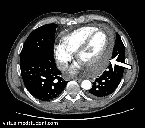
There are numerous causes of ventricular fibrillation. The most common cause of abnormal electrical activity occurs in diseased heart tissue that has lost its normal architecture. For example, muscle damage from heart attacks or disorganized heart structures seen in cardiomyopathies can serve as abnormal areas of electrical impulse formation; these irritated areas can predispose patients to develop ventricular fibrillation.

Other causes of ventricular fibrillation include electrolyte abnormalities. For example, hyperkalemia (ie: an elevated blood potassium level) can depolarize heart muscle cells and make them more likely to "fire" an action potential. In general, any electrolyte disturbance that makes the resting potential of the cardiac muscle fiber more positive (ie: more depolarized) can cause abnormal electrical impulse formation; these abnormal impulses can degenerate into ventricular fibrillation.

Signs and Symptoms

Ventricular fibrillation is a highly fatal rhythm because the heart fails to pump blood, and more specifically oxygen, to the bodies’ organs. As a result, every organ system in the body, including the heart becomes ischemic and dies.

The rapid decline in blood flow to the brain causes people to lose consciousness. If treatment is not sought quickly the patient will have anoxic brain injury (ie: a massive global stroke), which will lead to brain death.

Diagnosis

Ventricular fibrillation is diagnosed by looking at an electrocardiogram (ECG). The ECG will show disorganized and chaotic electrical activity.

Ventricular fibrillation ECG
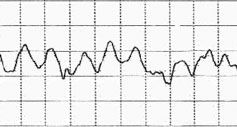
ECG of ventricular fibrillation

Treatment

Treatment of ventricular fibrillation is with immediate un-synchronized electrical cardioversion (ie: the paddle "thingies" they use to shock someone’s heart). The goal of shocking the heart with electricity is to reset (ie: repolarize) all the cardiac muscle fibers at the same time. From there the sinus node should theoretically take over, and reset the heart back into a normal rhythm.

If a patient survives their first episode of ventricular fibrillation they often have a cardiac defibrillator implanted. Implantable cardiac defibrillators shock the heart when they detect an abnormal rhythm.

Overview

Ventricular fibrillation is a rapidly fatal, disorganized, and inefficient “quivering” of heart muscle. It causes cardiogenic shock and organ death if left untreated. It is most commonly due to underlying heart disease seen in people with coronary artery disease, previous heart attacks, and cardiomyopathies, although other causes exist. Treatment is with immediate electrical cardioversion (ie: “shocking” the heart).

Related Articles

References and Resources

  • Marcus GM, Scheinman MM, Keung E. The year in clinical cardiac electrophysiology. J Am Coll Cardiol. 2010 Aug 17;56(8):667-76.
  • Dosdall DJ, Fast VG, Ideker RE. Mechanisms of defibrillation. Annu Rev Biomed Eng. 2010 Aug 15;12:233-58.
  • Braunwald E. Hypertrophic cardiomyopathy: the early years. J Cardiovasc Transl Res. 2009 Dec;2(4):341-8. Epub 2009 Oct 7.
  • Rea TD, Page RL. Community approaches to improve resuscitation after out-of-hospital sudden cardiac arrest. Circulation. 2010 Mar 9;121(9):1134-40.
  • Schaer B, Kühne M, Koller MT, et al. Therapy with an implantable cardioverter defibrillator (ICD) in patients with coronary artery disease and dilated cardiomyopathy: benefits and disadvantages. Swiss Med Wkly. 2009 Nov 14;139(45-46):647-53.
  • Callans DJ. Out-of-hospital cardiac arrest–the solution is shocking. N Engl J Med. 2004 Aug 12;351(7):632-4.
  • Lilly LS, et al. Pathophysiology of Heart Disease: An Introduction to Cardiovascular Medicine. Seventh Edition. Lippincott Williams and Wilkins, 2020.

Fluid Around the Heart: Pericardial Effusions

The heart is encased in a connective tissue capsule known as the pericardium. The pericardium contains two layers, known as the parietal and visceral pericardium. These layers are like two blankets enveloping the heart. The visceral pericardium sits adjacent to the heart muscle itself and the parietal pericardium sits on top of the visceral pericardium. Because of this arrangement there is a potential space between the two layers.

When fluid (ie: blood, pus, water, etc.) leaks into this space a pericardial effusion is present. Fluid can leak out quickly, in which case the effusion is said to be “acute”; or it can leak out gradually in which case it is said to be “chronic”.

Causes

There are numerous causes of pericardial effusion some of which are listed below:

  • Infections
    • Viral (including HIV, coxsackie, echo, adeno, ebstein-barr, and varicella viruses)
    • Bacterial (including pneumococcus, neisseria meningitides, staphylococcus aureus)
    • Tuberculosis
  • Malignancies (cancers)
  • Autoimmune conditions (including connective tissue disorders, vasculitis, and drug induced)
  • Uremia (from renal failure)
  • Cardiovascular (cardiac surgery, myocardial infarction, aortic dissection, congestive heart failure)
  • Hypothyroidism
  • Cirrhosis (liver problems)
  • Idiopathic (unknown)

Please note that by no means is this list exhaustive, but these are the most common causes of effusions!

Signs and Symptoms

The classic symptom of pericardial effusion is chest pain that is better when the patient sits up and leans forward. However, numerous other symptoms including light headedness, shortness of breath, cough, and palpitations can occur.

Depending on how quickly the effusion develops, patients may spiral into a condition known as “tamponade”. When this occurs the effusion effectively “chokes” the heart muscle causing decreased contractile function. This causes decreased cardiac output and even multi-organ failure if left untreated!

The classic signs of tamponade are hypotension (ie: decreased blood pressure), muffled heart sounds, and increased jugular venous pressures (you can see the jugular veins engorged with blood). These three signs are known as “Beck’s triad”, which is generally a late finding of tamponade (ie: the patient is almost dead!).

Diagnosis

CT scan of pericardial effusion
There are numerous tests that can support the diagnosis. Electrocardiogram may show decreased voltages, and a finding known as “electrical alternans” where the QRS complexes change amplitude and/or direction as a result of the heart “sloshing” around in the effusion. Chest x-ray may show an enlarged heart, but this is neither specific, nor sensitive, for effusion. CT scans can show fluid surrounding the heart. Finally, echocardiography (ie: ultrasound of the heart) can be very useful in delineating not only the presence of, but also the size, and location of the effusion.

Treatment

For chronic effusions with no significant symptoms patients can be treated for the underlying condition causing the effusion. This will sometimes cure the effusion. However, in patients with acute presentations who have signs of cardiovascular instability (ie: low blood pressure, evidence of organ dysfunction from decreased blood flow, etc.) emergent removal of the fluid is performed. The quickest way to do this is to insert a needle under the xyphoid process and aspirate the fluid. In less acute situations, or in recurrent cases, surgical “windows” in the pericardial tissue can be created to allow the effusion to drain.

Overview

Pericardial effusions occur when fluid accumulates between the visceral and parietal pericardial layers surrounding the heart. There are numerous causes. Rapidly expanding effusions can cause cardiac tamponade and lead to cardiovascular collapse. Signs and symptoms include chest pain, shortness of breath, cough, distant heart sounds, and decreased blood pressure. Diagnosis is made by characteristic ECG, echocardiography, and CT scan findings.

References and Resources

  • Imazio M, Brucato A, Mayosi BM, et al. Medical therapy of pericardial diseases: part II: Noninfectious pericarditis, pericardial effusion and constrictive pericarditis. J Cardiovasc Med (Hagerstown). 2010 Nov;11(11):785-94. Review.
  • Khandaker MH, Espinosa RE, Nishimura RA, et al. Pericardial disease: diagnosis and management. Mayo Clin Proc. 2010 Jun;85(6):572-93. Review.
  • Spodick DH. Pericarditis, pericardial effusion, cardiac tamponade, and constriction. Crit Care Clin. 1989 Jul;5(3):455-76.
  • Mookadam F, Jiamsripong P, Oh JK, et al. Spectrum of pericardial disease: part I. Expert Rev Cardiovasc Ther. 2009 Sep;7(9):1149-57.
  • Jiamsripong P, Mookadam F, Oh JK, et al. Spectrum of pericardial disease: part II. Expert Rev Cardiovasc Ther. 2009 Sep;7(9):1159-69.
  • Woo KM, Schneider JI. High-risk chief complaints I: chest pain–the big three. Emerg Med Clin North Am. 2009 Nov;27(4):685-712, x.