Colloid Cysts of the Third Ventricle

Colloid cysts of the third ventricle are slow-growing, benign cranial tumors. They are believed to be composed of an epithelial wall with either mucous or protein like material trapped inside a spherical structure. However, their exact etiology is still under debate.

They are typically found in the anterior portion of the third ventricle near the foramen of Monroe (the channels that connect the lateral ventricles to the third ventricle). The third ventricle is one of the spinal fluid filled cavities of the brain.

Colloid cysts are “benign” because they are not cancerous (ie: don’t invade other parts of the body); however, they have the potential to block the flow of cerebrospinal fluid, which can lead to acute hydrocephalus and brain herniation. Therefore, in this regards they are certainly not “benign” tumors!

Signs and Symptoms

The most common presenting symptom of a colloid cyst is headache and difficulty walking. Acute hydrocephalus (dilation of the ventricular system secondary to blocked cerebrospinal fluid) can occur if the cyst blocks the flow of cerebrospinal fluid; this can cause nausea, vomiting, headache, and lethargy. Changes in mental status may also be seen in patients with these lesions.

There are numerous reports of patients dying suddenly from colloid cysts of the third ventricle. This is believed to be due to rapid obstruction of cerebrospinal fluid at the foramen of Monroe. The fluid builds up behind the blockage which puts pressure on the brain. Too much pressure can cause the brain to herniate through the base of the skull (see the Monro-Kellie doctrine).

Diagnosis

Colloid Cyst of the 3rd Ventricle CT
MRI of colloid cyst of the third ventricle
Diagnosis can be made with MRI or CT scan. Head CT scans will reveal a hyperdense (ie: bright or white colored) lesion. MRI is beneficial because it provides a superior picture of the regional anatomy around the cyst. Lumbar puncture should never be performed in a patient with a colloid cyst due to the risk of brain herniation.

Treatment

Treatment of colloid cysts is surgical. There are numerous approaches including the use of an endoscope, or the use of stereotactic guidance systems. In patients with contraindications to surgery bilateral cerebrospinal fluid shunts can be placed to prevent acute hydrocephalus from developing.

Overview

Colloid cysts of the third ventricle are "benign" tumors. They have the potential to block the flow of cerebrospinal fluid leading to acute hydrocephalus. The most common symptom is headache followed by gait instability. Diagnosis is made with CT and MRI imaging. Treatment is surgical resection.

Related Articles

References and Resources

Hydrocephalus is Greek for “Water Head”

Hydrocephalus literally means "water head". It is a term used to describe a pathological increase in the amount of cerebrospinal fluid (CSF) within the ventricles (fluid filled cavities) of the brain.

In order to understand hydrocephalus we have to first appreciate the cerebrospinal fluid pathway and ventricular system of the brain. The brain has four ventricles: a pair of lateral ventricles, a third ventricle, and a fourth ventricle. They are connected to one another through narrow channels. The fourth ventricle drains into the subarachnoid space around the upper spinal cord. The spinal fluid travels down into the lumbar cistern and then back up again where it is absorbed by the arachnoid granulations overlying the cerebral hemispheres.

CSF pathway

Cerebrospinal fluid is created at a rate of roughly 500 mL per day. It is primarily secreted by specialized cells within the walls of the ventricles known as choroid plexus. As you can imagine, if it is secreted at 500 mL per day there must be an equal amount of re-absorption. This re-absorption occurs in the subarachnoid space by venous structures known as "arachnoid villi". Re-absorption does not occur in the ventricles themselves; this is an important point to keep in mind as we discuss the difference between communicating and non-communicating forms of the disease.

Hydrocephalus occurs when excess cerebrospinal fluid backs up. It is called "communicating" hydrocephalus if all of the ventricles are enlarged. Otherwise it is known as "non-communicating" hydrocephalus. There is another informal type of hydrocephalus that is known as "ex-vacuo"; it occurs when dilation of the ventricular system is a result of brain tissue loss, rather than a pathological increase in the amount of cerebrospinal fluid.

The term hydrocephalus usually implies an abnormally high pressure within the ventricular system. However, a different type of hydrocephalus known as "normal pressure" hydrocephalus defies this rule.

Communicating hydrocephalus usually develops when the arachnoid villi get "gunked up". Abnormal materials (such as blood in subarachnoid hemorrhage or proteins after meningitis) can block the arachnoid villi and prevent re-absorption. The cerebrospinal fluid then backs up and causes the entire ventricular system to enlarge.

Non-communicating hydrocephalus is slightly different because the re-absorption pathway is functioning properly, but CSF backs up behind a "road block" in one of the channels connecting the individual ventricles. "Road blocks" can be anything from tumors to developmental narrowing of the channel itself. For example, colloid cysts of the third ventricle can occlude the foramen of Monroe (the channel between the lateral and third ventricles); when this occurs CSF produced in the lateral ventricles is not able to flow into the third ventricle. CSF then backs up in the lateral ventricle(s) resulting in pathologic dilation.

Overall, there are crap tons of causes for both communicating and non-communicating hydrocephalus. Some patients have congenital forms secondary to improper development of the CSF pathways. Others may develop hydrocephalus later in life as a result of infection, tumor formation, or aneurysm rupture.

Signs and Symptoms

The signs and symptoms depend on the age of the patient and the rapidity of onset. In newborns hydrocephalus often presents as failure to thrive with an abnormally enlarging head.

If hydrocephalus develops rapidly, the increase in intracranial pressure (as a result of cerebrospinal fluid putting pressure on the brain) leads to nausea, vomiting, headache, and if severe enough, coma and potentially death!

Normal pressure hydrocephalus (NPH) is a unique type of hydrocephalus in which there is no elevation in pressure (hence the term "normal pressure"). The classic symptoms of NPH are difficulty walking, dementia, and urinary incontinence.

Diagnosis

Non-communicating hydrocephalus
Communicating Hydrocephalus

Diagnosis of hydrocephalus is made with CT scan coupled with clinical evidence of increased pressure within the ventricular system.

The scan will show an abnormally enlarged ventricular system. MRI scans with contrast are also routinely done to evaluate for tumors as a cause of hydrocephalus.

Treatment

Treatment consists of "shunting" the cerebrospinal fluid to another part of the body, usually the peritoneal cavity.

Other techniques such as third ventriculostomy, in which a surgically made "hole" is placed between the third ventricle and the subarachnoid space is also sometimes employed. This is very effective in cases of non-communicating hydrocephalus secondary to aqueductal stenosis (ie: the small passage between the third and fourth ventricles).

The ultimate treatment depends on the type of hydrocephalus and whether symptoms are severe enough to warrant surgical intervention.

Overview

Hydrocephalus is a pathological accumulation of cerebrospinal fluid. It can be communicating, non-communicating, or normal pressure. It can cause numerous signs and symptoms, but headache, nausea and vomiting are amongst the most common. Diagnosis is with CT scanning and clinical evidence of increased intracranial pressure. Treatment consists of shunting the CSF to another part of the body where it can be reabsorbed.

Related Articles

References and Resources

Vein of Galen Malformations: A Misnomer of Sorts

The vein of Galen is located deep within the brain. The internal cerebral veins, basal veins of Rosenthal, atrial veins, and precentral cerebellar veins join together to form the vein of Galen.

As expected, the vein of Galen drains blood from deeply located brain structures. The vein of Galen then connects with the inferior sagittal sinus to form the straight sinus; blood then drains from the straight sinus into the transverse and sigmoid sinuses, where it eventually finds its way into the internal jugular veins and back to the heart.

Interestingly, a "vein of Galen" malformation is not actually a malformation of the true vein of Galen; the term is a misnomer. It is actually a malformation of primitive fetal anatomical structures that normally regress during development. These primitive structures include a dilated venous structure, as well as "feeding" arteries. Therefore, vein of Galen malformations represent true arteriovenous fistulas; in other words, blood moves directly from an artery to a vein without an intermediary capillary bed.

Between the 3rd and 11th weeks of fetal development a large primitive vein known as the median prosencephalic vein of Markowitz drains the deepest parts of the brain. As the brain develops, the internal cerebral veins annex the territory normally drained by the anterior portion of this vein. As a result, this portion of the median prosencephalic vein regresses. The internal cerebral veins then plug in to the posterior portion of the median prosencephalic vein, which becomes the "true", or "normal", vein of Galen.

The most common arterial "feeders" of the malformation can also be explained by aberrant embryology. During early fetal brain development the distal branches of the anterior cerebral arteries (ie: the pericallosal branches) make connections with the posterior cerebral arteries. These connections usually regress to form the anterior and posterior circulations, which are connected to one another via the posterior communicating arteries.

So how does a vein of Galen malformation form? In some infants the median prosencephalic vein of Markowitz does not regress like it should. As a result, a large abnormal venous midline pouch remains. It also retains its primitive arterial blood supply from the distal branches of the anterior cerebral artery (ie: pericallosal branches), anterior choroidal arteries, posterior communicating arteries, and branches of the posterior cerebral arteries (ie: posterior choroidal arteries).

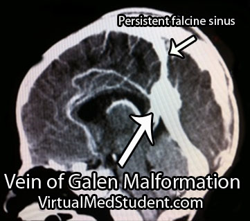
Vein of Galen malformations are also associated with other abnormalities in the venous structure of the brain. Not uncommonly, the straight sinus is absent or severely narrowed. As a result venous blood drains into a persistent falcine sinus, which is a structure that normally regresses in-utero.

To summarize, vein of Galen malformations are primitive direct arteriovenous fistulas. They are composed of a dilated venous pouch (ie: the median prosencephalic vein of Markowitz) with any combination of anterior and posterior circulation feeding arteries.

Signs and Symptoms

Signs and symptoms depend on the severity of the malformation. Severe malformations present in new borns with high output cardiac failure. This is because so much blood is being shunted into the malformation that the heart cannot keep pace!

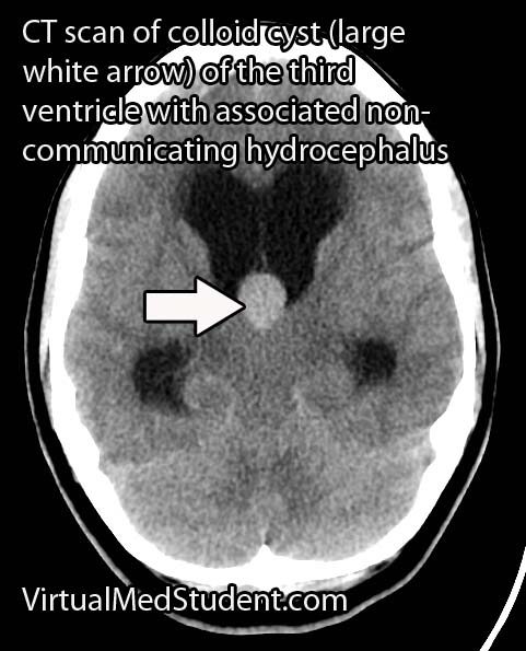
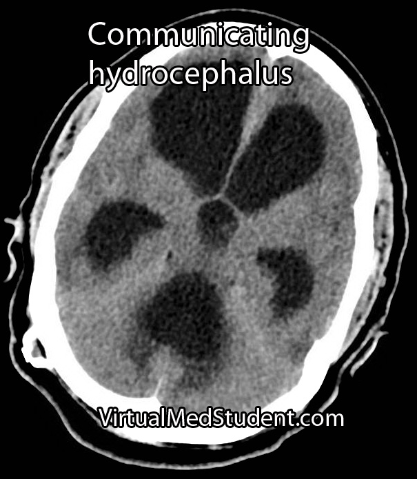
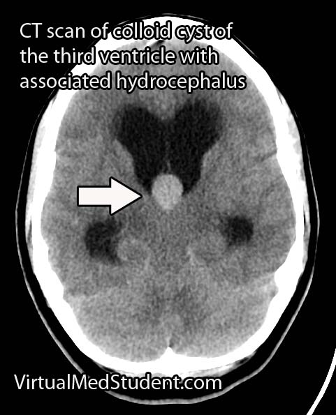
Less significant malformations present later in infancy with a rapidly enlarging head circumference secondary to hydrocephalus, developmental delay, and seizures. The increase in venous blood pressure within the head can cause a "melting brain" syndrome in which the white matter of the brain fails to develop properly. This can lead to severe mental retardation later in life if left untreated.

Diagnosis

Vein of Galen Malformations
Diagnosis is made with a combination of MRI, CT, and diagnostic angiograms. MR venograms can show the dilated venous pouch, as well as associated venous anomalies.

CT angiograms can show associated arterial feeding vessels. Formal diagnostic angiograms are the gold standard test; they delineate both the spatial and temporal relationship of the arterial feeding vessels to the venous pouch.

Formal catheter angiograms are also necessary to distinguish true vein of Galen malformations from arteriovenous malformations of the adjacent brain tissue.

Treatment

Treatment is dependent on the age of the child as well as the severity of the malformation. The most commonly used grading system developed by Dr. Lasjaunias is known as the Bicetre score. It takes into account the child’s cardiac, pulmonary, hepatic (liver), and renal (kidney) function. Lower scores indicate more severe disease with poorer outcomes.

The Bicetre score also dictates the optimal time for treatment. If the score is very low then aggressive treatment, even in the neo-natal period, may be indicated to try and prevent death and severe disability. Higher scores are typically treated later in life; however, worse outcomes, in terms of mental retardation, have been illustrated if treatment is delayed.

Most of these lesions are treated endovascularly (ie: from inside the blood vessels). The arterial feeders are embolized with a glue like material, which ultimately shuts down the fistula in an attempt to restore normal venous pressures. The vein itself may also be filled with tiny metal coils to help reduce flow through the fistula; this is known as trans-venous endovascular therapy.

Surgical ligation of the arterial feeders has mostly become a treatment of the past. Radiation therapy with Gamma Knife has also been used in some cases; it is showing some promise as an alternative treatment modality in select cases.

Overview

Vein of Galen malformations are fetal abnormalities in the brain’s normal venous drainage. They represent true arteriovenous fistulas. They are composed of a dilated median prosencephalic vein of Markowitz and numerous arterial feeding vessels. Feeders may come from the anterior cerebral arteries, posterior cerebral arteries, or posterior communicating arteries. Symptoms are usually from high output heart failure in the neonatal period; older infants and children suffer from increasing head circumference, seizures, and developmental delay. Treatment is usually with endovascular techniques.

References and Resources