The ACOMM Aneurysm: Balloons and Blood

In order to understand the anterior communicating artery, we have to first appreciate the anatomy of the anterior cerebral arteries. The anterior cerebral arteries are one of the two terminal branches of each internal carotid artery (the other being the middle cerebral artery). Each anterior cerebral artery has several sections from A1 to A5.

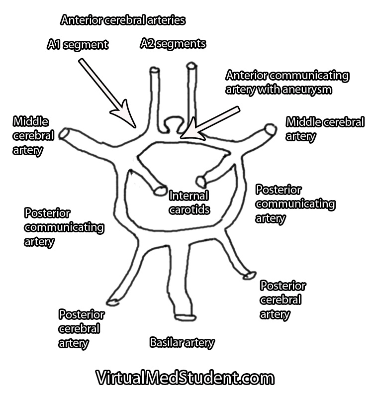
Each A1 segment branches into an A2 segment and into the anterior communicating artery. The anterior communicating artery connects each anterior cerebral arteries’ A1 segment together to form a circle (see schematic below).

The anterior communicating artery is one of the most common sites for intracranial aneurysm formation. Patients at risk for developing aneurysms include those with atherosclerosis, those with a family history of intracranial aneurysms, those with a history of hypertension or collagen vascular disease, and those with polycystic kidney disease. Smokers are also at a higher risk of developing aneurysms.

Basilar Tip Schematic Drawing
Anterior communicating artery aneurysms form when the lining of the vessel wall is thinned and the muscular layer of the blood vessel (tunica media) becomes weakened.

This thinning allows turbulent blood flow to form out-pouchings in the vessel wall. Typically these out-pouchings occur at points where blood vessels branch.

Signs and Symptoms

Anterior communicating artery aneurysms commonly present after a subarachnoid hemorrhage, which can cause a variety of signs and symptoms. The most common being a severe headache, although cranial nerve dysfunction, stroke, coma, and death can also occur.

Less commonly, aneurysms in this location can compress the optic chiasm or optic nerves leading to problems with vision.

How Do You Diagnose Aneurysms

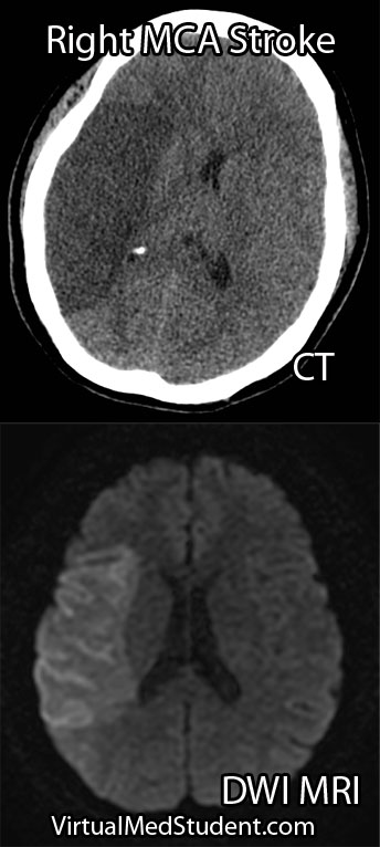
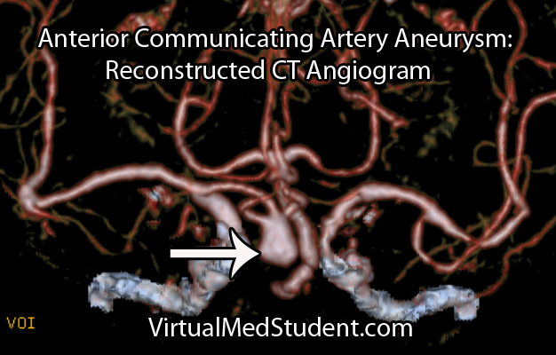
Anterior cerebral artery aneurysms are most commonly diagnosed after a subarachnoid hemorrhage when a patient presents with the "worst headache of their life". The best imaging methods for diagnosing these aneurysms are CT angiograms (see image below), MR angiograms, and formal cerebral angiograms.

Treatment

Like other intracranial aneurysms, anterior communicating artery aneurysms may be clipped or coiled. Clipping of an aneurysm involves an open surgical procedure where the surgeon dissects down to the aneurysm and places a clip across its neck. This excludes it from the circulation and prevents it from rupturing.

Anterior Communicating Artery Aneurysm CT Angiogram

Aneurysms may also be treated from inside the blood vessel. In this procedure a catheter is threaded from the femoral artery in the groin up towards the location of the aneurysm. Small metallic coils are placed within the dome of the aneurysm, which also excludes it from the normal circulation.

Regardless of how the aneurysm is treated – either with clipping or coiling – the end result is that the aneurysm is excluded from the normal circulation. This prevents it from rupturing.

The merits of clipping versus coiling are still under debate. Ultimately, the treatment depends on the size and location of the aneurysm, as well as other medical problems that the patient may have.

The Highlights…

Anterior communicating artery aneurysms are the most common intracranial aneurysm. They typically present after rupturing into the subarachnoid space and/or adjacent frontal lobes. They are diagnosed using CT angiograms or formal cerebral angiography. Treatment is with clipping and/or coiling.

Related Readings

Not Satisfied? More Reading on the Subject…

  • Bederson JB, Awad IA, Wiebers DO, et al. Recommendations for the management of patients with unruptured intracranial aneurysms. Stroke 2000;31:2742-2750.
  • li>Hunt WE, Hess RM. Surgical Risk as Related to Time of Intervention in the Repair of Intracranial Aneurysms. Journal of Neurosurgery 1968; 28:14-20.
  • Brisman JL, Song JK, Newell DW. Cerebral Aneurysms. NEJM 2006; 355:928-939.
  • Kumar V, Abbas AK, Fausto N. Robbins and Cotran Pathologic Basis of Disease. Seventh Edition. Philadelphia: Elsevier Saunders, 2004.
  • Frontera JA. Decision Making in Neurocritical Care. First Edition. New York: Thieme, 2009.
  • Greenberg MS. Handbook of Neurosurgery. Sixth Edition. New York: Thieme, 2006. Chapter 25.

Meningioma and the Arachnoid Cap Cell

A meningioma is a tumor that arises from the lining of the brain or spinal cord (ie: the “meninges”). They arise from cells known as “arachnoid cap cells”. Meningiomas are usually slow growing, “benign” tumors, which means that they are not usually considered cancerous in the strictest sense of the term.

When viewed under the light microscope, meningiomas can have many different appearances. The most common type is a dense sheet-like formation of cells interspersed with closely packed blood vessels. Sometimes the sheets of cells can be separated by connective tissue.

Frequently, meningiomas will have calcium deposits in them known as “psammoma bodies”. Uncommonly, meningioma cells may take on a malignant appearance characterized by increased cellular division (ie: “mitotic figures”) and invasion of the tumor cells into surrounding brain or bone.

Meningiomas test positive for epithelial membrane antigen (EMA) and vimentin (a marker of connective tissue). Ki-67 (a marker of proliferation) can be elevated in more aggressively behaving tumors.

The most common genetic abnormality seen in patients with meningiomas is found on chromosome 22. If present, a mutation in the NF2 gene on this chromosome causes type 2 neurofibromatosis. This disease predisposes individuals to developing multiple tumor types including meningiomas. Other less common genetic abnormalities can be seen on other chromosomes as well.

Signs and Symptoms

Due to their slow growing and benign nature, many meningiomas cause no symptoms. However, if they become too large or start to compress adjacent brain tissue they can cause headache, seizures, confusion, or visual problems. Spinal cord compression can result in myelopathy.

The most common location for a meningioma is in between the two hemispheres of the brain – the so called “parasagittal” location. Parasagittal meningiomas near the portion of the brain responsible for muscle movements may cause weakness of the opposite leg.

Diagnosis

Diagnosis of meningioma can reliably be made on characteristic findings seen on CT or MRI scans. Interestingly, many meningiomas are found incidentally when a CT or MRI is done for other reasons.

Meningioma MRIs

However, like any other tumor, meningiomas can only be truly diagnosed once a specimen is sent to the pathology lab for analysis. Pathologists can reliably make the diagnosis based on typical histological features.

Meningiomas must be distinguished from a more malignant tumor known as a hemangiopericytoma. Hemangiopericytomas can look similar to meningiomas on imaging studies.

Dish Me Up Some Treatment Sir

Many meningiomas can be watched over time with repeat imaging studies; this is especially true if they are small and not causing neurological signs or symptoms.

On the other hand, large or symptomatic meningiomas require surgical resection. Many meningiomas can be removed completely. However, some meningiomas may be near vital structures such as the carotid artery, cranial nerves, or venous draining systems of the brain where complete surgical removal may be very difficult without causing significant neurologic impairment. In these cases the tumor is debulked as much as possible. The residual tumor can be followed or irradiated depending on the grade of meningioma.

Residual tumor after incomplete surgical resection, or meningiomas in difficult to access locations are candidates for radiation therapy. Many studies have shown long term growth control rates.

Overview

Meningiomas are considered “benign” tumors of the brain. They arise from arachnoid cap cells, which are located in a layer of the meninges (ie: the covering of the brain) known as the arachnoid. Symptoms include headache, weakness, vision problems, paresthesias (ie: abnormal sensations), amongst many other possible symptoms. Diagnosis can be made reliably from imaging studies such as CT or MRI. If symptomatic, or large, treatment is surgical resection. Small asymptomatic meningiomas can be managed with repeat imaging to assess for growth over time.

Related Articles

Curated References for Your Pleasure…

Cerebral Cavernous Malformations: Leaky Vessels

Cavernous malformations (aka: cavernomas or cavernous hemangiomas) can be thought of as vascular tumors. They are composed of a capillary-like network of endothelial cells (the cells that normally line blood vessels). However, unlike normal capillaries throughout the body, the capillaries of cavernomas can leak.

Interestingly, cavernous malformations do not have any brain tissue within them. This helps distinguish them from another related vascular abnormality known as an arteriovenous malformation.

The genetics of cavernous malformations have been elucidated by studying familial forms of the disorder. There are at least three known genetic defects that predispose patients to develop cavernomas. These genes appear to be important in the formation of blood vessels (a process known as "angiogenesis") and the blood brain barrier. Therefore, mutations in these genes can cause the abnormal growth of vascular tissue.

What Havock Do These Guys Cause?

Cavernomas can cause numerous signs and symptoms depending on their location within the brain. Seizures are the most common symptom. However, progressive neurological impairment such as worsening weakness can also occur.

Sometimes cavernomas can block the flow of cerebrospinal fluid leading to hydrocephalus. Hydrocephalus can cause increased intracranial pressure leading to headaches, nausea, and vomiting. However, it is important to realize that many patients with cavernomas have no symptoms at all!

If symptoms are present, they tend to progress over time. This is because cavernous malformations bleed and re-bleed resulting in an expansion of its size over time. As the malformation increases in size it can push on adjacent brain tissue causing worsening symptoms.

Unlike arteriovenous malformations, life threatening and severe hemorrhages are rare. Cavernomas bleed at an initial yearly rate of anywhere between 1% to 5%. After the first bleed, the risk of re-bleeding increases to as high as 10% per year. In other words, if a cavernoma bleeds, it is more likely to re-bleed at a later date.

How Do You Diagnose These Buggers?

Cavernoma Marked

The diagnosis of cavernous malformations are made via imaging studies. They are usually detected via MRIs that are ordered for evaluation of neurological symptoms. Cavernomas are seen best on T2 and gradient echo MRI sequences. They typically look like a piece of popcorn.

Select patients undergo a more invasive imaging procedure known as angiography. Angiography is used to rule out another similar lesion known as an arteriovenous malformation. Since cavernomas are venous malformations they are not seen on angiograms.

How Are These Treated?

Depending on the location, most cavernous malformations in the brain or spinal cord are removed surgically. Some institutions offer radiation as a means of treatment, especially in difficult to access areas (ie: where the risk of surgical removal is very high).

Let’s Recap this MoFo

Cavernous malformations of the brain are abnormal vascular growths composed of capillary networks. They are likely the result of genetic mutations in genes responsible for blood vessel growth. Depending on their location they can cause numerous neurological symptoms such as seizures and weakness. MRI often shows the characteristic “popcorn” lesion. Treatment is usually with surgical resection, although some cavernomas may be radiated depending on their location.

Related Articles

Not Satisfied? More References and Resources…

Middle Cerebral Artery: A Common Site for Stroke

Middle cerebral artery (MCA) strokes occur when the MCA or its branches are occluded. With occlusion, blood, and along with it, oxygen and nutrients fail to reach the brain. If blood flow is not restored quickly the affected brain tissue dies leading to permanent neurological injury.

Risk factors for MCA strokes are the same for other strokes. They include hypertension, diabetes, smoking, atrial fibrillation, and a whole slew of hypercoagulable states (ie: pathologic increases in the bodies’ propensity to form blood clots).

In order to understand MCA strokes we have to first appreciate the anatomy of the MCAs, as well as the brain that they serve. The MCAs are subdivided into four parts. The M1 segment is one of the terminal branches of the internal carotid artery. The MCAs then become progressively more narrow and form more and more branches as they reach out towards the cortical surfaces of the brain. The most distal portions of the MCA are deemed the M4 branches. The lateral lenticulostriate arteries are small branches that branch from the M1 segments; these small arteries feed some of the deeper structures of the brain.

Importantly, the MCAs and their branches provide blood flow to an extremely large portion of the brain. These areas include the lateral and inferior frontal lobes, superior portion of the temporal lobes, insula, and lateral parietal lobes. Branches of the first portion of the MCAs (aka: the lateral lenticulostriate arteries) also provide blood flow to the deep sections of the brain including the putamen, head and body of the caudate, external globus pallidus, and parts of the posterior limb of the internal capsule.

The segmental anatomy of the MCAs is important because strokes that occur in the outer segments (ie: M3 and M4) cause less neurological injury than the inner MCA segments (ie: M1 and M2). This is because less total brain volume is affected by outer segment strokes.

The most common cause of MCA strokes are clots that break off and travel from the heart or the carotid arteries to the MCA (aka: emboli). Less commonly, a blood clot will form directly in the MCA itself (aka: thrombus). Atherosclerotic disease is the most common cause of thrombus formation in the MCA and atrial fibrillation (an abnormal heart rhythm) is the most common cause of emboli from the heart.

Signs and Symptoms

Middle cerebral artery strokes present with one of two types of syndromes depending on which MCA – right or left – is involved, as well as which segments of the MCA are involved.

In the worst case scenario, a stroke of the right or left M1 segment of the MCA causes weakness of the opposite side of the body. This is a result of cortical damage to the primary motor cortex as well as possible infarction of the posterior limb of the internal capsule (ie: the location of descending motor tracts). MCA strokes usually present with face and arm weakness that is worse than leg weakness. Remember that the motor cortex that controls leg function is served by the anterior cerebral arteries.

M1 strokes also cause decreased ability to feel sensation on the opposite side of the body as a result of damage to the parietal lobes.

Damage to the optic radiations, which course in the parietal (Baum’s loop) and temporal lobes (Meyer’s loop) can cause problems with vision. Finally, injuries to the frontal eye fields cause the eyes to deviate towards the side of the stroke (ie: the frontal eye fields normally allow you to make fast eye movements in the opposite direction, therefore damage prevents patients from looking to the non-affected side).

Right and left sided M1 strokes will give you all of the above symptoms; however, laterality can be determined based on specific symptoms caused by only a left or a right sided strokes.

The poor patients with left M1 strokes have a decreased ability to speak and/or understand language because of damage to Broca’s area in the left frontal lobe (speech production) and Wernicke’s area in the left temporal lobe (speech comprehension). Remember this is in addition to all the other stuff above.

Right M1 strokes can cause "anosognosia", in which the patient is unaware of certain deficits they may have; these patients also often fail to recognize the left side of their body (ie: they "neglect" or fail to appreciate the entire left side of the world) and may have difficulty appreciating people or objects presented in their left visual field.

Right MCA stroke
Less "severe" cases, in which M2, M3 or M4 branches are affected can produce a variety of signs and symptoms depending on the specific branches involved.

Diagnosis

Diagnosis of MCA strokes are based on symptoms, CT scans, and MRI images. CT and MR angiograms frequently show the blocked blood vessel causing the stroke. CT perfusion scans are a newer technology that give information regarding the amount of blood flow to affected brain tissue.

Treatment

Treatment depends on the timing of the stroke. If the patient presents within 3 hours of symptom onset, and the head CT reveals no bleeding, than intravenous tissue plasminogen activator (tPA) may be given to help "break" up the blood clot causing the stroke.

Other treatments using catheter based approaches are frequently used in patients who are unable to receive tPA. Such treatments include mechanical clot removal with special catheter and wire devices. In addition, in patients more than 3 hours, but less than 6 hours out from symptom onset intra-arterial (not to be confused with intravenous) tPA may be used.

Less commonly, large "malignant" MCA strokes may cause significant swelling, which can put pressure on the brainstem. These patients sometimes undergo an open surgical procedure known as a "craniectomy", in which the bone overlying the affected brain tissue is removed. This surgery allows the edematous brain tissue to swell outwards preventing it from herniating downwards towards vital brainstem structures.

Overview

Middle cerebral artery strokes are most commonly caused by blood clots that break off from the heart or carotid artery. Symptoms of MCA strokes depend on the segment involved, as well as which MCA (right versus left) is involved. Diagnosis is made with a combination of MRI, CT, and symptomatology. Treatment consists of intravenous or intra-arterial tPA and/or mechanical clot removal depending on the time frame of the symptoms.

Related Articles

References and Resources

Monro-Kellie and Their Doctrine: Blood, Brain, Spinal Fluid

The Monro-Kellie doctrine states that three things exist within the fixed dimensions of the skull: blood, cerebrospinal fluid, and brain. An increase in any one component must necessarily lead to a decrease in one (or both) of the other components, otherwise intracranial pressure will increase.

Increases in one of the three components can take many different shapes and sizes. For example, abnormal bleeding within the cranium such as in epidural and subdural hematomas are common examples, which typically occur after traumatic events. Bleeding within the brain tissue itself – known as an intraparenchymal or intracerebral hematoma – can also occur, especially in patients with untreated high blood pressure. Brain tumors of any type effectively increase the amount of brain tissue. And last, but not least, the cerebrospinal fluid can back up in a condition known as hydrocephalus.

Regardless of the cause, the end result is an abnormal increase in either blood, brain, or cerebrospinal fluid within the confines of the skull.

So what’s the big deal? If the abnormality becomes large enough, the pressure within the skull can increase rapidly. Eventually the pressure can become so great that the brain gets squished, and will pop over rigid boundaries and out the small holes within the skull.

This is known as “herniating” the brain tissue. It can occur in numerous places depending on where the pressure is greatest. However, the most important herniation clinically occurs at the base of the skull where a hole known as the foramen magnum exists.

When the brain herniates here it really pisses off a vital structure known as the brainstem. The brainstem is responsible for all the stuff we don’t consciously think about (heart rate, breathing, etc.), which ultimately keeps us alive. When herniation of the brainstem through the foramen magnum occurs it stretches all the “wires” that allow our brainstem to function properly. If severe enough, all those autopilot functions (ie: breathing, beating of the heart, etc.) stop working and brain death occurs.

Overview

The skull contains three components within it: blood, brain, and cerebrospinal fluid. An abnormal increase in any one of these components causes an increase in pressure, which if severe enough, can cause herniation of brain tissue out of the skull. This can lead to coma and brain death.

References and Resources

Vein of Galen Malformations: A Misnomer of Sorts

The vein of Galen is located deep within the brain. The internal cerebral veins, basal veins of Rosenthal, atrial veins, and precentral cerebellar veins join together to form the vein of Galen.

As expected, the vein of Galen drains blood from deeply located brain structures. The vein of Galen then connects with the inferior sagittal sinus to form the straight sinus; blood then drains from the straight sinus into the transverse and sigmoid sinuses, where it eventually finds its way into the internal jugular veins and back to the heart.

Interestingly, a "vein of Galen" malformation is not actually a malformation of the true vein of Galen; the term is a misnomer. It is actually a malformation of primitive fetal anatomical structures that normally regress during development. These primitive structures include a dilated venous structure, as well as "feeding" arteries. Therefore, vein of Galen malformations represent true arteriovenous fistulas; in other words, blood moves directly from an artery to a vein without an intermediary capillary bed.

Between the 3rd and 11th weeks of fetal development a large primitive vein known as the median prosencephalic vein of Markowitz drains the deepest parts of the brain. As the brain develops, the internal cerebral veins annex the territory normally drained by the anterior portion of this vein. As a result, this portion of the median prosencephalic vein regresses. The internal cerebral veins then plug in to the posterior portion of the median prosencephalic vein, which becomes the "true", or "normal", vein of Galen.

The most common arterial "feeders" of the malformation can also be explained by aberrant embryology. During early fetal brain development the distal branches of the anterior cerebral arteries (ie: the pericallosal branches) make connections with the posterior cerebral arteries. These connections usually regress to form the anterior and posterior circulations, which are connected to one another via the posterior communicating arteries.

So how does a vein of Galen malformation form? In some infants the median prosencephalic vein of Markowitz does not regress like it should. As a result, a large abnormal venous midline pouch remains. It also retains its primitive arterial blood supply from the distal branches of the anterior cerebral artery (ie: pericallosal branches), anterior choroidal arteries, posterior communicating arteries, and branches of the posterior cerebral arteries (ie: posterior choroidal arteries).

Vein of Galen malformations are also associated with other abnormalities in the venous structure of the brain. Not uncommonly, the straight sinus is absent or severely narrowed. As a result venous blood drains into a persistent falcine sinus, which is a structure that normally regresses in-utero.

To summarize, vein of Galen malformations are primitive direct arteriovenous fistulas. They are composed of a dilated venous pouch (ie: the median prosencephalic vein of Markowitz) with any combination of anterior and posterior circulation feeding arteries.

Signs and Symptoms

Signs and symptoms depend on the severity of the malformation. Severe malformations present in new borns with high output cardiac failure. This is because so much blood is being shunted into the malformation that the heart cannot keep pace!

Less significant malformations present later in infancy with a rapidly enlarging head circumference secondary to hydrocephalus, developmental delay, and seizures. The increase in venous blood pressure within the head can cause a "melting brain" syndrome in which the white matter of the brain fails to develop properly. This can lead to severe mental retardation later in life if left untreated.

Diagnosis

Vein of Galen Malformations
Diagnosis is made with a combination of MRI, CT, and diagnostic angiograms. MR venograms can show the dilated venous pouch, as well as associated venous anomalies.

CT angiograms can show associated arterial feeding vessels. Formal diagnostic angiograms are the gold standard test; they delineate both the spatial and temporal relationship of the arterial feeding vessels to the venous pouch.

Formal catheter angiograms are also necessary to distinguish true vein of Galen malformations from arteriovenous malformations of the adjacent brain tissue.

Treatment

Treatment is dependent on the age of the child as well as the severity of the malformation. The most commonly used grading system developed by Dr. Lasjaunias is known as the Bicetre score. It takes into account the child’s cardiac, pulmonary, hepatic (liver), and renal (kidney) function. Lower scores indicate more severe disease with poorer outcomes.

The Bicetre score also dictates the optimal time for treatment. If the score is very low then aggressive treatment, even in the neo-natal period, may be indicated to try and prevent death and severe disability. Higher scores are typically treated later in life; however, worse outcomes, in terms of mental retardation, have been illustrated if treatment is delayed.

Most of these lesions are treated endovascularly (ie: from inside the blood vessels). The arterial feeders are embolized with a glue like material, which ultimately shuts down the fistula in an attempt to restore normal venous pressures. The vein itself may also be filled with tiny metal coils to help reduce flow through the fistula; this is known as trans-venous endovascular therapy.

Surgical ligation of the arterial feeders has mostly become a treatment of the past. Radiation therapy with Gamma Knife has also been used in some cases; it is showing some promise as an alternative treatment modality in select cases.

Overview

Vein of Galen malformations are fetal abnormalities in the brain’s normal venous drainage. They represent true arteriovenous fistulas. They are composed of a dilated median prosencephalic vein of Markowitz and numerous arterial feeding vessels. Feeders may come from the anterior cerebral arteries, posterior cerebral arteries, or posterior communicating arteries. Symptoms are usually from high output heart failure in the neonatal period; older infants and children suffer from increasing head circumference, seizures, and developmental delay. Treatment is usually with endovascular techniques.

References and Resources