Enterobius Vermicularis and Itchy Butts

Enterobius vermicularis, better known as pinworm, is a nematode or roundworm. Like other worms it has a unique lifecycle that is quite interesting, albeit somewhat disgusting! It begins when an egg is ingested by a human. Eggs are usually ingested because of poor hygiene (ie: not washing your hands after doing number 2) and can sometimes be found in contaminated food. Once ingested the eggs hatch in the small intestine. From there the worms migrate to the large intestine where they mate. For unknown reasons the pregnant females head towards the anus at night where they lay their eggs.

The eggs are then shed in the feces and potentially picked up by another unlucky host. It is the most common worm related infection in the United States.

Signs and Symptoms

The eggs in the perianal area are extremely pruritic (ie: itchy). Scratching of the anus secondary to the intense pruritis can lead to skin breakdown and potential bacterial super infection. The symptoms are generally most severe at night because this is when the females migrate to the anus to lay their eggs. Systemic signs are generally not present although some patients can have malaise (ie: feeling "crappy").

Diagnosis

The traditional way of diagnosing is the "scotch tape" test. The clinician takes a piece of scotch tape and applies it to the patient’s perianal region. Eggs can be seen on the tape once its removed. The best time to perform this test is early in the morning before bathing or at night.

Treatment

Two anti-helminth medications are used to treat pinworm. The first is albendazole. Generally a single dose is enough to kill all worms, but since eggs may be present on clothing, bedding, etc. a second dose is often given two weeks later. Mebendazole is another medication that is also given as a single dose repeated two weeks later. These medications work by inhibiting microtubule polymerization in the cytoplasm of the worm’s cells.

Overview

Enterobius vermicularis (pinworm) is the most common worm infection in the United States. It causes an intense itch in the perianal area that is worst at night. It is diagnosed by the "scotch tape" test. Treatment is with albendazole or mebendazole.

Related Articles

References and Resources

Pyogenic Liver Abscesses: Pus, Needle Drainage, and Antibiotics

Pyogenic liver abscesses are localized collections of pus and bacteria. The initial infection occurs when bacteria travel through the portal vein. This most commonly occurs after bowel contents leak into the peritoneal cavity from the gut. Other causes of liver abscesses include direct spread of infection from the bile duct system, or from other bacterial infections of the blood. Rarely, penetrating injuries (gunshots, stab wounds, surgery) may directly introduce infection. There are numerous “bugs” that can cause pyogenic liver abscesses. They include, but are not limited to, streptococcus species, klebsiella pneumoniae, and staphylococcus species.

Signs and Symptoms

Similar to other infections, liver abscesses can cause fevers, chills, decreased appetite, abdominal pain, and a generalized sense of not feeling well (ie: malaise). Interestingly, hiccups may also be present if the abscesses are causing adjacent inflammation/irritation of the diaphragm.

Diagnosis

Pyogenic Liver Abscesses
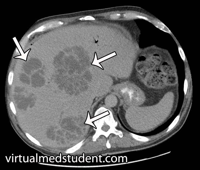
The diagnosis of pyogenic liver abscesses is made with imaging. The most commonly used method is a CT scan of the abdomen with and without contrast. If present, a liver abscess will look like a collection of fluid, with or without septated dividers.

Another commonly employed imaging modality is the use of ultrasound to detect the fluid filled pockets within the liver.

However, it is important to note that imaging studies alone cannot distinguish between the different types of liver abscesses. Imaging can also not tell you what bacteria is responsible for the abscess.

Treatment

Pyogenic abscesses must be drained and treated with antibiotics. Drainage is usually done under image guidance using a needle placed through the skin into the abscess. Although occasionally direct surgical evacuation of the abscess is necessary.

Draining the abscess is important for two reasons. First, it decreases the size of the abscess allowing antibiotic therapy to work more effectively. And secondly, it provides abscess fluid that can be sent to the lab for bacterial culture and gram stain.

The results of the culture help guide subsequent antibiotic therapy. Commonly used antibiotics include piperacillin-tazobactam (Zosyn®), vancomycin, metronidazole (Flagyl®), and ceftriaxone; once cultures confirm the causative bug antibiotic therapy can be narrowed.

Overview

Pyogenic liver abscesses are collections of pus and bacteria. They occur most commonly after the spilling of gut bacteria into the peritoneal space (ie: peritonitis). There are numerous causative bacteria. Symptoms include fever, chills, decreased appetite, and abdominal discomfort. Treatment is with drainage and antibiotics.

References and Resources

  • Hasper D, Schefold JC, Baumgart DC. Management of severe abdominal infections. Recent Pat Antiinfect Drug Discov. 2009 Jan;4(1):57-65.
  • Benedetti NJ, Desser TS, Jeffrey RB. Imaging of hepatic infections. Ultrasound Q. 2008 Dec;24(4):267-78.
  • Mortelé KJ, Segatto E, Ros PR. The infected liver: radiologic-pathologic correlation. Radiographics. 2004 Jul-Aug;24(4):937-55.
  • Kurland JE, Brann OS. Pyogenic and amebic liver abscesses. Curr Gastroenterol Rep. 2004 Aug;6(4):273-9.

Ixodus, Burgdorferi, and Lyme Oh My!

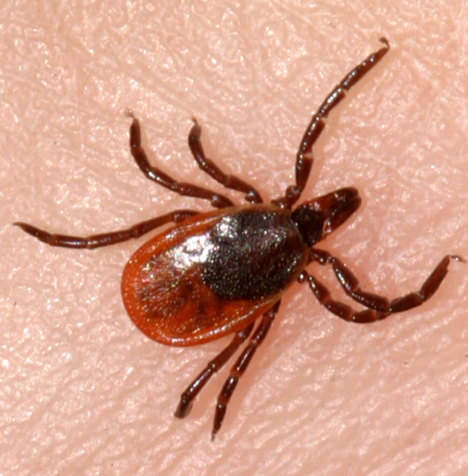
Borrelia burgdorferi is a gram negative spirochete bacterium. Its hosts are mice, deer, and humans. Spread from host to host is via a tick from the species Ixodes. Ixodes scapularis is the most common tick vector in the Northeastern United States, whereas Ixodes pacificus is the most common in the Western United States.

When an infected tick feeds from a human it can pass borrelia burgdorferi bacteria into the bloodstream. From there the bacterium is usually attacked by both the innate and adaptive immune systems. The innate immune system uses toll-like receptors on macrophages to bind to bacterial proteins. The adaptive immune response uses antibodies to bind to bacterial surface proteins, which results in complement activation and bacterial death.

Unfortunately, the immune response is often not enough to entirely eliminate this bacteria. This is because borrelia burgdorferi has the ability to evade the immune system by changing proteins on its cell surface.

Signs and Symptoms

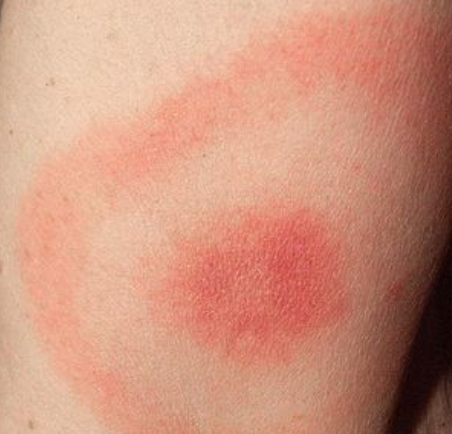
Borrelia burgdorferi causes Lyme disease. Lyme disease manifests in different ways depending on which stage of the disease the patient enters. The first stage of the disease presents with a rash known as erythema chronicum migrans. This rash appears as a red ring around a clear central area (ie: it resembles a bull’s eye). During this stage of the disease many patients also have flu-like symptoms.

If left untreated the disease can progress to stage two. During this stage neurological, joint, and cardiac symptoms can occur. The bacterium can cause cardiac arrhythmias, especially heart block. Facial drooping can be caused by damage to the facial nerve (cranial nerve seven). Meningitis, which ranges in severity, can occur. Finally, a migrating intermittent arthritis can also be seen during this stage of the disease.

If the disease continues to remain untreated, stage three ensues! This stage happens approximately a year or more after the initial tick bite. It is characterized by chronic arthritis, which can cause severe joint damage. Encephalitis (inflammation of the brain tissue) and myelitis (inflammation of the spinal cord) can also occur.

Diagnosis

The diagnosis of Lyme disease is a two step process. A blood sample is taken from the patient and a test known as an ELISA is performed. ELISA stands for “enzyme linked immunosorbent assay”. This test detects antibodies made by the patient against borrelia burgdorferi antigens (ie: it’s surface proteins). If the ELISA is positive, then a second confirmatory test called a "western blot" is performed. This test further separates the different antibodies to borrelia. If both the ELISA and western blot are "positive" the likelihood of Lyme disease is high.

Treatment

Treatment for early stage disease is with an antibiotic known as doxycycline. Children and pregnant women are often treated with amoxicillin. Other medications used include cefuroxime; erythromycin is sometimes used in patients who are allergic to pencillin. Later stages of the disease may need intravenous antibiotics.

If patients do not respond to initial antibiotic treatment it is important to think of co-existent infections with other tick-borne illnesses. Ticks may carry not only borrelia burgdorferi, but also other pathologic entities so treatment may need to be further tailored.

Overview

Lyme disease is caused by the bacterium borrelia burgdorferi. It is transmitted by the Ixodes tick and is common in the northeastern United States. Symptoms include a bull’s eye rash known as erythema migrans. Neurological, joint, and cardiac symptoms can also occur. Treatment for early stage disease is with oral antibiotics, usually doxycycline, although amoxicillin is sometimes used. Intravenous antibiotics may be necessary for later stages.

References and Resources