The Internal Capsule: Some Pricey Brain Real Estate

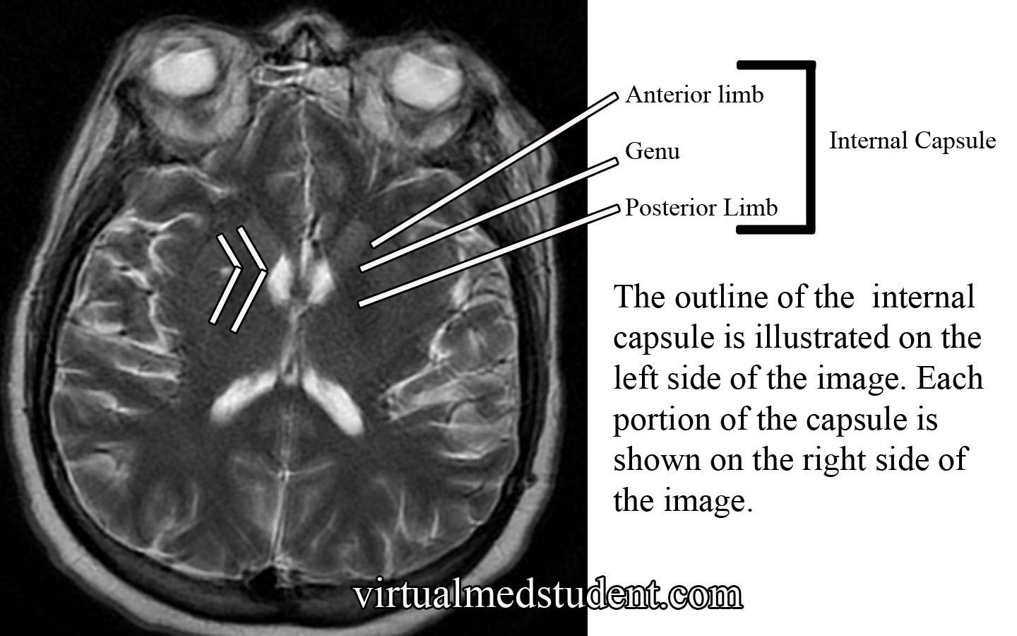
The internal capsule is one pricey piece of brain real estate! It contains all of the pathways that allow information to be transferred between the cerebral cortex and the spinal cord, brainstem, and subcortical structures (ie: thalamus, basal ganglia). It is divided into an anterior limb, posterior limb, and genu (ie: the area where the anterior and posterior limbs meet).

The anterior limb contains axons that send information between the thalamus and the cingulate gyrus and pre-frontal cortex. It also contains axons in the frontopontine pathway (ie: axons going from the frontal cortex to a portion of the brainstem known as the pons).

The genu contains the corticobulbar tract, which originate in the motor areas of the frontal lobes and extend to the cranial nerve nuclei in the brainstem. It also contains axons that connect the motor section of the thalamus (ie: VA and VL nuclei) with the motor areas of the frontal cortex.

The posterior limb contains the corticospinal tract, which are axons that come from the motor area of the frontal cortex and travel all the way to the anterior horns of the spinal cord where α-motor neurons are located. The posterior limb also contains sensory information coming from the body via the medial lemniscus and the anterolateral (aka: spinothalamic tract) systems.

Internal Capsule MRI

The blood supply to most of the internal capsule comes from the lenticulostriate arteries. These small arteries originate from the first portion of the middle cerebral artery. Two other important arteries also supply portions of the internal capsule: the anterior choroidal artery and the recurrent artery of Heubner. The anterior choroidal artery is a branch of the internal carotid. It supplies the inferior portion of the posterior limb. The recurrent artery of Heubner is a branch of the anterior cerebral artery. It supplies the inferior portions of the anterior limb and the genu.

Anatomy of the Internal Capsule
Division Major Communication Tracts Blood Supply
Anterior limb

– Tracts between the frontal lobe and pons (brainstem)

– Tracts between the thalamus and prefrontal cortex

– Tracts between the thalamus and cingulate gyrus

– Lenticulostriate arteries (branches of the middle cerebral artery)

– Recurrent artery of Heubner (branch of the anterior cerebral artery)

Genu – Tracts between the motor cortex in the frontal lobe and the cranial nerve nuclei in the brainstem (aka: corticobulbar tract)

– Lenticulostriate arteries (branches of the middle cerebral artery)

– Recurrent artery of Heubner (branch of the anterior cerebral artery)
Posterior limb

– Tracts between the motor cortex of frontal lobe and anterior horn of spinal cord (aka: corticospinal tract)

– Medial lemniscus tract (a continuation of the dorsal columns), which carries information about light touch, vibration, and pressure sensation from the body and spinal cord.

– Anterolateral (aka: spinothalamic) tract, which carries pain and temperature information

– Lenticulostriate arteries (branches of the middle cerebral artery)

– Anterior choroidal artery (branch of the internal carotid)

Importance in Disease

Thalamic Hemorrhage
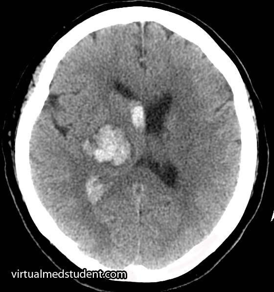
Thalamic intracerebral hematoma
compressing the posterior limb
of the internal capsule

Damage to the internal capsule can be devastating neurologically because it contains so many vital tracts.

For example, a stroke of the anterior choroidal artery can lead to posterior limb damage. This can cause paralysis of the contralateral (ie: opposite) arm and leg secondary to interruption of the corticospinal tract.

Posterior limb disruption can also cause co-existent sensory deficits including an inability to feel light touch, pain, and temperature due to damage of the spinothalamic and medial lemniscal pathways.

Hypertensive hemorrhages in the thalamus or basal ganglia can compress the adjacent fibers of the internal capsule leading to similar clinical findings. The head CT to the right shows a thalamic hemorrhage secondary to severely elevated blood pressure. The patient had compression of the posterior limb of the internal capsule. As a result she was unable to move her left arm and leg, and could not feel pain or light touch on the left side of her body.

Overview

The internal capsule is a vital structure. It contains many communication pathways between the brain’s cortex, brainstem, spinal cord, and subcortical nuclei (ie: thalamus, basal ganglia). Its blood supply comes from branches of the middle cerebral artery (ie: lenticulostriates), anterior cerebral artery (ie: recurrent artery of Heubner), and the internal carotid (ie: anterior choroidal artery). Lesions in this area caused by strokes or hypertensive hemorrhages can have devastating clinical consequences.

Other Pertinent Articles…

References and Resources

  • Greenberg MS. Handbook of Neurosurgery. 9th Edition. New York: Thieme, 2006. Chapter 25.
  • Chowdhury F, Haque M, Sarkar M, et al. White fiber dissection of brain; the internal capsule: a cadaveric study. Turk Neurosurg. 2010 Jul;20(3):314-22. doi: 10.5137/1019-5149.JTN.3052-10.2.
  • Simon RP, Aminoff MJ, Greenberg DA. Clinical Neurology, Seventh Edition (LANGE Clinical Medicine). Seventh Edition. New York: McGraw Hill, 2009.
  • Nolte J. The Human Brain: An Introduction to its Functional Anatomy. Sixth Edition. Philadelphia: Mosby, 2008.
  • Bickley LS, Szilagyi PG. Bates’ Guide to Physical Examination and History Taking. Ninth Edition. New York: Lippincott Williams and Wilkins, 2007.

Radial Nerve and the Saturday Night Palsy

Radial Nerve Course
In order to appreciate the radial nerve, we have to first understand the brachial plexus. The brachial plexus can be thought of as a massive highway intersection, in which numerous highways come together and then split apart again.

The "highways" merging into the brachial plexus are the 5th, 6th, 7th, and 8th cervical nerve roots, as well as the 1st thoracic nerve root. These nerve "highways" tangle together to form trunks, divisions, cords, and then branches. The radial nerve is one of the branches of the brachial plexus; it gets its input from the 5th, 6th, 7th and 8th cervical nerve roots.

The radial nerve courses along the humerus in the upper arm. It wraps around the humerus in a spot called the spiral groove. Just before wrapping around the humerus, it sends a branch that innervates the triceps muscle (long, medial, and lateral heads) in the upper arm.

After wrapping around the spiral groove, it sends additional branches to the brachioradialis, extensor carpi radialis longus, and extensor carpi radialis brevis muscles.

The first major branch in the forearm is known as the superficial radial nerve. This nerve courses along the medial/ulnar aspect of the forearm (ie: the ulnar or medial side of the forearm is closest to your body when your palms are facing forward) and heads straight for the hand. It relays sensory information from the lateral portion of the back of the hand.

The second major branch at the elbow can be thought of as the deep radial nerve, but it is formally known as the posterior interosseous nerve.

The posterior interosseus nerve sends branches to eight muscles in the forearm. They include the supinator (through which the nerve travels via a fibrous tunnel known as the arcade of Frohse), extensor digiti minimi, extensor carpi ulnaris, extensor digitorum, abductor pollicis longus, extensor pollicis longus, extensor pollicis brevis, and extensor indicis.

Muscles Innervated by the Radial Nerve and Its Branches
Muscle Action of Muscle
Triceps brachii – Extension of the forearm away from upper arm
Brachioradialis – Helps flex the forearm closer to the upper arm
– Helps supinate (ie: palm towards the sky)
– Helps pronate (ie: palm towards the floor)
Extensor carpi radialis
(longus and brevis)
– Helps extend the wrist
– Abducts the hand (ie: hand moves away from
the body when the palms are facing forward)
Extensor digiti minimi – Helps extend the little finger (5th digit)
Extensor carpi ulnaris – Helps extend the wrist
– Adducts the hand (ie: hand moves towards
the body when the palms are facing forward)
Supinator – Allows palms to face up towards the sky
Extensor digitorum – Helps with extension of the fingers
(specifically at the metacarpophalngeal joint)
Abductor pollicis longus – Helps abduct the thumb
Extensor pollicis
(longus and brevis)
– Help extend the thumb
Extensor indicis – Helps extend the index finger

Generally speaking, the radial nerve and its branches are involved in muscles that allow joints to extend (ie: widen or separate away from one another).

Importance in Disease

Damage to the radial nerve may take the form of compression or sheering injuries, typically after traumatic events.

The most common site of injury is at the spiral groove of the humerus. The nerve may be damaged if someone breaks their humerus, or if someone leans the back of their arm on something for an extended period of time (ie: a "Saturday night palsy" is the informal term given to a drunk who falls asleep with their arms draped over a chair… they end up waking up with a radial nerve palsy!).

Injury to the nerve at the spiral groove causes a wrist drop, in which the affected person cannot extend their wrist. The triceps are not affected because nerve branches to this muscle are proximal to the spiral groove. In addition, patients also complain of decreased sensation on the back of the hand (see image to right).

Radial Nerve Sensory Distribution in Hand
Additionally, the posterior interosseous branch of the radial nerve may get compressed as it passes through the supinator muscle in the forearm. This is referred to as posterior interosseous nerve syndrome.

The compression occurs at a fibrous portion of the supinator muscle known as the "arcade of Frohse", which causes an inability to extend the fingers at the metacarpophalangeal joints; this is due to dysfunction of the extensor digitorum muscle (extension at the interphalangeal – both distal and proximal – joints is controlled by the lumbricals and interossei muscles which are innervated by the median and ulnar nerves).

In posterior interosseous nerve syndrome it is still possible to extend the wrist because the branches of the nerve to the extensor carpi radialis muscle are unaffected (ie: they branch before the arcade). However, when the wrist is extended it deviates towards the radial side of the forearm because of the unopposed action of the extensor carpi radialis muscles (in other words, the extensor carpi ulnaris is affected and cannot keep the wrist extension neutral).

Sensation is entirely normal when the posterior interosseous nerve gets compressed because it contains no sensory fibers.

Overview

The radial nerve is a terminal branch of the brachial plexus. It sends branches to most of the extensor muscles of the arm and forearm. It also provides sensation to the back side of the hand. If injured it causes either a radial nerve palsy, or posterior interosseous syndrome, in which the affected patient has an inability to extend various joints at the wrist and/or fingers.

Related Anatomy You Should Know About…

References and Resources

  • Ducic I, Felder JM 3rd, Quadri HS. Common nerve decompressions of the upper extremity: reliable exposure using shorter incisions. Ann Plast Surg. 2012 Jun;68(6):606-9
  • Colbert SH, Mackinnon SE. Nerve compressions in the upper extremity. Mo Med. 2008 Nov-Dec;105(6):527-35.
  • Reddy MP. Peripheral nerve entrapment syndromes. Am Fam Physician. 1983 Nov;28(5):133-43.
  • Calfee RP, Wilson JM, Wong AH. Variations in the anatomic relations of the posterior interosseous nerve associated with proximal forearm trauma. J Bone Joint Surg Am. 2011 Jan 5;93(1):81-90.
  • Arle JE, Zager EL. Surgical treatment of common entrapment neuropathies in the upper limbs. Muscle Nerve. 2000 Aug;23(8):1160-74.