Maroteaux-Lamy Syndrome: GAGs and Dermatan Sulfate

Maroteaux-Lamy syndrome falls into a family of conditions called the “mucopolysaccharidoses”. This fancy term describes any disorder where patients are unable to metabolize long chains of repeating sugar units.

Another term for mucopolysaccharide is glycosaminoglycan, or “GAG”. GAGs are composed of long, repeating, un-branched sugar chains. Each repeating unit has a six carbon sugar (ie: the general term for a simple six carbon sugar is a “hexose”; glucose, mannose, galactose, etc. are specific examples) attached to another six carbon sugar that has a nitrogen group sticking off of it; the general name for a six carbon sugar with an attached nitrogen group is a “hexosamine”.

These two-sugar units, a hexose plus a hexosamine, connect to one another over and over again to form a very long chain called a glycosaminoglycan.

GAGs and mucopolysaccharides are important in the human body because they make up a significant proportion of connective tissue. Connective tissues provide support and structure to many of the organ systems. As such, the body is constantly forming, breaking down, and re-molding GAGs to provide the structural framework so that the rest of our cells can do their jobs well.

So what do GAGs have to do with Maroteaux-Lamy disease? A specific GAG known as dermatan sulfate is not broken down appropriately in patient’s suffering from this condition.

The reason these individuals are unable to break down dermatan sulfate is because they have a genetic mutation in the enzyme arylsulfatase B. In a normal person, arylsulfatase B degrades excess and unwanted dermatan sulfate; however, when the enzyme is defective it causes dermatan sulfate to build up to abnormally high levels within cells.

This excess dermatan sulfate eventually “gunks” up various organ systems and results in the characteristic signs and symptoms associated with the disease.

Signs and Symptoms

It is important to realize that there are numerous genetic mutations that can cause Maroteaux-Lamy syndrome and not all mutations are created equal! Therefore, there is a wide spectrum of signs and symptoms that may occur quickly, or slowly, depending on which particular mutation the patient has.

Most patients are short and have progressive changes in their facial features. Patients often develop early onset vision problems because their corneas become clouded by excess dermatan sulfate. Joint stiffness and skeletal abnormalities are also very common, as are heart and lung complications.

The lining of the brain and spinal cord can become excessively thick which can cause weakness secondary to spinal cord and nerve compression.

Patient’s also often have enlargement of the spleen – a condition called “splenomegaly”. It is also not uncommon for patients to have various types of hernias.

Unlike patients who suffer from some of the other mucopolysaccharidoses, people with Maroteaux-Lamy are of normal intelligence.

Diagnosis

Overall, Maroteaux-Lamy disease is more appropriately a “syndrome”, or constellation of signs and symptoms. By themselves, each sign or symptom is not diagnostic of the disorder, but when present together can support the diagnosis.

Genetic testing for mutations of the arylsulfatase B gene are done once the syndrome is suspected on clinical grounds. Additionally, urine samples contain elevated amounts of dermatan sulfate.

Treatment

Treatment consists of using a replacement enzyme known as Naglazyme® (aka: galsulfase). This enzyme performs the function of the deficient arylsulfatase B and helps in the degradation of dermatan sulfate.

Stem cell transplants can also provide the missing enzyme, but at significant risk. Unfortunately, few patients are candidates for stem cell transplant given difficulty with donor matching.

A very important component of treatment is monitoring for the development or progression of symptoms. This will allow specialists to step in and perform palliative procedures that can prevent worsening disability. For example, corneal transplants can be done to restore vision loss.

Overview

Maroteaux-Lamy is one of the mucopolysaccharidoses. It is caused by a genetic deficiency in the enzyme arylsulfatase-B, which prevents the degradation of dermatan sulfate. It presents with numerous signs and symptoms, which may be rapidly or slowly progressive depending on the specific genetic mutation. Treatment is with enzyme replacement therapies and palliative management of symptoms.

Related Articles

References and Resources

  • Calleja Gero ML, González Gutiérrez-Solana L, López Marín L, et al. Neuro-imaging findings in patient series with mucopolysaccharidosis. Neurologia. 2011 Dec 15.
  • Mtar A, Charfeddine B, Braham I, et al. Maroteaux-Lamy syndrome: a case report. Ann Biol Clin (Paris). 2011 Dec 1;69(6):693-697.
  • Golda A, Jurecka A, Tylki-Szymanska A. Cardiovascular manifestations of mucopolysaccharidosis type VI (Maroteaux-Lamy syndrome). Int J Cardiol. 2011 Jul 5.
  • Valayannopoulos V, Wijburg FA. Therapy for the mucopolysaccharidoses. Rheumatology (Oxford). 2011 Dec;50 Suppl 5:v49-v59.

Hans Krebs and His Cycle: Oxidation and Energy

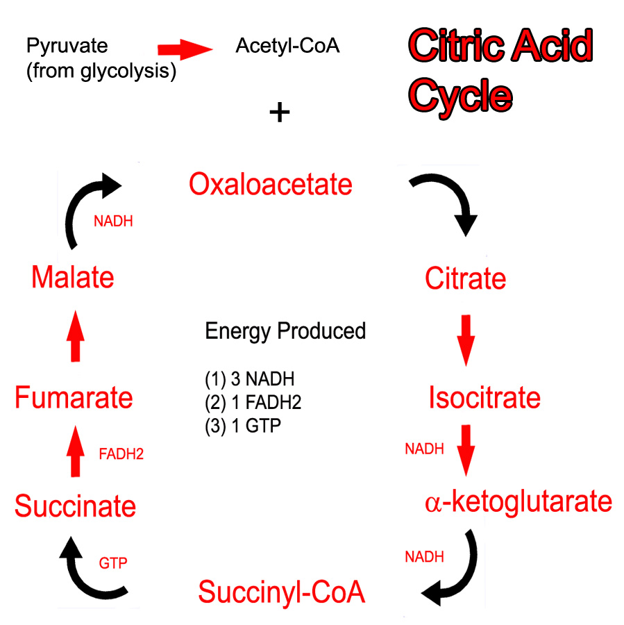
The main role of the citric acid cycle is to continue the oxidation (ie: energy "stripping") of acetyl-CoA. Acetyl-CoA can be obtained from the oxidation of pyruvate (the final molecule of glycolysis) or from the oxidation of long chain fatty acids.

The cycle begins when acetyl-CoA combines with oxaloacetate to form citrate. Several chemical reactions occur and citrate gets converted back to oxaloacetate (see cycle below). At this point, oxaloacetate re-combines with another acetyl-CoA to restart the cycle. Note that pyruvate and acetyl-CoA are NOT technically considered part of the cycle.

The Kreb’s cycle (aka: citric acid cycle) produces both energy and "waste". The waste are two molecules of carbon dioxide, which are formed from the two carbons present in the original acetyl-CoA molecule. The energy comes from removing electron pairs from the molecules in the cycle.

Citric Acid Cycle
These electrons are stored in the energy rich molecules NADH and FADH2. NADH and FADH2 donate the electrons to carrier molecules in the electron transport chain. The final result is the production of adenosine triphosphate (ATP), an energy rich molecule that can be used by cells for various purposes. In addition, the citric acid cycle also produces an ATP equivalent molecule known as guanosine triphosphate (GTP).

The cycle itself operates in the mitochondrial matrix. This is no mistake because the enzymes of the electron transport chain are located on the adjacent inner mitochondrial membrane; the NADH and FADH2 produced by the cycle do not have to travel far in order to donate their electron pairs.

Regulation

Two key enzymes control the citric acid cycle. The first is isocitrate dehydrogenase. This enzyme converts isocitrate to α-ketoglutarate. It is inhibited by ATP and NADH, and stimulated by ADP. If we think about it, this makes sense because if ATP and NADH are abundant (ie: the cell has adequate energy stores), then we want the cycle to slow down. ADP is the breakdown product of ATP and represents an energy depleted state. Thus, ADP stimulates the cycle so that more energy can be produced.

The second regulatory enzyme is α-ketoglutarate dehydrogenase. This enzyme converts α-ketoglutarate to succinyl-CoA. It is inhibited by succinyl-CoA (an example of feedback inhibition), NADH, and ATP. The rationale behind this inhibition is similar to isocitrate dehydrogenase.

These two steps are also regulatory because they are highly exergonic. This means that they release lots of energy, so much so, that they are effectively irreversible reactions.

Overview

The main goal of the citric acid cycle is to produce energy by oxidizing acetyl-CoA. The energy produced comes in the form of NADH and FADH2. These molecules donate their electrons (energy) to the electron transport chain, which ultimately drives oxidative phosphorylation. This allows the formation of energy rich ATP, which can be used by the cell for numerous processes.

References and Resources

  • Nelson DL, Cox MM. Lehninger Principles of Biochemistry. Fifth Edition. New York: Worth Publishers, 2008.
  • Champe PC. Lippincott’s Illustrated Reviews: Biochemistry. Second Edition. Lippencott-Ravens Publishers, 1992.
  • Le T, Bhushan V, Grimm L. First Aid for the USMLE Step 1. New York: McGraw Hill, 2009.

Phenylketonuria: A Musty Aminoacidopathy

Phenylketonuria is one of the “aminoacidopathies”. An aminoacidopathy is a fancy medical term for a malfunction in the bodies’ ability to handle specific amino acids, in this case, phenylalanine.

But before we get too crazy, first things first… What is an amino acid? Amino acids are the building blocks of proteins. Each amino acid consists of a carbon atom that has four “things” hanging off of it: an amine group, a carboxylic acid group, a hydrogen atom, and finally an “R” group. The “R” group varies depending on the specific amino acid being discussed. For example, the “R” in phenylalanine is a benzyl group.

Amino acids link up together via “peptide bonds”. A peptide bond is simply the carboxylic acid group of one amino acid “hooking” up to the amine group of another amino acid. This linking occurs over and over again resulting in a protein that can be thousands of amino acids in length.

With that background, let’s discuss one specific amino acid, phenylalanine, and what happens when the body has difficulty using it – a condition called phenylketonuria.

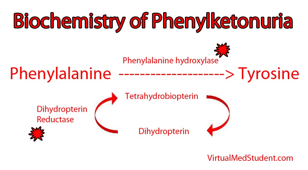
An enzyme in the liver called phenylalanine hydroxylase is necessary to transform phenylalanine into another amino acid called tyrosine. In certain individuals, the genes on chromosome 12 that encode the hydroxylase enzyme are deficient. Mutations in both of your hydroxylase genes (ie: the one you got from mom and the one you got from dad) must be deficient for phenylketonuria to develop; in other words, one adequate gene can produce enough normal hydroxylase to handle a normal dietary phenylalanine load. However, when both genes are deficient, phenylalanine can accumulate to abnormally high levels in the blood.

It is important to note that phenylketonuria can occur even if the hydroxylase enzyme is normal! How is this possible? The answer is because the hydroxylase enzyme requires another molecule called tetrahydrobiopterin (try saying that 3 times fast!) to function properly. When the enzyme (dihydrobiopterin reductase) that produces tetrahydrobiopterin is deficient, it gunks up the entire system and slows the ability of phenylalanine hydroxylase to convert phenylalanine to tyrosine.

Phenylketonuria

Either way you slice it, the excess phenylalanine interferes with the ability of other amino acids to enter the brain. This hinders brain development and, if left untreated, can cause mental retardation.

Signs and Symptoms

When excess phenylalanine backs up in the blood stream it blocks amino acid transporters in the brain. As a result, the brain does not get access to other essential amino acids, and patients that are left untreated, develop mental retardation and seizures.

Children affected by the condition also commonly have a musty odor. The odor is caused by phenylacetate, which is a breakdown product of excess phenylalanine.

Interestingly, patient’s who suffer from phenylketonuria are also usually blond haired and blue eyed. The reason for this is that tyrosine is necessary for melanin formation. Melanin is the pigment that gives hair, skin, and eyes their color. In phenylketonuria, phenylalanine is not converted to tyrosine and therefore melanin is not produced. The resultant lack of pigment causes blond hair and blue eyes.

Diagnosis

Many countries screen new babies for phenylketonuria because mental retardation can be prevented with strict dietary control. Diagnosis is based on finding an elevated phenylalanine to tyrosine ratio in the blood. Additionally, phenylpyruvate (a breakdown product of phenylalanine) will be elevated in urine samples of affected individuals.

Treatment

Treatment is, you guessed it, avoiding phenylalanine! This is done by ingesting a low protein diet, and in babies, using formula that is free of phenylalanine.

In phenylketonuria, tyrosine becomes an essential amino acid. Therefore, an important aspect of treatment is ensuring adequate intake of tyrosine via supplementation.

Overview

Phenylketonuria results when the body is unable to convert the amino acid phenylalanine into tyrosine. It can be caused by a primary genetic deficiency in the enzyme phenylalanine hydroxylase. It can also be caused by defects in the production of the hydroxylases’ necessary cofactor, tetrahydrobiopterin. Symptoms, if left untreated, include mental retardation. Patients with phenylketonuria are generally fair skinned and blue eyed because they have difficulty producing the melanin. Treatment is avoidance of phenylalanine and supplementation with tyrosine.

Related Articles

References and Resources

  • Cunningham GC. Phenylketonuria – early detection, diagnosis and treatment. Calif Med. 1966 July; 105(1): 1–7.
  • van Spronsen FJ, Huijbregts SC, Bosch AM, et al. Cognitive, neurophysiological, neurological and psychosocial outcomes in early-treated PKU-patients: A start toward standardized outcome measurement across development. Mol Genet Metab. 2011;104 Suppl:S45-51. Epub 2011 Oct 6.
  • Mitchell JJ, Trakadis YJ, Scriver CR. Phenylalanine hydroxylase deficiency. Genet Med. 2011 Aug;13(8):697-707.
  • Werner ER, Blau N, Thöny B. Tetrahydrobiopterin: biochemistry and pathophysiology. Biochem J. 2011 Sep 15;438(3):397-414. Review.

Sugar Strip Down: Glycolysis and Energy Formation

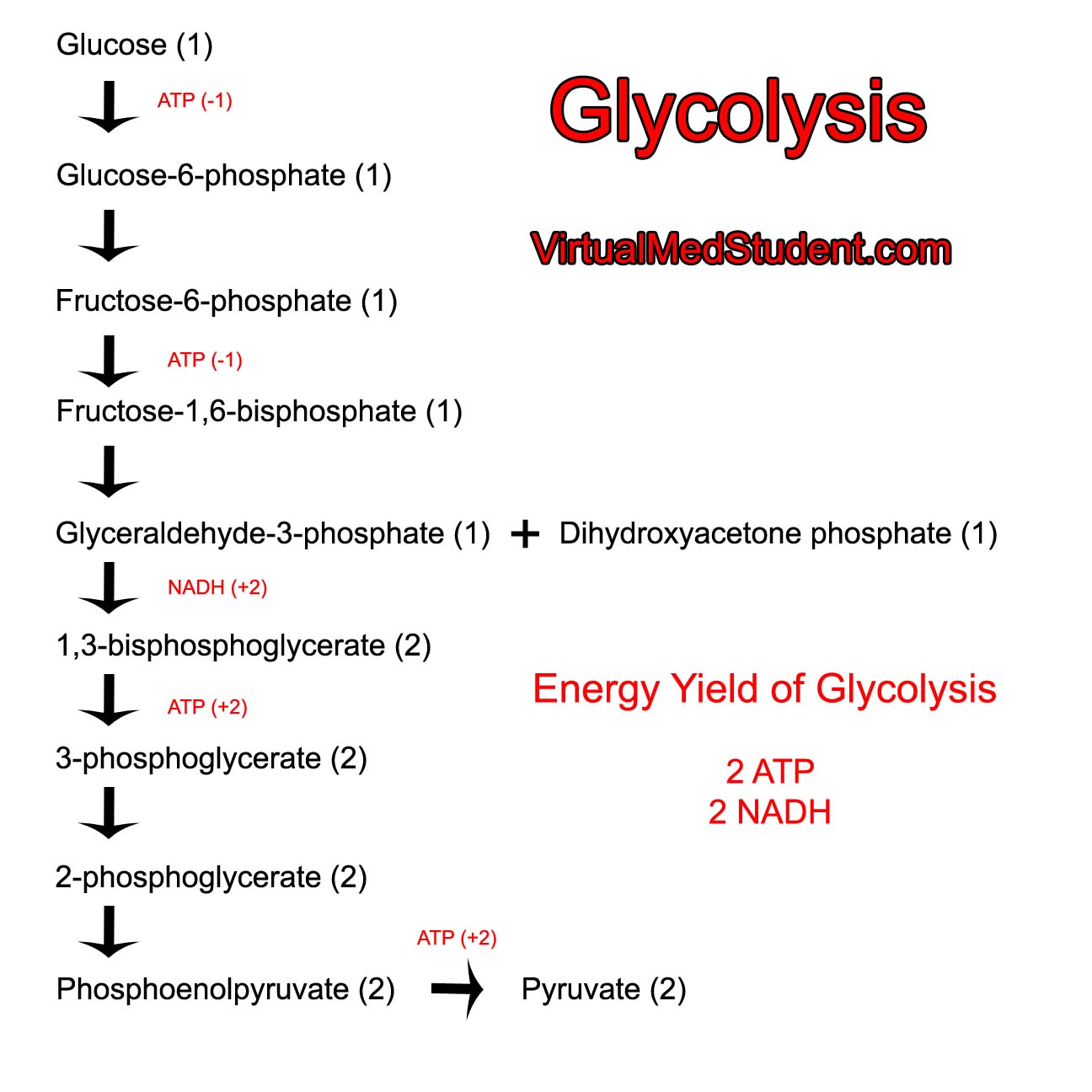
Glycolysis is the biochemical pathway that strips glucose of its energy. It starts with glucose and ends with pyruvate. Pyruvate is converted to acetyl-CoA, which can be stored as fat, or further metabolized in the citric acid cycle. The fate of pyruvate depends on the energy needs of the cell.

The net yield of energy from glycolysis is two adenosine triphosphates (ATP) and two nicotinamide adenine dinucleotide (NADH) molecules per glucose "burned" by the pathway. Each NADH molecule from glycolysis eventually generates 1.5 ATPs from the electron transport chain. Therefore, a total of 5 ATP molecules are produced per molecule of glucose consumed by the glycolysis pathway.

The net energy used or created (in the form of ATP or NADH) are shown in the schematic below:

Glycolysis

Want to know what happens to the NADH? Check out the article on the electron transport chain.

Regulation

Glycolysis is regulated at several steps in order to ensure that glucose molecules are used appropriately by the cell. The first point of regulation is at the conversion of glucose to glucose-6-phosphate (G6P). This reaction is catalyzed by an enzyme known as hexokinase. This enzyme is inhibited by its own product – G6P. This is known as "feedback inhibition". Once glucose is converted to G6P it becomes "trapped" inside the cell. Therefore, when there are adequate G6P levels any particular cell can shut off the flow of glucose into glycolysis so that it can be sent to other cells that may need it.

Phosphofructokinase 1
(PFK1) is the key
regulatory enzyme in the
glycolytic pathway.
The second, and key, regulatory point of glycolysis is at the enzyme phosphofructokinase-1 (PFK1). PFK1 converts fructose-6-phosphate (F6P) to fructose-1,6-bisphosphate. This enzyme is inhibited by ATP and citrate, and activated by ADP, AMP, and fructose-2,6-bisphosphate (note that this is a different molecule than fructose-1,6-bisphosphate). In other words, when the body is in an energy depleted state (ie: low levels of ATP and high levels of ADP and AMP) glycolysis is activated; under conditions of high energy (ie: high levels of ATP) glycolysis is inhibited. It is important to regulate the enzyme PFK1 closely because once F6P is converted to fructose-1,6-bisphosphate it MUST continue down the glycolytic pathway.

Side note: fructose-2,6-bisphosphate is produced by the enzyme phosphofructokinase-2 (PFK2) and degraded by the enzyme fructose bisphosphatase-2 (FBPase2). PFK2 becomes active under conditions of satiety (ie: well fed states in which lots of glucose/sugar is being absorbed from the gut). When PFK2 is active the concentration of fructose-2,6-bisphosphate increases. This activates PFK1 and increases the activity of the glycolytic pathway.

The final point of regulation is at the enzyme pyruvate kinase. This enzyme converts phosphoenolpyruvate into pyruvate. It is inhibited by ATP, acetyl-CoA, and long chain fatty acids, which are all markers of high energy levels.

In general, the overall regulation of glycolysis is related to how much energy the cell has. In energy rich states (ie: high ATP and low ADP/AMP) the cell slows glycolysis so that it can store glucose for use at a latter time. In energy depleted states (ie: low ATP and high ADP/AMP) the rate of glycolysis increases so that more energy can be formed by "burning" more glucose molecules per unit time.

Overview

Glycolysis breaks down glucose molecules. In the process the energy rich molecules ATP and NADH are formed. It is regulated at several enzymatic steps, most importantly at the enzyme phosphofructokinase-1. Pyruvate can be further metabolized to acetyl-CoA.

References and Resources

  • Nelson DL, Cox MM. Lehninger Principles of Biochemistry. Fifth Edition. New York: Worth Publishers, 2008.
  • Champe PC. Lippincott’s Illustrated Reviews: Biochemistry. Second Edition. Lippencott-Ravens Publishers, 1992.
  • Le T, Bhushan V, Grimm L. First Aid for the USMLE Step 1. New York: McGraw Hill, 2009.

Electron Transport Chain in All Its Glory

The electron transport chain is a series of interconnected proteins and carrier molecules that are embedded in the inner mitochondrial membrane. In order to understand the function of the electron transport chain in all its glory, we have to first appreciate two important molecules: NADH and FADH2. These two molecules can be thought of as energy dump trucks that strip sugars, fats, and proteins of their energy rich electrons (see the articles on glycolysis and the Kreb’s cycle for more details on how sugars and fats get stripped of their energy).

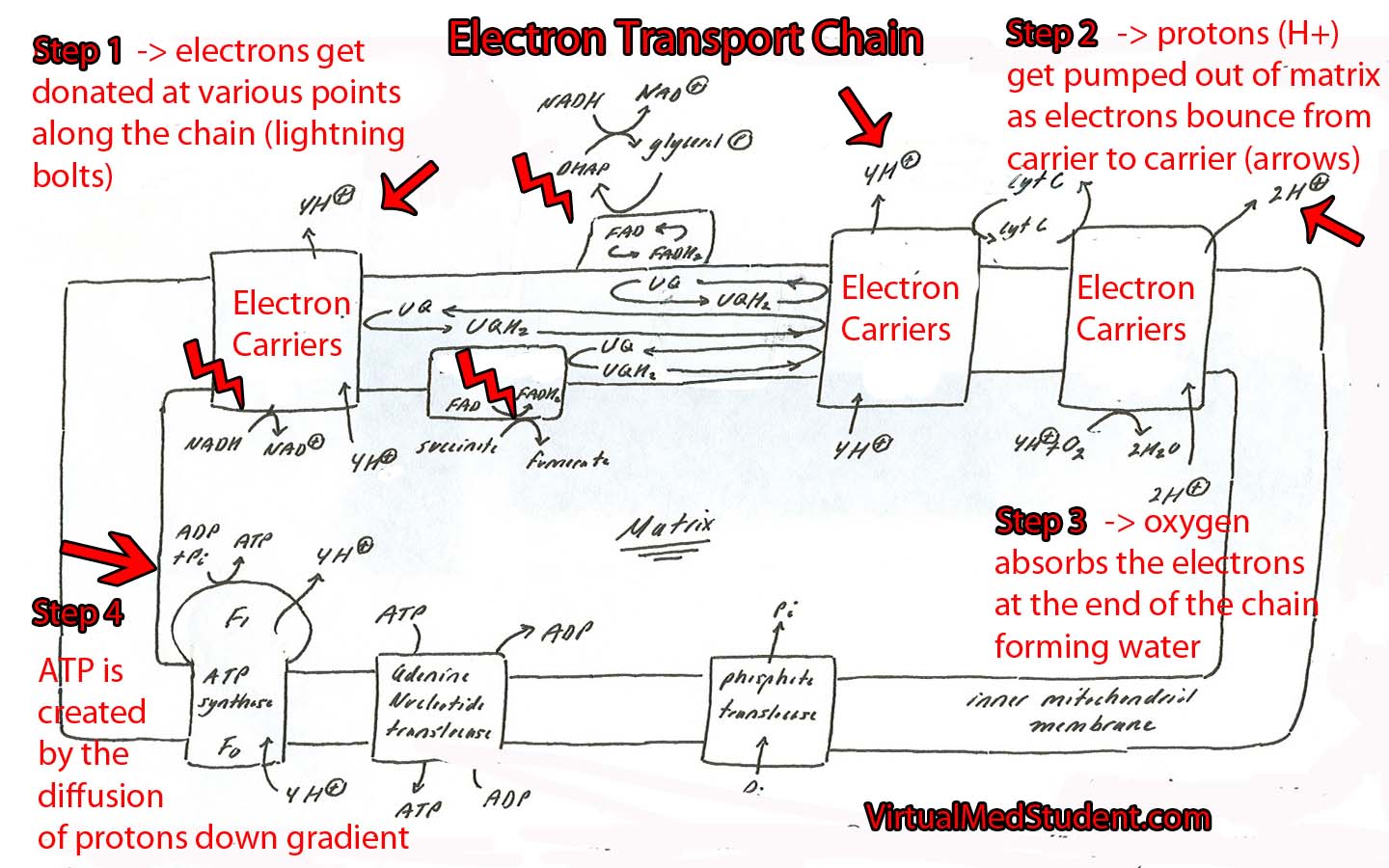
NADH and FADH2 then transfer their electrons to specific carrier molecules that constitute the “electron transport chain”. From there, the electrons "bounce" from carrier molecule to carrier molecule as if they were falling down an energy waterfall. Each time they bounce between carrier molecules energy is emitted; this energy is used to pump protons out of the mitochondrial matrix.

As protons get pumped out of the matrix they form an electrochemical gradient. As more electrons bounce down the chain, more protons get pumped out of the matrix. Eventually the concentration of protons outside the mitochondrial matrix is much higher than the concentration inside the matrix.

This is where the plot thickens… At this point all of the protons outside the matrix want to diffuse down their concentration gradient to their "home" in the matrix. However, the membrane that lines the matrix is impermeable to protons… Except at one specific point!

That point is a specialized enzyme embedded in the membrane called ATP synthase. The sole function of this enzyme is to rejuvenate a molecule known as adenosine triphosphate (ATP).

As the protons flow down their concentration gradient through the ATP synthase they release energy. The synthase uses this energy, via a wonderfully complicated molecular gear shifter, to form ATP from ADP (adenosine diphosphate) and free phosphate. This process is formally known as oxidative phosphorylation.

ATP is an energy rich molecule that can be thought of as the gasoline used to run a car. In simple terms, if a cell is building a molecule (ie: protein, DNA, RNA, etc.) it is likely using the energy in ATP to do it. When ATP runs out, the cell stops producing molecules and is in jeopardy of dying.

Electron Transport Chain Schematic

The whole point of the electron transport chain is to produce more ATP by using the energy obtained from the electron pillaging of molecules like glucose, long chain fatty acids, and proteins!

So what happens to the poor old electrons? Oxygen absorbs them at the end of the chain and forms water! This is why humans require oxygen to live! Without oxygen, electrons back up in the chain and eventually the whole transport system grinds to a halt.

Regulation

Like every other biochemical pathway in the body, the electron transport chain is under strict regulation. When ATP is abundant in the cell the chain slows down (ie: why drop electrons down the chain and strip sugars, fats, and proteins of their energy when you don’t have to?).

However, when the cell has little ATP around, then the chain springs in to action as the body burns sugars, proteins, and fats to create the electron carriers necessary to run the chain.

There are several points in the chain where things can turn awry. The first is if there is no oxygen. Oxygen is the last point in the electron "waterfall". Once the electrons get to the last carrier in the chain they have no where to go but to reduce oxygen to form water.

Without oxygen the electrons back up like lemmings. Eventually the whole chain becomes saturated with electrons and causes the whole system to stop, which can be disastrous to cellular function.

Overview

So let’s bring it all together… Cells strip sugars, proteins, and fats of their energy rich electrons. These electrons hitch a ride on special molecules like NADH and FADH2. NADH and FADH2 donate their electrons to special carrier proteins embedded in the inner mitochondrial membrane. The electrons bounce from carrier to carrier releasing energy. The released energy is used to pump protons out of the matrix. The resulting energy rich proton gradient is then used to create ATP as the protons flow down their concentration gradient back into the matrix through the ATP synthase unit… Now go drink another cup of coffee!

References and Resources

  • Le T, Bhushan V, Grimm L. First Aid for the USMLE Step 1. New York: McGraw Hill, 2009.
  • Kumar V, Abbas AK, Fausto N. Robbins and Cotran Pathologic Basis of Disease. Seventh Edition. Philadelphia: Elsevier Saunders, 2004.
  • Champe PC. Lippincott’s Illustrated Reviews: Biochemistry. Second Edition. Lippincott-Ravens Publishers, 1992.
  • Nelson DL, Cox MM. Lehninger Principles of Biochemistry. Fifth Edition. New York: Worth Publishers, 2008.