Diabetes Mellitus: Sugar Pee and A1C

Diabetes mellitus, or “sugar” diabetes, occurs when the body inappropriately manages its blood sugar levels. The key player in diabetes mellitus is a hormone called insulin. For simplicity we will use the term “glucose” as a relatively generic term for “sugar” going forward, but remember that glucose is actually one type of sugar.

Insulin is a protein secreted by specialized cells in the pancreas known as β-cells. When the body senses a high level of glucose floating around in the blood it causes the pancreas to secrete insulin. Many individual insulin molecules then travel throughout the body where it binds to receptors on many different tissue types. For example, if insulin binds to hepatocytes (liver cells) it instructs those cells to remove glucose from the blood and store it in a polymerized form known as glycogen. Insulin’s goal is to bring blood sugar levels to within normal limits.

The problem in diabetes mellitus is that insulin is either not secreted by the pancreas, or it does not perform its functions appropriately. Either way, glucose is not removed from the blood stream. The result is a higher than normal blood sugar concentration.

There are two distinct forms of diabetes mellitus: type 1 diabetes (sometimes referred to as juvenile onset or insulin dependent diabetes) and type II diabetes (sometimes referred to as adult onset or insulin independent diabetes).

Type 1 diabetes is an auto-immune disorder, which means that the bodies’ immune system attacks itself. In this case, the body attacks the β-cells in the pancreas. Once enough β-cells are destroyed the pancreas can no longer secrete insulin.

The pathology of type 2 diabetes mellitus is more complicated. There is no single "cause" of type 2 diabetes, and as such, it is believed to be the result of a combination of factors. Genetics, lifestyle, and diet all play an important role in the development of this form of the disease. I like to think of type 2 diabetes as a slew of factors that have beaten down the bodies ability to properly manage blood sugar levels.

In type 2 diabetes the pancreas is still able to produce insulin. However, the problem is that insulin does not cause its normal effect on body tissues. This is known as "insulin resistance." In an effort to combat this resistance the pancreas secretes more insulin. Once the insulin resistance becomes too severe the pancreas can not keep up, and blood sugar levels begin to rise. If the levels rise beyond a specific laboratory threshold the diagnosis of diabetes is made.

How Does Diabetes Mellitus Present?

High blood sugar levels lead to the symptoms of diabetes mellitus. One of the most common symptoms of diabetes is a frequent need to urinate. The reason this occurs is because the excess blood sugar exceeds the kidney’s ability to re-absorb it. As a result, glucose leaks into the urine. The body is forced to dilute this excess solute load by secreting more water into the urine. Hence more urine production –> increased trips to the bathroom!

Since diabetics have difficulty using sugar as a fuel they often lose weight (although most type II diabetics are overweight or obese secondary to excessive caloric intake). Diabetes, in metabolic terms, is similar to slowly starving. The body is unable to utilize sugar appropriately, which is the main “fuel” for most people. Normally, excess sugar gets converted to fat (ie: for you “need-to-know it all types” glucose is converted into acetyl-CoA fragments. If these are not “burned” by the Krebs cycle they get polymerized into long chain fatty acids, which get stored in fat tissue). However, in diabetics the sugar is "lost" in the urine and not used for metabolic purposes.

Diabetes, in metabolic terms,
is similar to slowly starving.
If blood sugar levels become ridiculously high severe complications, and potentially death can result. These patients can become very dehydrated and will often have significant electrolyte abnormalities. These factors can lead to coma, heart arrhythmias, and death if left untreated. These severe complications usually occur during periods of "stress." Conditions like infections and certain drugs (both prescription and illicit) can cause the "stress" needed to induce a diabetic crisis.

When a diabetic crisis occurs in type 1 diabetics it is referred to as "diabetic ketoacidosis". It is known as "non-ketotic hyperosmolar syndrome" if it occurs in a patient with type 2 diabetes.

What Else Can Happen to People with Diabetes?

There are many complications of diabetes mellitus. These complications are the result of years of high sugar levels in the blood. Over time the excess sugar undergoes a metabolic transformation known as "glycosylation". Glycosylated sugar is toxic to nerves and blood vessels.
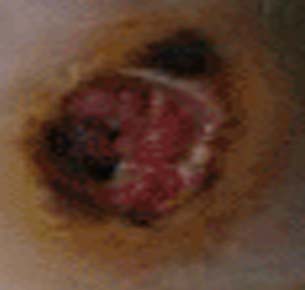
Diabetic Ulcer
Diabetic foot ulcer
If damage occurs to the nerves that innervate the stomach a condition known as "gastric paresis" can occur, which is a fancy term for the stomach not being able to contract as well as it used to.

In addition, nerves that carry sensory information can also be damaged, especially at the finger tips and toes. Patients with this type of nerve damage may not feel cuts and blisters on their toes. These areas can become secondarily infected resulting in gangrenous digits that are often amputated (see image to left). This is part of the reason why all diabetics, especially poorly controlled diabetics, should see a foot specialist regularly.

Perhaps the most frightening complication of diabetes is the havoc it wreaks on the cardiovascular system. Diabetics have a much larger risk of heart attack and stroke.

Patients with diabetes are at risk for retinopathy, or damage to the retina of the eye. This can lead to blindness. Damage to the kidney’s filtering area known as the glomerulus can also occur; this can lead to chronic kidney disease. If severe enough, patients may need permanent dialysis or kidney transplantation.

Diagnosis

The diagnosis of diabetes is made by measuring the amount of sugar in the blood. There are three common ways to diagnose diabetes today. The first is by checking a molecule known as hemoglobin A1C. Diabetes can also be diagnosed with fasting blood sugar levels or by a glucose tolerance test. There are three possible results of these tests: normal, impaired (pre-diabetic), or diabetic.

Hemoglobin A1C levels are drawn from the blood. Two separate results above 6.5% indicate diabetes. Levels between 5.7 and 6.4% indicate pre-diabetes. Levels below 5.7% are normal.

"Fasting" blood glucose levels are usually taken in the morning before breakfast. It is considered "normal" if the blood glucose levels are between 60 and 100 mg/dL. Impaired is between 100 and 126 mg/dL. Diabetes is diagnosed if there are two fasting blood glucose levels greater than 126 mg/dL.

If fasting is not an option, or the patient ate breakfast, the clinician can do a "glucose tolerance test." In this test the patient drinks a liquid that is rich in sugar (75 grams of sugar is used for most adults). The blood sugar levels are then tested two hours later. If the blood sugar level at two hours is less than 140 mg/dL the test is "normal." If the level falls between 140 and 200 mg/dL the patient is impaired or pre-diabetic. And if the levels are greater than 200 mg/dL the diagnosis of diabetes is made.

Diagnosing Diabetes Mellitus

  Hemoglobin A1C Fasting blood glucose levels Glucose tolerance test levels at 2 hours after a 75g sugar load
Normal Less than 5.7% < 100 mg/dL < 140 mg/dL
Impaired 5.7% to 6.4% 100-126 mg/dL 140-200 mg/dL
Diabetes 6.5% or greater > 126 mg/dL > 200 mg/dL

Heal Me Doctor

Treatment of diabetes involves either replacing insulin or improving insulin sensitivity.

Type 1 diabetics require insulin since their bodies no longer produce it. This is why type 1 diabetes was previously referred to as “insulin dependent diabetes”. Insulin as a treatment is discussed in a separate article.

Treatment Options:
(1) Replace insulin
(2) Improve insulin
       sensitivity
A healthy diet and exercise are very important treatments in managing diabetes. However, many patients require medications.

There are numerous medicines used to control blood sugar levels in type two diabetics. One option is a class of medications known as the sulfonylureas. These medications work by increasing pancreatic production of insulin.

A second category of medications, known as the thiazolidinediones, improve the ability of insulin to act on peripheral tissues like fat, muscle, and liver.

The third category of medications, known as the biguanides, perform a similar function to the thiazolidinediones, and also inhibit liver cells from secreting stored sugar into the blood stream.

Regardless of which medications are used, the ultimate goal of medical therapy is to maintain the blood glucose levels within a specified range. Fasting blood glucose levels should ideally be kept between 90 and 130 mg/dL; post-meal glucose levels should ideally be less than 180 mg/dL. Maintaining glucose values within these ranges helps keep glycosylated hemoglobin (aka: hemoglobin A1C) levels below 7%, which decreases the risk of the diabetic complications.

In addition to replacing insulin, or increasing insulin sensitivity, patients with diabetes should also be treated for other co-morbid conditions. For example, statins are often started to control hyperlipidemia. Dialysis might be necessary to control severe kidney disease. Frequent foot and neurological exams are also necessary to prevent complications of diabetic neuropathy (ie: foot ulcers, gastric paresis, etc).

It is currently recommended that patients with diabetes have a blood pressure goal of 130/80 or better. In addition the LDL cholesterol level should be maintained below 100 mg/dL, HDL should be above 40 mg/dL, and triglycerides should be kept below 150 mg/dL.

The Replay…

Diabetes is classified as type 1 or type 2. In type 1 diabetes destruction of the pancreatic cells responsible for insulin secretion occurs through an auto-immune process. In type 2 diabetes, insulin sensitivity is lost by peripheral body tissues causing the pancreas to secrete more and more insulin until it “burns out”. Diagnosis is based off of blood sugar levels, usually measured when the patient is fasting (although other methods exist). Treatment is with insulin or insulin-sensitizing medications.

Just Keep Learning, Just Keep Learning (That’s a Dory Reference)…

References and Resources

  • Swanson A, Watrin K, Wilder L. Clinical Inquiries: How can we keep impaired glucose tolerance and impaired fasting glucose from progressing to diabetes? J Fam Pract. 2010 Sep;59(9):532-3.
  • Judge EP, Phelan D, O’Shea D. Beyond statin therapy: a review of the management of residual risk in diabetes mellitus. J R Soc Med. 2010 Sep;103(9):357-62.
  • Fowler GC, Vasudevan DA. Type 2 diabetes mellitus: managing hemoglobin A(1c) and beyond. South Med J. 2010 Sep;103(9):911-6.
  • Kumar V, Abbas AK, Fausto N. Robbins and Cotran Pathologic Basis of Disease. Seventh Edition. Philadelphia: Elsevier Saunders, 2004.
  • Le T, Bhushan V, Grimm L. First Aid for the USMLE Step 1. New York: McGraw Hill, 2009.
  • Flynn JA. Oxford American Handbook of Clinical Medicine (Oxford American Handbooks of Medicine). First Edition. Oxford University Press, 2007.
  • Champe PC. Lippincott’s Illustrated Reviews: Biochemistry. Second Edition. Lippincott-Ravens Publishers, 1992.
  • American Diabetes Association. Standards of medical care in diabetes. Diabetes Care. 2004 Jan;27 Suppl 1:S15-35.

Brain Boo-Boos: Cerebrovascular Accidents (Stroke)

MCA Stroke CT Scan

Stroke - ADC Map

Stroke - Diffusion Weighted
A cerebrovascular accident, commonly known as a stroke, occurs when blood flow stops reaching brain tissue. If the entire brain is involved it is referred to as a "global" stroke; if a specific region of brain is involved it is referred to as a "focal" or "territorial" stroke. There are three broad causes of territorial strokes: thrombotic, embolic, and hemorrhagic.

A thrombotic stroke occurs when a blood clot forms in a blood vessel that supplies brain tissue. This is similar to what happens in cardiac infarction (ie: heart attacks). Thrombi are most commonly caused by atherosclerotic disease of the cerebral blood vessels. Thrombi usually form at areas of turbulent blood flow and at locations where vessels form branch points.

Embolic strokes are similar because they are technically blood clots. However, an embolus is a fragment of a clot (thrombus) that formed in another part of the body. Those fragments break free from the original clot and travel to blood vessels in the brain. They get lodged at some point and prevent blood from flowing resulting in a stroke if treatment is not obtained quickly.

Strokes can also be caused by bleeding into brain tissue. These type of strokes are called “hemorrhagic stroke”. Bleeding can occur in people with long standing untreated high blood pressure, or in those that have underlying structural disorders of the blood vessels in the brain (ie: aneurysms or arteriovenous malformations).

Diagnosis

Speedy diagnoses of stroke is extremely important because brain tissue dies quickly if it doesn’t receive adequate oxygen.

The first test that is done in cases of suspected stroke is a CT scan of the head. The purpose of the CT scan is not necessarily to "see" the stroke, but rather to rule out some other cause (ie: tumor, subdural hematoma, etc) for the symptoms. If bleeding is present on the CT scan the treatment algorithm becomes much different. If no bleeding is seen on CT then the second scan is usually an MRI.

An MRI takes longer than a CT scan, but it gives a much more detailed picture of the brain. In addition, it can pick up ischemia (ie: cell death related to decreased blood flow) much earlier than CT.

The best sequences to detect a stroke on an MRI are the diffusion weighted images and apparent diffusion coefficient maps. Stroked brain tissue will appear “bright” on diffusion weighted imaging and “dark” on the apparent diffusion coefficient map (see images to the left).

In addition, the carotid arteries are scanned using ultrasound in order to detect potential narrowing from atherosclerotic disease. Atherosclerotic carotid arteries are a potential source of emboli.

Sometimes a procedure known as transcranial doppler, which also uses ultrasound technology, is used to detect blood flow in the individual blood vessels of the brain. This can sometimes help determine the specific location of the thrombus/embolus.

Cerebral angiograms are much more invasive tests, but give a detailed view of which vessels are blocked. Cerebral angiograms can also be used to treat some strokes by directly removing clot from the affected blood vessel.

Most patients should undergo a thorough work up for atherosclerotic disease including a fasting lipid panel and hemoglobin A1C levels (a marker of diabetes).

If the heart is a suspected source of emboli than transthoracic echocardiography (ie: an ultrasound of the heart) is often done as well.

Signs and Symptoms

Cerebrovascular accidents present with a wide variety of signs and symptoms. It is entirely dependent on the blood vessel, and therefore, region of the brain involved. For example, strokes in the left middle cerebral artery will often cause significant language impairments if left untreated. Middle cerebral artery strokes usually cause contralateral paresis as well (usually the face and arm are more affected than the leg). Strokes in the frontal lobes caused by blockage of the anterior cerebral arteries can cause personality changes, as well as paresis/paralysis of the contralateral lower extremity.

Suffice it to say that there are a variety of possible clinical presentations in patients suffering from stroke. These presentations generally correlate with our understanding of brain anatomy and function.

Treatment

Prompt treatment of stroke is critical for preserving viable brain tissue. If a stroke is due to a blood clot (ie: thrombus or embolus) the treatment is with a drug known as tissue plasminogen activator (tPA). tPA is a medication that helps break up the clot.

It can be a dangerous medication because it can cause serious bleeding, but if given early enough, and in the right patient, it can completely prevent brain tissue death. There are numerous contraindications to giving tPA so caution must be used. The traditional teaching is that is should be given within three hours of symptom onset (this is the FDA approved indication); however, up to 4.5 hours from symptom onset has become common in clinical practice (but this is not FDA approved).

Endovascular therapies that mechanically remove the clot are becoming more common, especially for large vessel disease. However, this type of treatment requires specialized interventional neuro-radiologists and is not available in all medical centers. Endovascular therapy with a clot retrieving device is usually indicated up to 6 hours post symptom onset for large vessel occlusions. More distal (ie: further out) occlusions are not candidates for this type of procedure yet.

If a patient survives their first stroke, they are often started on medications to decrease their risk of having a second stroke. One of the most common medications used to prevent a second stroke is aspirin.

However, other medications like ticlopidine and clopidogrel (Plavix®) are also frequently used. All three of these medications prevent platelets (ie: one of the bodies natural ways of forming blood clots) from clumping together. In addition, aspirin is often mixed with another medication called dipyridamole (dipyridamole + aspirin = Aggrenox® in the United States). Patients who have suffered a minor stroke or have high risk transient ischemic attacks should be started on aspirin and clopidogrel and then transitioned to aspirin alone at 21 days.

If atherosclerosis is believed to be the cause of the stroke patients are often started on a statin. This helps slow the process of atherosclerosis and can help prevent another stroke from occurring.

If an embolus was the cause of the stroke patients are often started on an anticoagulant. The most common one used is warfarin (although there are many others). Warfarin is also used to treat a common cause of embolic stroke, an abnormal heart rhythm known as atrial fibrillation.

Overview

Strokes can be caused by thrombi or emboli which are blood clot that block blood flor, or from hemorrhage into brain tissue. Diagnosis is made by CT and MRI scans. Additional studies including carotid ultrasound, cerebral angiography, echocardiography, fasting lipid profiles, and tests for diabetes are also frequently performed.

Treatment depends on the etiology. Tissue plasminogen activator (tPA) is given if thrombi or emboli are the cause, and symptoms began less than 3 hours prior to presentation (4.5 hours is becoming the standard of care). Mechanical endovascular removal of the clot is also possible in some medical centers with specialized equipment.

Prevention of secondary strokes involve the use of anti-platelet (ie: aspirin, clopidogrel), anti-coagulant (ie: warfarin), and anti-atherosclerotic medications depending on the etiology of the previous stroke.

Related Articles

References and Resources

Ossified Posterior Longitudinal Ligament: Its Clinical Significance

The posterior longitudinal ligament is a long ligament that runs from the base of the skull to the sacrum. It provides a significant amount of mechanical integrity to the spinal column. The ligament runs down the back of the vertebral bodies just in front of the spinal cord itself.

In some individuals this ligament becomes ossified, which means that it takes on a bone like quality. As a result, its overall size and hardness increase. Given the ligament’s location adjacent to the spinal cord, any increase in the girth or flexibility of the ligament can cause injury to the cord.

Ossification of the posterior longitudinal ligament can occur at multiple spots along the spinal column. The most common spot is in the cervical spine (usually C2 to C6), but ossification can also occur in the thoracic (usually T4 to T7) and lumbar spine as well.

Exactly why the longitudinal ligament ossifies in some people is unknown. Current thinking is that a combination of genetic and lifestyle factors play a role in its pathology. For example, family studies have shown increased rates of OPLL in first degree relatives of people known to have the disorder. OPLL is also more common in people of Japanese and Korean descent.

Both familial and racial linkage usually indicate a genetic component to the disease. In fact, patients with OPLL have higher rates of dysfunctional collagen gene regulation (specifically, type XI and VI collagens). Linkage to specific human leukocyte antigen haplotypes on chromosome 6 have also been implemented.

Lifestyle factors such as diet have also been shown to increase risk. Patient’s who are diabetic or pre-diabetic have higher rates of OPLL compared to the rest of the population. High protein diets seems to decrease the risk, whereas high salt diets seem to increase risk.

Overall, the reasons why the posterior longitudinal ligament ossifies in some people, but not others remains an area of ongoing debate and research.

Signs and Symptoms

When an ossified posterior longitudinal ligament pushes on the spinal cord it causes numerous signs and symptoms. The constellation of clinical findings seen in patient’s with symptomatic ossified posterior longitudinal ligaments is known as myelopathy.

Myelopathic patients present with a combination of weakness, clumsiness (ie: decreased ability to hold objects), bowel or bladder dysfunction, spasticity – which is manifested as increased reflexes, as well as changes in sensation (ie: numbness, tingling, etc.).

Ossified Posterior Longitudinal Ligament
Myelopathy can be subtle at first, but can become debilitating depending on how much compression of the cord is present.

Diagnosis and Classification

Diagnosis of an ossified posterior longitudinal ligament is usually made when a patient presents with myelopathic features, or after neurological injury from a traumatic event.

Imaging studies such as xrays and CT scans illustrate the bony quality of the ligament. MRIs are frequently ordered to assess how "squashed" the spinal cord is. CT myelograms also provide excellent detail of cord compression when MRI is not feasible.

The ossification is classified according to its anatomic location and continuity. There are four distinct patterns. They include a continuous pattern, in which there is ossification behind both the vertebral bodies and the disc spaces. The second pattern is known as segmental; in this type the ossification is only present behind the vertebral bodies and does not span the disc spaces. The third pattern is localized, which means that the ossified ligament is present and localized behind only one vertebral body. Finally, there is a mixed type, which is a combination of continuous and segmental.

Treatment

The problem with ossification of the posterior longitudinal ligament is that it progresses over time. Slowly, the ligament will compress the spinal cord. Therefore, surgical treatment is typically offered at the first sign of spinal cord compression (ie: myelopathy).

The best surgical treatment is controversial. Approaching the spine from the front (ie: anterior approach) allows direct removal of the ossified ligament. However, it is important to note that the ligament is often stuck to the dura mater overlying the spinal cord which can make dissection extremely difficult. A cerebrospinal fluid leak is not uncommon when an anterior approach is taken, and there is an increased risk of inadvertent injury to the spinal cord. That being said, when successful, surgery from the front offers several distinct advantages. The first is that myelopathic symptoms seem to respond better to this type of approach; in addition, removal of the ossified ligament can retard further progression of the disease.

The second surgical option is to approach the spine from the back (ie: posterior approach). Removing the bone behind the spinal cord allows the cord to "drift" backwards away from the ossified ligament. This approach is considered more safe because there is no direct removal of the ossified ligament, and therefore there is a decreased risk of inadvertent cord injury. That being said, progression of the ossification can still occur and myelopathic symptoms do not respond as well to this type of surgery.

Ultimately, the type of surgery offered – anterior versus posterior – is dictated by the severity of the ossification and symptoms present, as well as the preferred approach of the surgeon.

Overview

Ossification of the posterior longitudinal ligament is seen more commonly in patients of Japanese descent. There are genetic factors that appear to increase risk. Symptoms are related to compression of the spinal cord by the ossified ligament. Ossification is progressive in nature. Treatment consists of surgery from either an anterior, posterior, or combined approach.

Related Articles

References and Resources