Hemangiopericytoma: A Tumor of Pericytes

In order to understand hemangiopericytomas we have to define a few terms. The first term is mesenchyma. Mesenchyma is a word used to describe the different tissues that provide structure to the bodies’ organ systems. A type of mesenchymal cell known as a “pericyte” provides structural support to blood vessels. When pericytes go haywire they form hemangiopericytomas.

Hemangiopericytomas can occur anywhere blood vessels are located, but are most commonly located in the lower extremities, pelvis, head, and neck.

Intracranial hemangiopericytomas are uncommon. They represent less than 1% of tumors within the confines of the skull. They typically arise from blood vessels adjacent to the dura (ie: lining of the brain) and often form dural attachments. They are therefore commonly lumped into the category of “dural-based tumors”, but should be distinguished from their more benign meningeal cousins (ie: meningiomas).

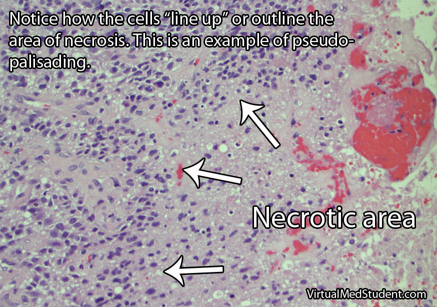
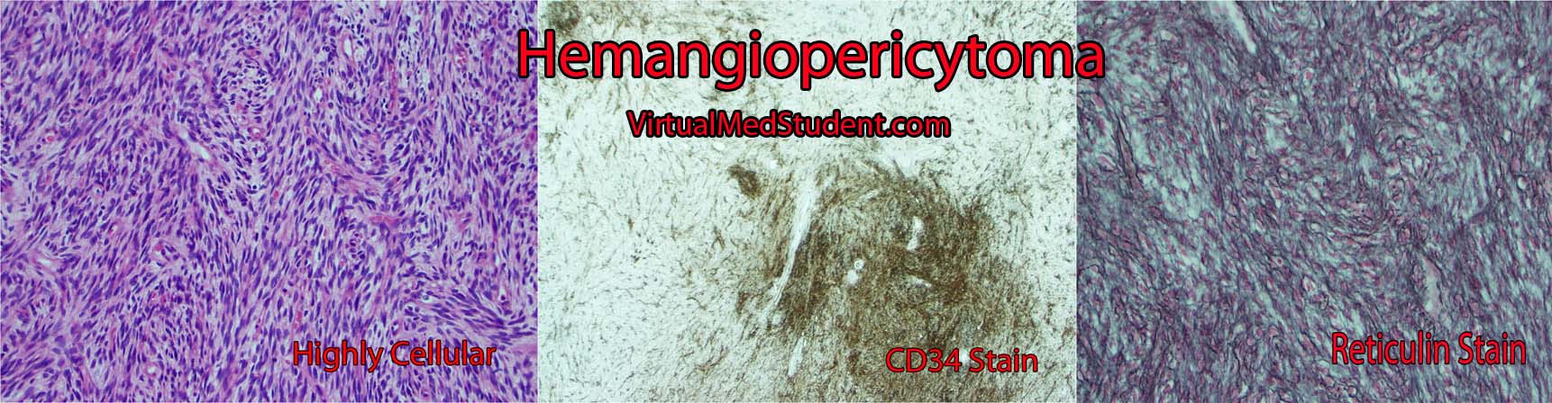
Since hemangiopericytomas are mesenchymal in origin, they typically have lots of reticulin (a collagen fiber) that envelopes individual cells (see pathology slide). They are highly cellular tumors, and vascular channels in the shape of "staghorns", may be seen under the microscope. Actively dividing cells (aka: "mitotic" figures) are commonly seen and are a testament to their more malignant nature. Unlike meningiomas, calcifications are absent.

Hemangiopericytomas test positive for vimentin (a marker of connective tissue), Ki-67 (a marker of proliferation), vascular endothelial growth factor (VEGF, a marker of blood vessel proliferation), CD34 (a marker of blood and vascular cell lineage), and reticulin (a collagen fiber). These tumors do not stain positive for epithelial membrane antigen. Genetic mutations have been found on different chromosomes , but the importance of these abnormalities is not well understood.

Intracranial hemangiopericytomas are considered malignant tumors. This means that they can spread to other areas of the body. In addition, hemangiopericytomas that have been removed surgically have a high recurrence rate.

Hemangiopericytoma

Signs and Symptoms

Hemangiopericytomas are relatively slow growing and often do not cause symptoms until they become quite large. However, once they start to compress adjacent brain tissue they may cause headaches, seizures, confusion, weakness, or visual problems.

Diagnosis

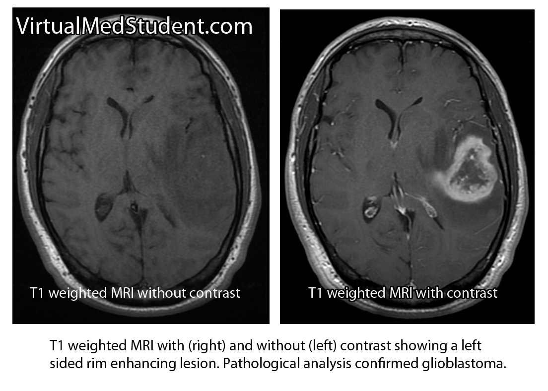
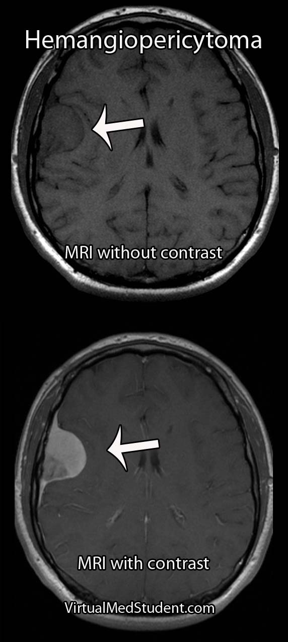
MRIs and CT scans of the brain typically reveal a contrast enhancing dural-based lesion. Cerebral angiograms show a highly vascular tumor with blood supply coming from the dura, as well as the underlying brain tissue.

Based on imaging alone, hemangiopericytomas are often mistaken for meningiomas. Subtle characteristics such as a lack of calcification seen on CT scans may help distinguish one from the other, but this is not reliable.

The only reliable way to diagnose hemangiopericytoma is to look at a specimen of the tumor under a microscope. Special stains and features of the tumor can help delineate it from a meningioma (see pathology section above).

Did I Hear Someone Say “Treatment”?

Intracranial hemangiopericytomas should be surgically resected when feasible. Unfortunately, even after complete resection, they frequently recur and/or spread to other areas of the body.

Because of their aggressive nature, patients with hemangiopericytomas should also have adjuvant radiation therapy. Radiation treatment after surgical removal of the tumor has been shown to lengthen survival and slows (but doesn’t appear to prevent) the time to recurrence.

The role of chemotherapy is less clear and is still being investigated. At this point, chemotherapy is typically used in patients where radiation and surgery have failed to control the disease.

Let’s Recap It…

Intracranial hemangiopericytomas are malignant dural-based tumors that arise from pericytes. They are highly vascular tumors that enhance on MRI and CT scans. Symptoms are variable and depend on the size and location of the tumor. Treatment is with surgical removal followed by radiation therapy. Recurrence rates are high despite optimal treatment.

You Should Learn About This Stuff Too…

Want Some More?

Glioblastoma: A Real Beast of a Tumor

In order to understand what a glioblastoma is we have to first appreciate the different cell types that compose healthy brain tissue. Brain tissue has both neurons and glia. Neurons are the “action” cells of the brain. Glia are the “helper” cells of the brain. They ensure that neurons stay healthy.

A glioblastoma is a malignant brain tumor that arises from a specific type of glial cell known as an astrocyte. Glioblastomas are not only the most common astrocytic tumor, but they are also the most common primary brain tumor!

It is important to realize that there are less malignant tumors that arise from astrocytes (discussed in other articles). Many of these tumors have a much better prognosis, which is why it is important to distinguish glioblastoma from less malignant behaving tumors.

The first distinguishing characteristic is neovascularization. Neovascularization is a fancy medical term used to describe the proliferation of blood vessels within the tumor. As the tumor grows, it requires new vessels to feed it oxygen and nutrients; the process of neovascularization allows the tumor to obtain these essential factors so that it can continue to grow.

Interestingly, as the tumor expands, sections of it will get choked off from its own blood supply. The end result is that part(s) of the tumor actually dies. This is referred to as "necrosis", which is a common finding in glioblastoma.

One of the most distinguishing features of glioblastomas is when cancerous astrocytes "line up" and outline areas of necrosis in a process known as pseudopalisading (see image below).

Given the malignant nature of glioblastoma it is common to see many mitotic figures. Mitotic figures are cells in various states of cell division; these figures indicate a relatively rapidly growing tumor type.

Glioblastoma Pathology - Pseudopalisading

Glibolastoma is therefore characterized by the following pathological characteristics: prominent microvascular proliferation (ie: development of new blood vessels within the tumor), mitosis (ie: an indicator of cell division/growth), and necrosis (ie: areas of dead tumor); pseudopalisading necrosis is a specific form of necrosis shown in the above image that is a hallmark of glioblastomas.

Signs and Symptoms

Glioblastomas may present with any number of signs and/or symptoms depending on their location within the brain. Lesions that are located on the left side of the brain may cause problems with speech if they involve the Broca or Wernicke areas. Tumors in the areas of the brain that control motor movement may cause weakness. Additionally, tumors that arise in the frontal lobes may cause odd behavioral changes. Some patients present with seizures, and others with only a dull headache.

Diagnosis

An official diagnosis of glioblastoma can only be made when a pathologist looks at a sample of the tumor under a microscope. These samples are typically obtained by a neurosurgeon who resects or biopsies the tumor.

However, glioblastomas also have typical features seen on imaging studies such as MRI. For example, these tumors will “rim-enhance” when a contrast material such as gadolinium is infused into the patient during the scan. Rim enhancement is a result of the contrast material leaking out of all of the blood vessels present within the tumor. It is important to note that other diseases such as abscesses, lymphomas, and other infections can also cause rim-enhancement.

MRI of glioblastoma

Another useful study known as MR spectroscopy measures the relative amounts of different molecules present within the tumomr. In a glioblastoma the amount of lactate, choline, and lipid are all increased. Lactate is a marker of brain tissue that is not receiving enough oxygen, which is common in necrotic tumor areas. Choline is a molecule that is present in cell membranes. When neurons are rapidly dividing, which is what occurs in glioblastoma, the amount of choline present also increases. A different molecule known as N-acetyl aspartate (NAA) is present in mature cells. Therefore, unlike lactate and choline levels, NAA is decreased in glioblastoma because these cells are "immature" (ie: poorly differentiated).

Treatment

Treatment combines a mixture of surgery, radiation therapy, and different chemotherapeutic drugs, the most common being temozolomide (Temodar®). Surgery is only useful when a significant amount of the tumor can be removed. Despite optimal treatment the prognosis for patients with glioblastoma remains extremely poor.

It is also highly important to treat the edema that frequently surrounds the tumor. Steroids, most commonly dexamethasone (Decadron®), are used to decrease the amount of edema, which usually improves symptoms.

Patients are often started on an anti-seizure medication such as levetiracetam (Keppra®) or phenytoin (Dilantin®).

Overview

Glioblastoma is a malignant astrocytic tumor. It is the most common primary brain tumor. It has unique characteristics that distinguish it from more benign brain tumors that also arise from astrocytes. It is treated with a combination of surgery, radiation, and chemotherapy.

References and Resources