Chordoma and that Pesky Notochord

In order to understand chordomas we have to first learn a little bit about how the nervous system develops. Enter the notochord.

The notochord is a midline structure in the developing fetus that sends out various molecules (the most well known of which is “sonic hedgehog”). These molecules influence the development of the layers of embryonic cells that surround the notochord. One of these layers, the ectoderm, which is immediately behind (ie: posterior) the notochord eventually forms the brain and spinal cord. The mesoderm, which is immediately adjacent to the notochord forms the vertebral column and axial skeleton (amongst other things).

The purpose of the notochord is to ensure that each layer of tissue forms what it is supposed to. Once this occurs, the notochord ultimately becomes the nucleus pulposus of the intervertebral discs.

In some people, nests of cells that composed the fetal notochord remain (unnaturally) after birth. These collections of cells are known formally as ecchordoses physaliphora (try saying that 5 times fast). These cells can divide and turn into a slow growing tumor… What is that tumor called? You guessed it! A chordoma!

Chordomas are slow growing tumors that are most commonly located at the ends of the vertebral column. The most common place to see them is in the sacrococcygeal region, followed in frequency by a bone known as the clivus at the base of the skull. However, chordomas can occur anywhere along the vertebral column. The reason they occur most frequently at the "ends" of the vertebral column (ie: skull base and sacrum) is because these are the last areas to fuse in-utero.

Additionally, since the notochord is a midline structure in the fetus, chordomas are almost always midline in location.

Microscopically chordomas contain large polygonal shaped cells embedded in a mesh of long repetitive sugar and nitrogen containing molecules known as mucopolysaccharides.

Chordoma Highlights:
– Arise from notochord cells
– S-100 positive
– Cytokeratin positive
– Polygonal cells
– Slow growing
– Midline location
– Sacrum and clivus most
   common locations
– Worse prognosis than
   chondrosarcomas
Less than a third of chordomas will show cartilage like features. These chordomas are classically called “chondroid” chordomas because of the chondrocyte-like (ie: cartilage-like) cells and extracellular material within them. Chondroid chordomas must be distinguished from a similar looking tumor known as a chondrosarcoma.

Distinguishing chondrosarcomas from chordomas is possible with immunocytologic staining techniques. Chondromas and chondrosarcomas stain positive for a protein known as S-100. S-100 proteins have numerous intracellular functions and are commonly present in cells such as adipocytes (fat cells), chondrocytes (cartilage forming cells), melanocytes (pigment producing cells), and Schwann cells, amongst others.

So if chondrosarcomas and chordomas can look alike, and both stain positive for S-100, how the heck do we distinguish between the two? Using another molecule known as cytokeratin! Cytokeratin is a molecule that forms part of the intracellular frame for many cells. It is present in chordomas, but not in chondrosarcomas.

On to the Clinic I Say…

Chordomas are slow growing tumors and will usually start to cause symptoms in mid-adulthood. Symptoms are based on the location of the tumor.

If the tumor is located in the sacrum or coccyx then pain is the most frequent presenting symptom. If undiscovered this may progress to bowel and bladder problems as the tumor slowly envelopes the sacral nerves that go to the bowel and bladder. Additionally, patients may present with radicular symptoms such as numbness, tingling, or sharp pain in the distribution of the sacral nerves.

Clivus Chordoma
Chordomas of the clivus, the second most common location, can present with headache and if large enough symptoms of brainstem or upper cervical spinal cord compression. These symptoms may include vertigo, difficulty moving the tongue, double vision, hearing problems, spastic gait, increased reflexes, clumsiness, amongst other symptoms.

Diagnose Me McCoy

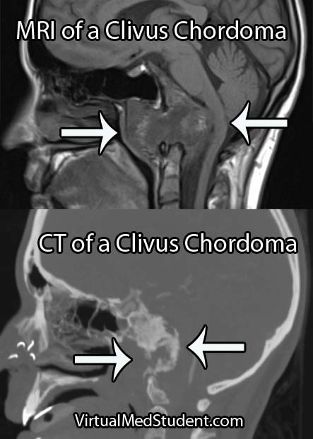
Diagnosis of chordoma can only be officially made by looking at the specimen under a microscope. However, imaging studies with x-rays, CT scans, and MRI imaging can support the diagnosis. Imaging studies will typically show a midline lytic lesion centered in the bone. Invasion of adjacent anatomical structures can occur, but is a late manifestation of the disease course.

Treating These Ugly Tumors

The gold-standard treatment for chordomas is en-bloc surgical resection with wide margins followed by radiation therapy. Complete resection is difficult, if not impossible to achieve in the skull base, but may be possible in the spine and/or sacrum with very skilled surgical teams.

Without an en-bloc surgical resection the risk of tumor re-growth and recurrence is very high. If you are planning to biopsy of a sacral lesion you should mark the biopsy tract with methylene blue so that the tract can also be resected during surgery as tumor cells can seed the tract as the needle is being pulled out.

Let’s Remix This Overview

Chordomas are slow growing, but malignant tumors that arise from notochord cells that fail to regress after birth. They are most frequently found in the sacrum and clivus (one of the bones constituing . Diagnosis is made with CT, MRI, and x-rays, as well as via tissue diagnosis at the time of surgical removal. Chordomas stain positive for S-100 and cytokeratin proteins, which helps distinguish them from chondrosarcomas that only stain positive for S-100. Symptoms are based on where the lesion is located (skull base or spine). Treatment is surgery followed by radiation therapy.

Just Warming Up…

Want to Research More About Chordomas?

Cerebral Cavernous Malformations: Leaky Vessels

Cavernous malformations (aka: cavernomas or cavernous hemangiomas) can be thought of as vascular tumors. They are composed of a capillary-like network of endothelial cells (the cells that normally line blood vessels). However, unlike normal capillaries throughout the body, the capillaries of cavernomas can leak.

Interestingly, cavernous malformations do not have any brain tissue within them. This helps distinguish them from another related vascular abnormality known as an arteriovenous malformation.

The genetics of cavernous malformations have been elucidated by studying familial forms of the disorder. There are at least three known genetic defects that predispose patients to develop cavernomas. These genes appear to be important in the formation of blood vessels (a process known as "angiogenesis") and the blood brain barrier. Therefore, mutations in these genes can cause the abnormal growth of vascular tissue.

What Havock Do These Guys Cause?

Cavernomas can cause numerous signs and symptoms depending on their location within the brain. Seizures are the most common symptom. However, progressive neurological impairment such as worsening weakness can also occur.

Sometimes cavernomas can block the flow of cerebrospinal fluid leading to hydrocephalus. Hydrocephalus can cause increased intracranial pressure leading to headaches, nausea, and vomiting. However, it is important to realize that many patients with cavernomas have no symptoms at all!

If symptoms are present, they tend to progress over time. This is because cavernous malformations bleed and re-bleed resulting in an expansion of its size over time. As the malformation increases in size it can push on adjacent brain tissue causing worsening symptoms.

Unlike arteriovenous malformations, life threatening and severe hemorrhages are rare. Cavernomas bleed at an initial yearly rate of anywhere between 1% to 5%. After the first bleed, the risk of re-bleeding increases to as high as 10% per year. In other words, if a cavernoma bleeds, it is more likely to re-bleed at a later date.

How Do You Diagnose These Buggers?

Cavernoma Marked

The diagnosis of cavernous malformations are made via imaging studies. They are usually detected via MRIs that are ordered for evaluation of neurological symptoms. Cavernomas are seen best on T2 and gradient echo MRI sequences. They typically look like a piece of popcorn.

Select patients undergo a more invasive imaging procedure known as angiography. Angiography is used to rule out another similar lesion known as an arteriovenous malformation. Since cavernomas are venous malformations they are not seen on angiograms.

How Are These Treated?

Depending on the location, most cavernous malformations in the brain or spinal cord are removed surgically. Some institutions offer radiation as a means of treatment, especially in difficult to access areas (ie: where the risk of surgical removal is very high).

Let’s Recap this MoFo

Cavernous malformations of the brain are abnormal vascular growths composed of capillary networks. They are likely the result of genetic mutations in genes responsible for blood vessel growth. Depending on their location they can cause numerous neurological symptoms such as seizures and weakness. MRI often shows the characteristic “popcorn” lesion. Treatment is usually with surgical resection, although some cavernomas may be radiated depending on their location.

Related Articles

Not Satisfied? More References and Resources…

Cerebral Ateriovenous Malformations: A Disease of Eloquence

A cerebral arteriovenous malformation is an abnormal tangle of blood vessels within the brain.

In order to understand these tangles we have to first understand normal blood flow. Blood flows from arteries to smaller arteries and then into capillary beds. In the capillary beds, gas, nutrients, and "wastes" are exchanged between the blood and adjacent body tissue. Once past the capillaries, the blood drains into successively larger veins where it eventually returns to the heart to be re-oxygenated.

In arteriovenous malformations there are no capillaries. Because of this, blood is shunted from the high pressure arterial system directly into the low pressure venous system. The "shunted" blood is unable to deliver its nutrients or oxygen to the nearby brain.

The risk of an arteriovenous malformation rupturing is relatively high because the pressure of arterial blood is "banging" into the walls of low pressure veins. The body tries to compensate for this by "arterializing" the blood vessels associated with the AVM.

The term "nidus" is often used to describe the center of the malformation. This is the point where the arterial feeding vessels meet the draining venous structures.

In addition, any brain tissue around, or within the AVM is usually gliotic (a term used to describe scarring within the brain). Macrophages are sometimes present and are usually there to "gobble up" hemosiderin (a breakdown product of blood).

Signs and Symptoms

The signs and symptoms of cerebral arteriovenous malformations are dependent on the location of the malformation.

Most patients discover they have an AVM after it bleeds into the surrounding brain tissue. Patients can present with everything from a mild headache to a severe neurological deficit depending on the location and size of the malformation.

In addition, AVMs may cause transient neurological symptoms. These transient symptoms are caused by blood being shunted away from the surrounding normal brain tissue. Again, the location of the AVM dictates what symptoms may develop (ie: weakness if near the motor strip, difficulty with speech if located near Wernicke’s or Broca’s area, balance problems if in the cerebellum, disturbances in sensation if in the parietal lobe, etc., etc.).

Patients may also present with seizures as a result of irritation of the surrounding cortex by hemosiderin (a breakdown product of blood). In fact, seizures are the second most common presenting symptom.

Interestingly, headache is an uncommon symptom of arteriovenous malformations.

Diagnosis and Classification

Cerebral Arteriovenous Malformation
Diagnosis is made with special imaging studies like CT angiography, MR angiography, and formal catheter angiography (formal angiography is the gold standard).

AVMs are characterized by an abnormal tangle of blood vessels. The tell tale sign of an AVM on an angiogram is that both arterial and venous structures are seen at the same time (normally the venous phase follows the arterial phase).

The Spetzler-Martin grading system helps guide treatment decisions. This system takes into account the size, location, and type of venous drainage (see the first reference below).

Treatment

Treatment is highly individualized. There are currently three accepted treatment strategies: surgery, radiation, and embolization.

Surgery is still the treatment of choice, especially for AVMs near the surface of the brain or in non-eloquent cortex. Surgery is also considered "definitive" therapy (ie: the AVM is removed all at once), which is ideal for lesions considered high risk for rupture. Patient’s with deep seated lesions (ie: basal ganglia, thalamus, etc.), or those located in very "eloquent" cortical areas may be better treated with radiation or embolization.

Radiation works by causing changes in the vessels of the AVM. Over the course of several months to years the vessels are "cooked" by the radiation. This effectively eliminates blood flow into the AVM. Since the effects of radiation take months to years to shut down the AVM, the patient remains at risk for rupture. In addition, side effects from radiation may be permanent in a small percentage of patients.

Embolization is usually used as an adjunct to surgical resection. During embolization, various substances are injected into the AVM. These substances deprive the AVM of its arterial blood flow. This can be very useful prior to surgery to help with intra-operative blood loss (especially for very large AVMs!). Embolization is less commonly used as a stand alone treatment.

Overview

Arteriovenous malformations are abnormal tangles of blood vessels within the brain tissue. They have no intervening capillary bed so arterial blood flows directly into dilated veins. The main risk of an arteriovenous malformation is when it ruptures and bleeds into the surrounding brain. They can cause numerous signs and symptoms depending on their location. They are diagnosed with CT angiograms, MR angiograms, or formal catheter angiograms. Treatment is with surgery, radiation, and/or embolization depending on the risk of rupture and the location of the lesion.

Other Interesting Neurovascular Diseases…

References and Resources

  • Spetzler RF, Martin NA. A proposed grading system for arteriovenous malformations. J Neurosurg. 1986 Oct;65(4):476-83.
  • Ding D, Yen CP, Xu Z, et al. Radiosurgery for patients with unruptured intracranial arteriovenous malformations. J Neurosurg. 2013 May;118(5):958-66
  • Fokas E, Henzel M, Wittig A, et al. Stereotactic radiosurgery of cerebral arteriovenous malformations: long-term follow-up in 164 patients of a single institution. J Neurol. 2013 May 28.
  • Albuquerque FC, Ducruet AF, Crowley RW, et al. Transvenous to arterial Onyx embolization. J Neurointerv Surg. 2013 Mar 6.
  • Nataraj A, Mohamed MB, Gholkar A, et al. Multimodality Treatment of Cerebral Arteriovenous Malformations. World Neurosurg. 2013 Feb 20.

Glioblastoma: A Real Beast of a Tumor

In order to understand what a glioblastoma is we have to first appreciate the different cell types that compose healthy brain tissue. Brain tissue has both neurons and glia. Neurons are the “action” cells of the brain. Glia are the “helper” cells of the brain. They ensure that neurons stay healthy.

A glioblastoma is a malignant brain tumor that arises from a specific type of glial cell known as an astrocyte. Glioblastomas are not only the most common astrocytic tumor, but they are also the most common primary brain tumor!

It is important to realize that there are less malignant tumors that arise from astrocytes (discussed in other articles). Many of these tumors have a much better prognosis, which is why it is important to distinguish glioblastoma from less malignant behaving tumors.

The first distinguishing characteristic is neovascularization. Neovascularization is a fancy medical term used to describe the proliferation of blood vessels within the tumor. As the tumor grows, it requires new vessels to feed it oxygen and nutrients; the process of neovascularization allows the tumor to obtain these essential factors so that it can continue to grow.

Interestingly, as the tumor expands, sections of it will get choked off from its own blood supply. The end result is that part(s) of the tumor actually dies. This is referred to as "necrosis", which is a common finding in glioblastoma.

One of the most distinguishing features of glioblastomas is when cancerous astrocytes "line up" and outline areas of necrosis in a process known as pseudopalisading (see image below).

Given the malignant nature of glioblastoma it is common to see many mitotic figures. Mitotic figures are cells in various states of cell division; these figures indicate a relatively rapidly growing tumor type.

Glioblastoma Pathology - Pseudopalisading

Glibolastoma is therefore characterized by the following pathological characteristics: prominent microvascular proliferation (ie: development of new blood vessels within the tumor), mitosis (ie: an indicator of cell division/growth), and necrosis (ie: areas of dead tumor); pseudopalisading necrosis is a specific form of necrosis shown in the above image that is a hallmark of glioblastomas.

Signs and Symptoms

Glioblastomas may present with any number of signs and/or symptoms depending on their location within the brain. Lesions that are located on the left side of the brain may cause problems with speech if they involve the Broca or Wernicke areas. Tumors in the areas of the brain that control motor movement may cause weakness. Additionally, tumors that arise in the frontal lobes may cause odd behavioral changes. Some patients present with seizures, and others with only a dull headache.

Diagnosis

An official diagnosis of glioblastoma can only be made when a pathologist looks at a sample of the tumor under a microscope. These samples are typically obtained by a neurosurgeon who resects or biopsies the tumor.

However, glioblastomas also have typical features seen on imaging studies such as MRI. For example, these tumors will “rim-enhance” when a contrast material such as gadolinium is infused into the patient during the scan. Rim enhancement is a result of the contrast material leaking out of all of the blood vessels present within the tumor. It is important to note that other diseases such as abscesses, lymphomas, and other infections can also cause rim-enhancement.

MRI of glioblastoma

Another useful study known as MR spectroscopy measures the relative amounts of different molecules present within the tumomr. In a glioblastoma the amount of lactate, choline, and lipid are all increased. Lactate is a marker of brain tissue that is not receiving enough oxygen, which is common in necrotic tumor areas. Choline is a molecule that is present in cell membranes. When neurons are rapidly dividing, which is what occurs in glioblastoma, the amount of choline present also increases. A different molecule known as N-acetyl aspartate (NAA) is present in mature cells. Therefore, unlike lactate and choline levels, NAA is decreased in glioblastoma because these cells are "immature" (ie: poorly differentiated).

Treatment

Treatment combines a mixture of surgery, radiation therapy, and different chemotherapeutic drugs, the most common being temozolomide (Temodar®). Surgery is only useful when a significant amount of the tumor can be removed. Despite optimal treatment the prognosis for patients with glioblastoma remains extremely poor.

It is also highly important to treat the edema that frequently surrounds the tumor. Steroids, most commonly dexamethasone (Decadron®), are used to decrease the amount of edema, which usually improves symptoms.

Patients are often started on an anti-seizure medication such as levetiracetam (Keppra®) or phenytoin (Dilantin®).

Overview

Glioblastoma is a malignant astrocytic tumor. It is the most common primary brain tumor. It has unique characteristics that distinguish it from more benign brain tumors that also arise from astrocytes. It is treated with a combination of surgery, radiation, and chemotherapy.

References and Resources