Cardiac Output: Pump, Pump, Squeeze

The cardiac output (CO) measures how much blood the heart pumps per minute. It is directly related to the stroke volume (SV) and heart rate (HR). The stroke volume is the amount of blood in the left ventricle of the heart just before it contracts. The cardiac output is calculated by multiplying the heart rate by the stroke volume (CO = HR x SV).

The cardiac output is related to Ohm’s law, which in electrical terms states that the change in voltage of a circuit is equal to the flow of current, I, multiplied by the resistance, R, of the circuit (ΔV = I x R).

Like electrical current in a circuit, blood flows in a circular pathway from the left ventricle, through the body, where it eventually ends up in the right ventricle. We can change Ohm’s law to govern hemodynamics by stating that the change in voltage is equivalent to the change in pressure between the aorta and right atrium (mean arterial pressure and central venous pressures, respectively), flow of current is equal to the amount of blood pumped per unit time (ie: cardiac output), and resistance is equal to the resistance the blood sees as it travels through the vessels of the body (aka: the systemic vascular resistance).

Therefore, if we re-write Ohm’s law for the hemodynamics of cardiac output we get: central venous pressure (CVP, measured in mmHg) subtracted from the mean arterial pressure (MAP, measured in mmHg) is equal to cardiac output (CO, measured in liters/minute) multiplied by the systemic vascular resistance (SVR, measured in dynes-s / cm5), or:

(MAP – CVP) = (CO x SVR) / 80
(ΔV = I x R)

Doing a simple mathematical rearrangement of the equation above we get:

CO = [(MAP – CVP) / SVR] x 80
*** The 80 in the equation is a conversion factor to convert Wood to metric units ***.

In essence, the cardiac output is directly proportional to the difference in blood pressure between the left (arterial side) and right (venous side) sides of the body, and inversely proportional to the vascular resistance. In other words, if the pressure difference between the arterial and venous side of the body decreases the cardiac output will fall. Along the same lines, if the systemic vascular resistance increases then cardiac output will decrease.

Clinical Measurements

How do you clinically measure the components that constitute the cardiac output?

The most reliable, but invasive way is to use a pulmonary artery catheter, also known as a "Swan Ganz" catheter. The catheter is inserted into the pulmonary artery and advanced until it “wedges” in a small branch of the pulmonary arterial tree. From there a balloon in the tip of the catheter is inflated to keep it in place. At this location the tip of the catheter is effectively measuring the pressure of blood in the left atrium (ie: the "pulmonary artery wedge pressure"). Using the catheter, and various methods such as the thermodilution technique, the cardiac output and/or stroke volume can be measured directly.

Unfortunately, the use of Swan-Ganz catheters are fraught with serious problems such as arrhythmias and infection. Therefore, other less invasive techniques such as echocardiography with doppler (ie: an ultrasound of the heart) can be used to estimate cardiac output and stroke volume.

The other elements of cardiac output can also be measured clinically. The mean arterial pressure is calculated from the systolic and diastolic blood pressures, which are measured from a cuff or arterial catheter. Central venous pressure can be measured with a central venous catheter (ie: a "central line").

Role in Disease

A normal cardiac output is roughly 5.5 L/min for an average sized male and 5.0 L/min for an average sized female. However, in individuals with diseased heart muscle the pumping ability of the heart is reduced. The result is a decrease in cardiac output.

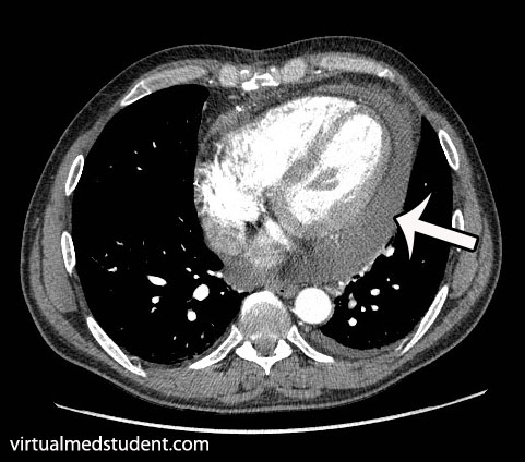
So what’s the big deal? A fall in cardiac output means that the body sees less oxygenated blood per minute. Every organ in the body requires oxygen to function properly. If cardiac output is decreased the organs may not receive enough oxygen to do their job. In super severe cases, when cardiac output approaches zero, cardiogenic shock occurs. If no oxygen reaches the organs multi-organ failure occurs and the patient may die.

Overview

Cardiac output is related to the stroke volume, heart rate, systemic vascular resistance, and difference in pressure between the arterial and venous sides of the body. When cardiac output drops the bodies’ organs receive less oxygen per unit time. In severe cases this may lead to organ death.

Related Articles

References and Resources

Fluid Around the Heart: Pericardial Effusions

The heart is encased in a connective tissue capsule known as the pericardium. The pericardium contains two layers, known as the parietal and visceral pericardium. These layers are like two blankets enveloping the heart. The visceral pericardium sits adjacent to the heart muscle itself and the parietal pericardium sits on top of the visceral pericardium. Because of this arrangement there is a potential space between the two layers.

When fluid (ie: blood, pus, water, etc.) leaks into this space a pericardial effusion is present. Fluid can leak out quickly, in which case the effusion is said to be “acute”; or it can leak out gradually in which case it is said to be “chronic”.

Causes

There are numerous causes of pericardial effusion some of which are listed below:

  • Infections
    • Viral (including HIV, coxsackie, echo, adeno, ebstein-barr, and varicella viruses)
    • Bacterial (including pneumococcus, neisseria meningitides, staphylococcus aureus)
    • Tuberculosis
  • Malignancies (cancers)
  • Autoimmune conditions (including connective tissue disorders, vasculitis, and drug induced)
  • Uremia (from renal failure)
  • Cardiovascular (cardiac surgery, myocardial infarction, aortic dissection, congestive heart failure)
  • Hypothyroidism
  • Cirrhosis (liver problems)
  • Idiopathic (unknown)

Please note that by no means is this list exhaustive, but these are the most common causes of effusions!

Signs and Symptoms

The classic symptom of pericardial effusion is chest pain that is better when the patient sits up and leans forward. However, numerous other symptoms including light headedness, shortness of breath, cough, and palpitations can occur.

Depending on how quickly the effusion develops, patients may spiral into a condition known as “tamponade”. When this occurs the effusion effectively “chokes” the heart muscle causing decreased contractile function. This causes decreased cardiac output and even multi-organ failure if left untreated!

The classic signs of tamponade are hypotension (ie: decreased blood pressure), muffled heart sounds, and increased jugular venous pressures (you can see the jugular veins engorged with blood). These three signs are known as “Beck’s triad”, which is generally a late finding of tamponade (ie: the patient is almost dead!).

Diagnosis

CT scan of pericardial effusion
There are numerous tests that can support the diagnosis. Electrocardiogram may show decreased voltages, and a finding known as “electrical alternans” where the QRS complexes change amplitude and/or direction as a result of the heart “sloshing” around in the effusion. Chest x-ray may show an enlarged heart, but this is neither specific, nor sensitive, for effusion. CT scans can show fluid surrounding the heart. Finally, echocardiography (ie: ultrasound of the heart) can be very useful in delineating not only the presence of, but also the size, and location of the effusion.

Treatment

For chronic effusions with no significant symptoms patients can be treated for the underlying condition causing the effusion. This will sometimes cure the effusion. However, in patients with acute presentations who have signs of cardiovascular instability (ie: low blood pressure, evidence of organ dysfunction from decreased blood flow, etc.) emergent removal of the fluid is performed. The quickest way to do this is to insert a needle under the xyphoid process and aspirate the fluid. In less acute situations, or in recurrent cases, surgical “windows” in the pericardial tissue can be created to allow the effusion to drain.

Overview

Pericardial effusions occur when fluid accumulates between the visceral and parietal pericardial layers surrounding the heart. There are numerous causes. Rapidly expanding effusions can cause cardiac tamponade and lead to cardiovascular collapse. Signs and symptoms include chest pain, shortness of breath, cough, distant heart sounds, and decreased blood pressure. Diagnosis is made by characteristic ECG, echocardiography, and CT scan findings.

References and Resources

  • Imazio M, Brucato A, Mayosi BM, et al. Medical therapy of pericardial diseases: part II: Noninfectious pericarditis, pericardial effusion and constrictive pericarditis. J Cardiovasc Med (Hagerstown). 2010 Nov;11(11):785-94. Review.
  • Khandaker MH, Espinosa RE, Nishimura RA, et al. Pericardial disease: diagnosis and management. Mayo Clin Proc. 2010 Jun;85(6):572-93. Review.
  • Spodick DH. Pericarditis, pericardial effusion, cardiac tamponade, and constriction. Crit Care Clin. 1989 Jul;5(3):455-76.
  • Mookadam F, Jiamsripong P, Oh JK, et al. Spectrum of pericardial disease: part I. Expert Rev Cardiovasc Ther. 2009 Sep;7(9):1149-57.
  • Jiamsripong P, Mookadam F, Oh JK, et al. Spectrum of pericardial disease: part II. Expert Rev Cardiovasc Ther. 2009 Sep;7(9):1159-69.
  • Woo KM, Schneider JI. High-risk chief complaints I: chest pain–the big three. Emerg Med Clin North Am. 2009 Nov;27(4):685-712, x.

A Simple Approach to Hemodynamic Instability

What is “hemodynamic instability”? Hemodynamics is the study of blood movement; when this movement is compromised in some way you get hemodynamic instability. If left untreated it can cause multi-organ failure and death.

You can think of hemodynamic instability as the collapse of the cardiovascular system. This collapse causes a significant drop in blood pressure.

There are many causes of hemodynamic instability and fortunately you don’t need to know all of them to manage a patient who is acutely experiencing cardiovascular collapse. A few basic tenets of physiology, when examined in the right order, will help you manage a patient who is unstable.

The tenets are as follows: preload (volume status), afterload (systemic vascular resistance), heart rate, heart rhythm, and contractility.

Preload and Volume Status

The first component of cardiovascular physiology that should be assessed in a hemodynamically unstable patient is preload and intravascular volume. Intravascular volume measures how much blood is present in the circulatory system. If a patient is volume deplete (ie: has a decreased intravascular volume) then hemodynamic instability can occur. An easy way to visualize this is to imagine someone bleeding. If the bleeding is not stopped, the cardiovascular system will eventually collapse. In the hospital this is often referred to as a patient being "dry".

Preload is one way to estimate a patient’s intravascular volume. Measuring preload is easy if you have a central venous catheter, and even easier if you have a Swan Ganz catheter, although these are used less frequently nowadays. These types of catheters can measure central venous and pulmonary artery pressures, which provide a good estimate of intravascular volume status.

If a patient does not have a central line you have to rely on clinical clues. One way to assess intravascular volume is to measure urine production. If urine output is less than normal, then the patient is trying to “hold on” to fluid, and therefore may be volume deplete. Urine that is dark amber in color is another clue. Decreased skin turgor is another clinically useful sign. Examining a patient’s neck veins (ie: jugular venous pulsations) can also be useful under certain circumstances.

In essence, if a patient’s preload, and therefore intravascular volume status is decreased it can be quickly corrected by giving the patient intravenous fluids. The type of fluid given is dictated by the clinical situation, but the most commonly used fluids for hemodynamic resuscitation are normal saline, lactated Ringer’s solution, and good old fashioned blood (especially when blood loss is responsible for the decreased volume!).

That’s great! But what do we do if we assess the patient’s volume status and come to the conclusion that it is sufficient, and the patient is STILL hemodynamically unstable? Not to fear… Let’s move on to the next tenet – systemic vascular resistance.

Systemic Vascular Resistance (SVR) and Afterload

The systemic vascular resistance is the resistance that blood sees as it travels through the arterial system. The vascular resistance varies depending on the etiology and time frame of the hemodynamic instability.

For example, patients in the initial stages of hypovolemic shock (ie: acute blood loss) the SVR will be elevated as the arteries constrict to maintain blood pressure; however, once the blood volume reaches critically low levels even constriction of the arteries is not enough to maintain the SVR. At this point the blood pressure will drop precipitously.

In contrast, patients in septic shock have a decreased systemic vascular resistance as the blood vessels dilate pathologically in response to infection. The result is the same as the above example: the blood pressure drops.

So how do we treat a decrease in systemic vascular resistance? Assuming intravascular volume (ie: preload) is adequate we sometimes need to "help" the patient constrict their blood vessels so they can maintain an adequate blood pressure. We do this using medications known as "pressors". The most commonly used pressors are norepinephrine (Levophed®), phenylephrine (Neosynephrine®), vasopressin, and dopamine.

Pressors should only be used once a patient has been treated with volume resuscitation. In other words, it does not make sense to help someone constrict their blood vessels when they have no intravascular volume to constrict around! This is why SVR is the 2nd (and not the 1st) tenet of managing someone with hemodynamic instability.

Heart Rate

Once intravascular volume status and vascular resistance have been dealt with, the next step is to monitor the heart rate. Patients who are excessively tachycardic (ie: high heart rate) or bradycardic (ie: low heart rate) can also be hemodynamically unstable. Let’s explain…

During tachycardia the left ventricle does not have as much time to fill with blood. Therefore, each beat of the heart ejects less blood than normal. If the tachycardia is severe enough this can cause hemodynamic instability and decreased blood flow. Likewise, in patients who are excessively bradycardic the amount of blood ejected over a specific unit of time (aka: cardiac output) may not be enough to sustain the bodies need for blood.

Patients who are excessively tachycardic can be treated with beta blockers assuming there is no other cause readily identified. It is also important to remember that tachycardia may be a sign of decreased intravascular volume! Therefore, the patient’s excessive heart rate may correct when the preload is corrected (again, always start at tenet number 1!).

Bradycardia can be treated with atropine. Atropine is a molecule that stimulates heart rate by blocking the actions of acetylcholine. It is also important to note that bradycardia is sometimes a "physiologic" response to the administration of pressors. For example, phenylephrine often causes a "reflex" bradycardia.

Heart Rhythm

Just like abnormal heart rates, abnormal heart rhythms can also cause hemodynamic instability. The most commonly encountered abnormal rhythms in hemodynamically unstable patients are atrial fibrillation, atrial flutter, heart block, ventricular fibrillation, and ventricular tachycardia.

Abnormal heart rhythms contribute to hemodynamic instability by decreasing the filling capacity of the heart. For example a patient in rapid atrial fibrillation will not have enough time between heart beats to load their left ventricle with blood.

The treatment of the various cardiac arrhythmias depends on the type of rhythm. It is important to note that abnormal rhythms may not have a significant impact on hemodynamic instability, and may need no additional treatment. This is most commonly encountered in people who are in atrial fibrillation, but their rate is well controlled.

Contractility

You’ve fixed intravascular volume, corrected the systemic vascular resistance, examined the heart rate and rhythm and you still can not stabilize the patient. Under these circumstances you have to think about the heart itself. It may not be ejecting blood efficiently or effectively enough to ensure cardiovascular stability.

Under these circumstances you need to order an echocardiogram to assess the heart’s function. If the heart is not holding its own it may need a little help. Medications such as epinephrine, dopamine, dobutamine, milrinone, and digoxin can help give the heart a little extra “squeezing power” to improve cardiac output.

Overview

The management of hemodynamic instability always begins with an assessment of intravascular volume (preload). If repleting intravascular volume does not fix the problem then the next step is to look at systemic vascular resistance. Does the patient need a little help constricting those blood vessels? If not, than you have to move quickly to assessing heart rate and rhythm. Finally, when all else fails, we have to assess the contractility of the heart. Maybe it isn’t pumping as hard or as efficient as it should be.

Correcting hemodynamic instability in the above order is both a quick and efficient framework for thinking about the unstable patient. Often times the physician will drop quickly down the list as they recognize a problem immediately (ie: rapid atrial fibrillation) and this is ok!!! The important thing is to have a framework for thinking about cardiovascular instability.

References and Resources

  • Stiell IG, Macle L. Canadian Cardiovascular Society atrial fibrillation guidelines 2010: management of recent-onset atrial fibrillation and flutter in the emergency department. CCS Atrial Fibrillation Guidelines Committee. Can J Cardiol. 2011 Jan-Feb;27(1):38-46.
  • Bozza FA, Carnevale R, Japiassú AM, et al. Early fluid resuscitation in sepsis: evidence and perspectives. Shock. 2010 Sep;34 Suppl 1:40-3. Review.
  • Price S, Uddin S, Quinn T. Echocardiography in cardiac arrest. Curr Opin Crit Care. 2010 Jun;16(3):211-5.
  • Naeem N, Montenegro H. Beyond the intensive care unit: a review of interventions aimed at anticipating and preventing in-hospital cardiopulmonary arrest. Resuscitation. 2005 Oct;67(1):13-23.
  • Sevransky J. Clinical assessment of hemodynamically unstable patients. Curr Opin Crit Care. 2009 Jun;15(3):234-8.

The Basics of Myocardial Infarction (Heart Attack)

Pathology

An acute coronary syndrome refers to any process where heart muscle receives less oxygen than it needs. It encompasses three separate, but overlapping pathologies: unstable angina, non-ST elevation myocardial infarction (NSTEMI), and ST elevation myocardial infarction (STEMI). Therefore, acute coronary syndrome represents a continuum of severity, which if severe enough, can lead to irreversible cardiac muscle death.

NSTEMI and STEMI are distinguished from unstable angina by the presence of heart muscle death. NSTEMI occurs when the inner most portion of the heart wall dies, and is generally due to a partially occlusive thrombus (blood clot) in one of the coronary arteries or their branches. An NSTEMI can, in some ways, be thought of as a "partial" heart attack.

On the other hand, STEMI occurs when a coronary blood vessel becomes fully blocked by thrombus. If untreated, the result is transmural (full wall) death of the heart muscle that the occluded vessel serves. A STEMI, in non-technical terms, can be thought of as a "full blown" heart attack.

Coronary thrombi (blood clots) form after rupture of an atherosclerotic plaque. The exposed plaque surface is highly thrombogenic, which means that blood clots form easily on it. The exposed surface causes platelet and coagulation cascade activation, which results in a thrombus that can occlude the entire vessel diameter resulting in decreased, or in the worst case scenario, no blood flow to the heart muscle. This is the basis of a "heart attack" or myocardial "infarction".

Signs, Symptoms, and Complications

Symptoms of acute coronary syndrome are consistent with anginal chest pain. If angina occurs at rest, or with less activity than it normally does, this is referred to as "unstable" angina, and signifies increased narrowing (ie: decreased diameter) of a coronary blood vessel.

Substernal “crushing” chest pain is the classic description of a heart attack (either NSTEMI or STEMI) and is usually accompanied by diaphoresis (ie: excessive sweating). In addition, nausea and vomiting can also occur. Pain from the chest can sometimes radiate into the arm (usually the left arm) or the jaw. Numbness and tingling in the arm and jaw are also common features. Shortness of breath is another symptom of heart attack, and is usually due to the acute back up of blood into the vessels of the lung, which leads to pulmonary edema.

Complications of infarction are due to mechanical changes in the heart. Damage of the electrical conduction system can lead to arrhythmias (abnormal heart rhythms), which are common in the first few days after an infarction.

If enough heart muscle is destroyed cardiogenic shock can occur. This happens when the heart is no longer able to pump enough blood to the rest of the body. Decreased blood flow to the other organs can lead to ischemia of these organs and multi-organ failure.

Myocardial infarction
and "heart attack"
refer to the same thing –
death of heart muscle.
Other complications are a result of the scarring process that occurs hours to days after the initial heart attack. Dead heart muscle is slowly replaced by scar tissue in a process called fibrosis. During the scarring process there are periods in which the scar and adjacent tissue is weaker than normal. This weakness can cause various complications such as rupture of the heart wall, interventricular septum rupture, or papillary muscle rupture. All of these complications occur roughly 5 to 10 days after the initial heart attack, and are due to structural instability in the newly formed fibrotic area(s). In addition, aneurysm (ie: a ballooning out of the heart) formation may occur several weeks after myocardial infarction.

Finally pericarditis, or inflammation of the sack that the heart sits in, can also occur. A condition known as Dressler’s syndrome is pericarditis that occurs after a heart attack. It usually occurs two weeks to several months after the infarction.

Long term complications of myocardial infarction include heart failure. Ultimately the damaged heart is unable to beat as well as it did in the past. This leads to the back up of blood and fluid in the lungs causing shortness of breath, amongst other symptoms.

Diagnosis

Diagnostic studies can confirm NSTEMI or STEMI. An electrocardiogram (ECG) is the gold standard. It will show ST segment depression in NSTEMI, and ST segment elevation in STEMI. Q-waves are also seen, but may represent previous infarction.

STEMI
Image (left) – An ECG of someone with a STEMI would look like the image to the left. Note how the ST segment is elevated relative to the baseline.

Blood tests can also show elevations in specific proteins released by the dying heart muscle. Cardiac troponin I is one of these markers, and is the most specific for heart muscle injury. It begins to rise 4 hours after the initial thrombus formation and stays elevated for 7-10 days. CK-MB (creatine kinase myocardial band) is another protein released by damaged heart tissue; it is useful in that it is cleared relatively quickly compared to troponin; this is useful in that it can aid in detection of early re-infarction.

Image (right) – An ECG of someone with an NSTEMI. Notice how the NSTEMI ECG
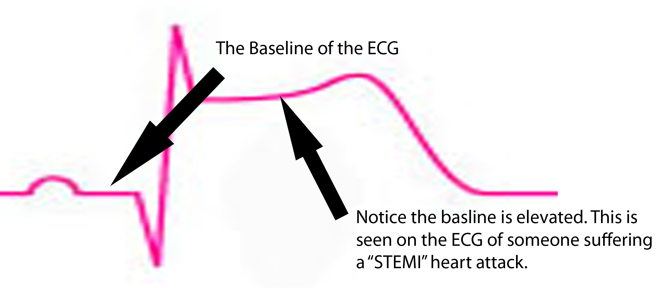
ST segment is depressed below baseline.
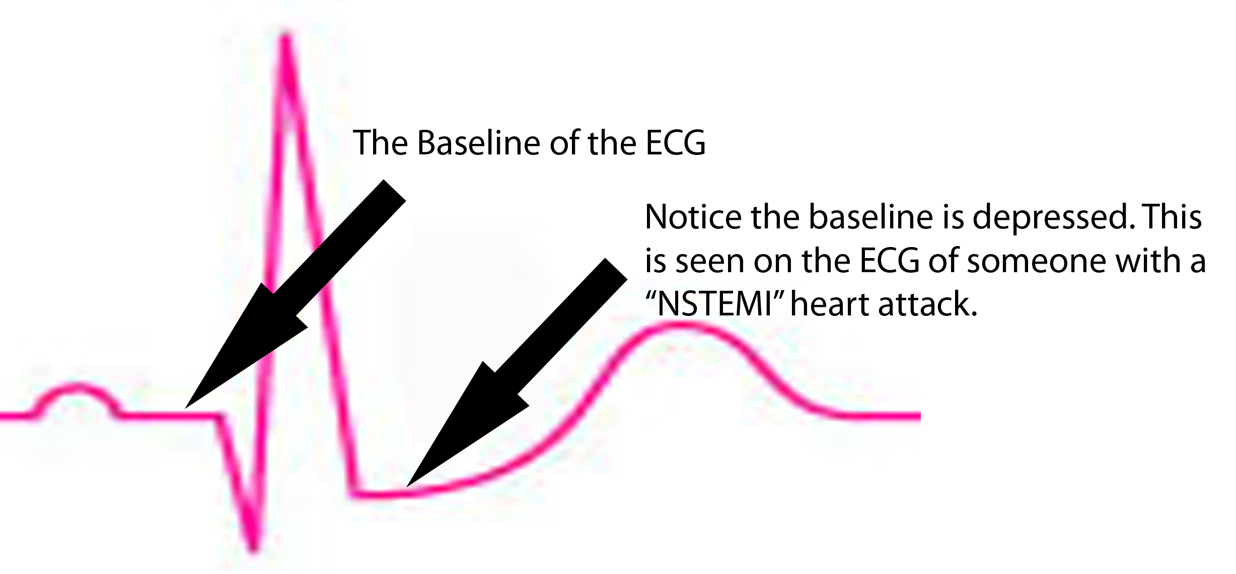
Treatment

The main goals of treatment are to restore blood flow, and to decrease the amount of work the heart is doing. Both of these measures help provide oxygen to the dying heart muscle. One of the main problems in myocardial infarction is that the heart is working harder than usual, and is doing so under decreased oxygen delivery (ie: decreased blood flow). This deadly combination increases the rate of heart muscle death. Therefore, treatment is designed to increase oxygen flow (ie: by restoring blood flow), or by decreasing the work of the heart, and therefore the amount of oxygen the heart needs to survive.

Treatment of any patient with suspected acute coronary syndrome should include pain control. For example, morphine controls the pain associated with heart attacks, and this indirectly lowers the heart rate. Lowering the heart rate decreases the amount of work the heart muscle is doing, and therefore decreases the amount of oxygen that must be delivered for muscle survival.

In addition, nitroglycerin is given because it decreases preload. Preload refers to the amount of blood inside the left ventricle just before contraction of the heart. A larger preload means that the heart must work harder because, in essence, it is pumping more blood per beat. Nitroglycerin dilates blood vessels (mostly veins) in the body and effectively decreases the amount of blood within the heart itself. Less blood to pump, equals less work the heart must do.

Oxygen therapy should also be started to help improve blood oxygen levels. This will ensure that any blood getting to the myocardium will have as much oxygen as possible.

There are several methods for restoring blood flow. The preferred method is through percutaneous coronary interventions (PCI). In this procedure a small catheter is inserted into blood vessels in the groin and threaded up towards the heart; once in the blood vessels of the heart the catheter can be used to mechanically remove any blockage. This is known as "angioplasty" or "thrombectomy". Percutaneous coronary intervention should occur in a reasonable amount of time. There is debate about the exact timing and depends on the type of acute coronary syndrome (ie: STEMI vs NSTEMI).

If the hospital does not have the capacity to do PCI then medications can be used to "break up" the blood clot/thrombus. All patients with suspected infarction should receive an aspirin. Aspirin works by inhibiting platelet plug formation. This helps slow the rate of thrombus formation. First line medications for restoring flow in myocardial infarction are intravenous unfractionated heparin or subcutaneous enoxaparin. Fibrinolytics like tissue plasminogen activator (tPa), reteplase, streptokinase, and urokinase are also sometimes used to try and restore blood flow.

Finally, revascularization operations (ie: coronary artery bypass grafting) may also be necessary depending on the level of disease in the other coronary vessels.

Overview

Acute coronary syndrome refers to a continuum of coronary blood vessel blockage. On the least extreme end is unstable angina; on the most extreme (ie: most dangerous) end is ST elevation myocardial infarction. Symptoms are usually crushing chest pain that radiates to the jaw and arm, but may also include nausea, vomiting, dizziness/fainting, and sweating. Diagnosis is based of ECG and blood tests for heart muscle damage. Treatment is aimed at increasing blood flow, and therefore oxygen delivery, to the heart muscle. This is done by decreasing the amount of work the heart is doing, increasing the amount of oxygen present in blood, and breaking up the blood clot via mechanical or medical means.

References and Resources

  • Husted SE, Nielsen HK. Unfractionated heparin and low molecular weight heparin for acute coronary syndromes–assessment of a Cochrane review. Ugeskr Laeger. 2010 Sep 13;172(37):2522-6.
  • Lepor NE, McCullough PA. Differential diagnosis and overlap of acute chest discomfort and dyspnea in the emergency department. Rev Cardiovasc Med. 2010;11 Suppl 2:S13-23.
  • >
  • Kumar V, Abbas AK, Fausto N. Robbins and Cotran Pathologic Basis of Disease. Seventh Edition. Philadelphia: Elsevier Saunders, 2004.
  • >
  • Lilly LS, et al. Pathophysiology of Heart Disease: A Collaborative Project of Medical Students and Faculty. Fourth Edition. Lippincott Williams and Wilkins, 2006.
  • Flynn JA. Oxford American Handbook of Clinical Medicine (Oxford American Handbooks of Medicine). First Edition. Oxford University Press, 2007.
  • Ferguson JJ, Califf RM, Antman EM, et al. Enoxaparin vs unfractionated heparin in high-risk patients with non-ST-segment elevation acute coronary syndromes managed with an intended early invasive strategy: primary results of the SYNERGY randomized trial. JAMA. 2004 Jul 7;292(1):45-54.