Middle Cerebral Artery: A Common Site for Stroke

Middle cerebral artery (MCA) strokes occur when the MCA or its branches are occluded. With occlusion, blood, and along with it, oxygen and nutrients fail to reach the brain. If blood flow is not restored quickly the affected brain tissue dies leading to permanent neurological injury.

Risk factors for MCA strokes are the same for other strokes. They include hypertension, diabetes, smoking, atrial fibrillation, and a whole slew of hypercoagulable states (ie: pathologic increases in the bodies’ propensity to form blood clots).

In order to understand MCA strokes we have to first appreciate the anatomy of the MCAs, as well as the brain that they serve. The MCAs are subdivided into four parts. The M1 segment is one of the terminal branches of the internal carotid artery. The MCAs then become progressively more narrow and form more and more branches as they reach out towards the cortical surfaces of the brain. The most distal portions of the MCA are deemed the M4 branches. The lateral lenticulostriate arteries are small branches that branch from the M1 segments; these small arteries feed some of the deeper structures of the brain.

Importantly, the MCAs and their branches provide blood flow to an extremely large portion of the brain. These areas include the lateral and inferior frontal lobes, superior portion of the temporal lobes, insula, and lateral parietal lobes. Branches of the first portion of the MCAs (aka: the lateral lenticulostriate arteries) also provide blood flow to the deep sections of the brain including the putamen, head and body of the caudate, external globus pallidus, and parts of the posterior limb of the internal capsule.

The segmental anatomy of the MCAs is important because strokes that occur in the outer segments (ie: M3 and M4) cause less neurological injury than the inner MCA segments (ie: M1 and M2). This is because less total brain volume is affected by outer segment strokes.

The most common cause of MCA strokes are clots that break off and travel from the heart or the carotid arteries to the MCA (aka: emboli). Less commonly, a blood clot will form directly in the MCA itself (aka: thrombus). Atherosclerotic disease is the most common cause of thrombus formation in the MCA and atrial fibrillation (an abnormal heart rhythm) is the most common cause of emboli from the heart.

Signs and Symptoms

Middle cerebral artery strokes present with one of two types of syndromes depending on which MCA – right or left – is involved, as well as which segments of the MCA are involved.

In the worst case scenario, a stroke of the right or left M1 segment of the MCA causes weakness of the opposite side of the body. This is a result of cortical damage to the primary motor cortex as well as possible infarction of the posterior limb of the internal capsule (ie: the location of descending motor tracts). MCA strokes usually present with face and arm weakness that is worse than leg weakness. Remember that the motor cortex that controls leg function is served by the anterior cerebral arteries.

M1 strokes also cause decreased ability to feel sensation on the opposite side of the body as a result of damage to the parietal lobes.

Damage to the optic radiations, which course in the parietal (Baum’s loop) and temporal lobes (Meyer’s loop) can cause problems with vision. Finally, injuries to the frontal eye fields cause the eyes to deviate towards the side of the stroke (ie: the frontal eye fields normally allow you to make fast eye movements in the opposite direction, therefore damage prevents patients from looking to the non-affected side).

Right and left sided M1 strokes will give you all of the above symptoms; however, laterality can be determined based on specific symptoms caused by only a left or a right sided strokes.

The poor patients with left M1 strokes have a decreased ability to speak and/or understand language because of damage to Broca’s area in the left frontal lobe (speech production) and Wernicke’s area in the left temporal lobe (speech comprehension). Remember this is in addition to all the other stuff above.

Right M1 strokes can cause "anosognosia", in which the patient is unaware of certain deficits they may have; these patients also often fail to recognize the left side of their body (ie: they "neglect" or fail to appreciate the entire left side of the world) and may have difficulty appreciating people or objects presented in their left visual field.

Right MCA stroke
Less "severe" cases, in which M2, M3 or M4 branches are affected can produce a variety of signs and symptoms depending on the specific branches involved.

Diagnosis

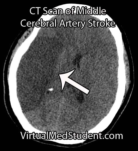
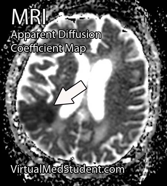
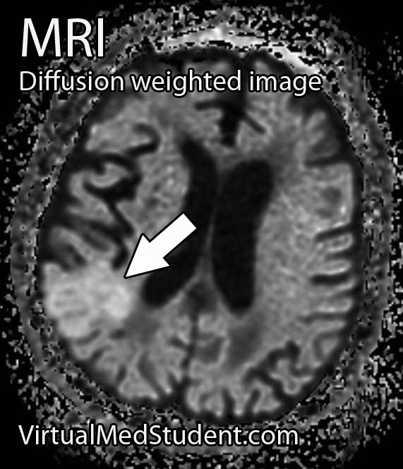
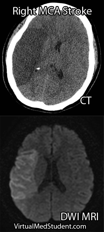
Diagnosis of MCA strokes are based on symptoms, CT scans, and MRI images. CT and MR angiograms frequently show the blocked blood vessel causing the stroke. CT perfusion scans are a newer technology that give information regarding the amount of blood flow to affected brain tissue.

Treatment

Treatment depends on the timing of the stroke. If the patient presents within 3 hours of symptom onset, and the head CT reveals no bleeding, than intravenous tissue plasminogen activator (tPA) may be given to help "break" up the blood clot causing the stroke.

Other treatments using catheter based approaches are frequently used in patients who are unable to receive tPA. Such treatments include mechanical clot removal with special catheter and wire devices. In addition, in patients more than 3 hours, but less than 6 hours out from symptom onset intra-arterial (not to be confused with intravenous) tPA may be used.

Less commonly, large "malignant" MCA strokes may cause significant swelling, which can put pressure on the brainstem. These patients sometimes undergo an open surgical procedure known as a "craniectomy", in which the bone overlying the affected brain tissue is removed. This surgery allows the edematous brain tissue to swell outwards preventing it from herniating downwards towards vital brainstem structures.

Overview

Middle cerebral artery strokes are most commonly caused by blood clots that break off from the heart or carotid artery. Symptoms of MCA strokes depend on the segment involved, as well as which MCA (right versus left) is involved. Diagnosis is made with a combination of MRI, CT, and symptomatology. Treatment consists of intravenous or intra-arterial tPA and/or mechanical clot removal depending on the time frame of the symptoms.

Related Articles

References and Resources

Brain Boo-Boos: Cerebrovascular Accidents (Stroke)

MCA Stroke CT Scan

Stroke - ADC Map

Stroke - Diffusion Weighted
A cerebrovascular accident, commonly known as a stroke, occurs when blood flow stops reaching brain tissue. If the entire brain is involved it is referred to as a "global" stroke; if a specific region of brain is involved it is referred to as a "focal" or "territorial" stroke. There are three broad causes of territorial strokes: thrombotic, embolic, and hemorrhagic.

A thrombotic stroke occurs when a blood clot forms in a blood vessel that supplies brain tissue. This is similar to what happens in cardiac infarction (ie: heart attacks). Thrombi are most commonly caused by atherosclerotic disease of the cerebral blood vessels. Thrombi usually form at areas of turbulent blood flow and at locations where vessels form branch points.

Embolic strokes are similar because they are technically blood clots. However, an embolus is a fragment of a clot (thrombus) that formed in another part of the body. Those fragments break free from the original clot and travel to blood vessels in the brain. They get lodged at some point and prevent blood from flowing resulting in a stroke if treatment is not obtained quickly.

Strokes can also be caused by bleeding into brain tissue. These type of strokes are called “hemorrhagic stroke”. Bleeding can occur in people with long standing untreated high blood pressure, or in those that have underlying structural disorders of the blood vessels in the brain (ie: aneurysms or arteriovenous malformations).

Diagnosis

Speedy diagnoses of stroke is extremely important because brain tissue dies quickly if it doesn’t receive adequate oxygen.

The first test that is done in cases of suspected stroke is a CT scan of the head. The purpose of the CT scan is not necessarily to "see" the stroke, but rather to rule out some other cause (ie: tumor, subdural hematoma, etc) for the symptoms. If bleeding is present on the CT scan the treatment algorithm becomes much different. If no bleeding is seen on CT then the second scan is usually an MRI.

An MRI takes longer than a CT scan, but it gives a much more detailed picture of the brain. In addition, it can pick up ischemia (ie: cell death related to decreased blood flow) much earlier than CT.

The best sequences to detect a stroke on an MRI are the diffusion weighted images and apparent diffusion coefficient maps. Stroked brain tissue will appear “bright” on diffusion weighted imaging and “dark” on the apparent diffusion coefficient map (see images to the left).

In addition, the carotid arteries are scanned using ultrasound in order to detect potential narrowing from atherosclerotic disease. Atherosclerotic carotid arteries are a potential source of emboli.

Sometimes a procedure known as transcranial doppler, which also uses ultrasound technology, is used to detect blood flow in the individual blood vessels of the brain. This can sometimes help determine the specific location of the thrombus/embolus.

Cerebral angiograms are much more invasive tests, but give a detailed view of which vessels are blocked. Cerebral angiograms can also be used to treat some strokes by directly removing clot from the affected blood vessel.

Most patients should undergo a thorough work up for atherosclerotic disease including a fasting lipid panel and hemoglobin A1C levels (a marker of diabetes).

If the heart is a suspected source of emboli than transthoracic echocardiography (ie: an ultrasound of the heart) is often done as well.

Signs and Symptoms

Cerebrovascular accidents present with a wide variety of signs and symptoms. It is entirely dependent on the blood vessel, and therefore, region of the brain involved. For example, strokes in the left middle cerebral artery will often cause significant language impairments if left untreated. Middle cerebral artery strokes usually cause contralateral paresis as well (usually the face and arm are more affected than the leg). Strokes in the frontal lobes caused by blockage of the anterior cerebral arteries can cause personality changes, as well as paresis/paralysis of the contralateral lower extremity.

Suffice it to say that there are a variety of possible clinical presentations in patients suffering from stroke. These presentations generally correlate with our understanding of brain anatomy and function.

Treatment

Prompt treatment of stroke is critical for preserving viable brain tissue. If a stroke is due to a blood clot (ie: thrombus or embolus) the treatment is with a drug known as tissue plasminogen activator (tPA). tPA is a medication that helps break up the clot.

It can be a dangerous medication because it can cause serious bleeding, but if given early enough, and in the right patient, it can completely prevent brain tissue death. There are numerous contraindications to giving tPA so caution must be used. The traditional teaching is that is should be given within three hours of symptom onset (this is the FDA approved indication); however, up to 4.5 hours from symptom onset has become common in clinical practice (but this is not FDA approved).

Endovascular therapies that mechanically remove the clot are becoming more common, especially for large vessel disease. However, this type of treatment requires specialized interventional neuro-radiologists and is not available in all medical centers. Endovascular therapy with a clot retrieving device is usually indicated up to 6 hours post symptom onset for large vessel occlusions. More distal (ie: further out) occlusions are not candidates for this type of procedure yet.

If a patient survives their first stroke, they are often started on medications to decrease their risk of having a second stroke. One of the most common medications used to prevent a second stroke is aspirin.

However, other medications like ticlopidine and clopidogrel (Plavix®) are also frequently used. All three of these medications prevent platelets (ie: one of the bodies natural ways of forming blood clots) from clumping together. In addition, aspirin is often mixed with another medication called dipyridamole (dipyridamole + aspirin = Aggrenox® in the United States). Patients who have suffered a minor stroke or have high risk transient ischemic attacks should be started on aspirin and clopidogrel and then transitioned to aspirin alone at 21 days.

If atherosclerosis is believed to be the cause of the stroke patients are often started on a statin. This helps slow the process of atherosclerosis and can help prevent another stroke from occurring.

If an embolus was the cause of the stroke patients are often started on an anticoagulant. The most common one used is warfarin (although there are many others). Warfarin is also used to treat a common cause of embolic stroke, an abnormal heart rhythm known as atrial fibrillation.

Overview

Strokes can be caused by thrombi or emboli which are blood clot that block blood flor, or from hemorrhage into brain tissue. Diagnosis is made by CT and MRI scans. Additional studies including carotid ultrasound, cerebral angiography, echocardiography, fasting lipid profiles, and tests for diabetes are also frequently performed.

Treatment depends on the etiology. Tissue plasminogen activator (tPA) is given if thrombi or emboli are the cause, and symptoms began less than 3 hours prior to presentation (4.5 hours is becoming the standard of care). Mechanical endovascular removal of the clot is also possible in some medical centers with specialized equipment.

Prevention of secondary strokes involve the use of anti-platelet (ie: aspirin, clopidogrel), anti-coagulant (ie: warfarin), and anti-atherosclerotic medications depending on the etiology of the previous stroke.

Related Articles

References and Resources

The Anterior Choroidal Artery: Small but Mighty

The anterior choroidal arteries are small, but vital blood vessels in the brain. They are branches of the internal carotid arteries. They arise proximal to the splitting of the internal carotid into the anterior and middle cerebral arteries.

The anterior choroidal arteries deliver blood to vital brain structures. These structures include the posterior limbs of the internal capsules, portions of the thalami, optic tracts, middle third of the cerebral peduncles, portions of the temporal lobes (ie: parts of the pyriform cortex, uncus, and amygdala), substantia nigra, portions of the globus pallidus, as well as the choroid plexus in the lateral ventricles.

The anterior choroidal artery forms connections (anastamoses) with the posterior lateral choroidal arteries. Cerebral angiograms are the best way to visualize the anterior choroidal arteries.

Importance in Disease

Blockage of the anterior choroidal artery can cause a stroke. The most common symptoms of an anterior choroidal stroke are hemiparesis (weakness on the opposite side of the body), hemianesthesia (decreased sensation on the opposite side of the body), and a homonymous hemianopsia (loss of a portion of the visual field of both eyes). High blood pressure is the most common underlying factor in people with anterior choroidal artery strokes.

The hemiparesis is a result of damage to the posterior limb and genu of the internal capsule. The posterior limb contains the corticospinal tracts, which send information about movement from the brain to the spinal cord.

The hemianesthesia is a result of damage to the ventral posterolateral nucleus of the thalamus. This nucleus contains neurons that receive information from the spinal cord about sensation from the body. This symptom is less common than weakness, and occurs in roughly half of patients with an anterior choroidal stroke.

Cerebral angiogram showing anterior choroidal artery.

The final symptom, homonymous hemianopsia, is caused by damage to the optic tracts and lateral geniculate nucleus of the thalamus. Patients lose the ability to see objects on the left or right side (depending on which anterior choroidal artery is involved) in both eyes. This is an even more uncommon symptom, which occurs in less than 10% of patients with an anterior choroidal artery stroke.

Strokes of the anterior choroidal artery rarely cause all three symptoms. This is because the brain tissue served by the anterior choroidals also receives blood flow from other arteries.

Aneurysms of the anterior choroidal arteries are rare and will not be discussed in this article.

Overview

The anterior choroidal arteries are paired structures that arise from the internal carotid arteries. They supply blood to many important structures within the brain. Stroke is the most common pathological disease related to this blood vessel and frequently causes weakness of the opposite side of the body. High blood pressure is the most common underlying disease seen in people who have a stroke in this vascular distribution.

Other Stuff Worth Looking At…

References and Resources

  • Pezzella FR, Vadalà R. Anterior choroidal artery territory infarction. Front Neurol Neurosci. 2012;30:123-7. Epub 2012 Feb 14.
  • Bruno A, Graff-Radford NR, Biller J, et al. Anterior choroidal artery territory infarction: a small vessel disease. Stroke. 1989 May;20(5):616-9.
  • Baehr M, Frotscher M. Duus’ Topical Diagnosis in Neurology: Anatomy, Physiology, Signs, Symptoms. Fourth Edition. Stuttgart: Thieme, 2005.
  • Nolte J. The Human Brain: An Introduction to its Functional Anatomy. Sixth Edition. Philadelphia: Mosby, 2008.
  • Baskaya MK, Coscarella E, Gomez F, et al. Surgical and angiographic anatomy of the posterior communicating and anterior choroidal arteries. Neuroanatomy (2004) v3:38-42.

Aortic Dissection: Intima, Media, DeBakey

Pathology

Aortic dissection occurs when blood flows into a “false” pathway created by damage to a layer of the aorta. In order to understand dissections, we have to first appreciate the layers of the aorta. Most blood vessels have three layers: the intima, media, and adventitia (see image below). You can think of these layers as insulation around a pipe. The adventitia is the outermost portion of the “pipe”; it helps connect the blood vessel to adjacent structures in the body. The media is the middle most layer, and is composed of smooth muscle; it helps control the diameter, and therefore pressure within the vessel. The intima is the layer that is immediately adjacent to blood flow.

Blood Vessel Layers
It is this intimal layer where all the “action” in aortic dissection occurs. Damage to the intima is the initiating event in a dissection, and can be caused by many different things. The most common cause is years and years of untreated high blood pressure (ie: hypertension). The hypertension “beats” down the intimal layer making it more likely to tear. In addition, other less common causes such as Marfans Disease or Ehlers-Danlos Syndrome can also cause aortic dissection. These genetic disorders result in weak connective tissue that can predispose the intimal layer to tearing.

And this is precisely what happens in aortic dissection. The intimal layer of the vessel tears. As a result blood can now take one of two possible pathways:

(1) Down into the aorta (ie: its normal path).

(2) Into the “false” space between the intimal and medial layer (aka: a "false lumen").

If blood takes the later pathway it ultimately “dissects” the intimal and medial layer away from one another, hence the name aortic “dissection”. Ultimately, the classification and treatment of aortic dissections depends on where along the aorta the dissection occurs.

Classification Systems

Aorta Schematic
There are two different systems for classifying aortic dissections based on their location along the aorta. The first system is the DeBakey system (named after a world renowned heart surgeon, Michael DeBakey). It is divided into 3 subtypes:

– DeBakey type 1 -> involves the ascending and descending aorta.
– DeBakey type 2 -> involves the ascending aorta only.
– DeBakey type 3 -> involves the descending aorta only.

The second classification system is more simple. It is called the Stanford classification, and is divided into 2 subtypes:

– Stanford type A -> Involves the ascending aorta (may or may not involve the descending aorta)

– Stanford type B -> involves the descending aorta (does not involve the ascending aorta)

The reason these classification systems exist is because the location of the dissection dictates treatment. Ascending dissections are treated much differently than descending dissections.

Signs and Symptoms

The symptoms of aortic dissection depend on its location. If the dissection affects the ascending aorta the classical presentation is a tearing or ripping chest pain. If the descending aorta is involved the pain is often referred to the upper back between the scapulae. In addition, patients usually come in with elevated blood pressures (which is usually the precipitating cause in most cases). However, the blood pressure readings can be different between arms. Pulses from blood vessels that originate before the dissection are often stronger. This can lead to pulse asymmetry on physical exam.

New heart murmurs can also occur. The most common one is the murmur of aortic regurgitation. New murmurs occur when the aortic root is involved in the dissection. This can cause mechanical damage to the aortic valve leading to abnormal function. Blood is then able to regurgitate (ie: flow backwards) into the heart due to the deficient valve.

Since the aortic arch (the portion between the ascending and descending sections) contains vessels that eventually go to the brain, patients sometimes have neurological symptoms as well, although this is a relatively uncommon finding in clinical practice.

Diagnosis

The diagnosis of aortic dissection is based on clinical suspicion combined with imaging studies. Chest x-ray will sometimes show a “widened mediastinum”. This occurs because the enlarged aorta casts a larger shadow on the x-ray detector. If this is seen, and there is a high clinical suspicion of a dissection, a CT scan of the chest with intravenous contrast is ordered (see image).

Aortic Dissection

The CT scan will show the true and false lumens associated with dissection. Transesophageal echocardiography (TEE) is another way of visualizing the aorta and is highly sensitive and specific for detecting dissections.

Treatment

Treatment is dependent on the location of the dissection along the aorta. Dissections of the ascending aorta (DeBakey type 1 and 2, and Stanford class A) are surgical emergencies and require operative repair.

Descending aortic dissections (DeBakey type 3, and Stanford class B) are often managed medically through blood pressure control.

With the advent of endovascular techniques, many dissections can be treated endovascularly (ie: "through the groin"), which may be superior to open operative techniques for certain types of dissections.

Overview

Aortic dissections occur after damage to the intimal layer of the blood vessel. There are different classification systems depending on what part of the aorta is involved. Symptoms are usually a severe ripping chest pain that can radiate to the back. Pulses and blood pressures may vary between the right and left arms. In addition, neurological symptoms may occur if the dissections includes the vessels leading to the head. If the ascending aorta is involved surgery is indicated; descending aortic dissections are usually managed with aggressive blood pressure control.

References and Resources

  • Kumar V, Abbas AK, Fausto N. Robbins and Cotran Pathologic Basis of Disease. Seventh Edition. Philadelphia: Elsevier Saunders, 2004.
  • Lilly LS, et al. Pathophysiology of Heart Disease: A Collaborative Project of Medical Students and Faculty. Fourth Edition. Lippincott Williams and Wilkins, 2006.
  • Flynn JA. Oxford American Handbook of Clinical Medicine (Oxford American Handbooks of Medicine). First Edition. Oxford University Press, 2007.
  • Blackbourne LH. Surgical Recall, Fifth North American Edition (Recall Series). Fifth Edition. Philadelphia: Lippincott Williams and Wilkins, 2009.
  • Hata M, Sezai A, Yoshitake I, et al. Clinical trends in optimal treatment strategy for type a acute aortic dissection. Ann Thorac Cardiovasc Surg. 2010 Aug;16(4):228-35.
  • Nordon IM, Hinchliffe RJ, Loftus IM, et al. Management of Acute Aortic Syndrome and Chronic Aortic Dissection. Cardiovasc Intervent Radiol. 2010 Nov 12. [Epub ahead of print].

Hypertension: Understanding and Managing High Blood Pressure

The pathology of essential hypertension is not well understood. However, there are numerous theories, each with supporting evidence.

Genetic causes are supported by the fact that children of hypertensive parents have an increased risk of developing the disease. However, specific known genetic mutations are not common. When mutations are responsible for essential hypertension they often involve the sodium and chloride channels in kidney cells, or mutations in the genes responsible for producing the proteins of the renin-angiotensin-aldosterone system. Sodium, renin, angiotensin, and other molecules play a vital role in blood pressure. In some hypertensive patients these systems are abnormal and can lead to elevated blood pressure.

Some patients with essential hypertension appear to be “salt sensitive”. Salt, or more specifically sodium, appears to play a role in the development of hypertension. Some patients likely have a genetic predisposition to retain sodium. Excess sodium enters the blood stream where it exerts an osmotic pull on water in adjacent tissues. Fluid from body tissues is "sucked" into the blood stream resulting in increased blood volumes. The expanded blood volume increases the pressure within the blood vessel causing hypertension. Although salt plays a role in hypertension, new research has shown its importance may not be as clear as once thought.

There are other known causes of hypertension, but they constitute a relatively small proportion of cases. They are discussed in separate articles.

Signs and Symptoms

Essential hypertension in its earliest stages does not cause any signs or symptoms. Many people do not know they have high blood pressure. However, if left untreated hypertension can cause serious long term consequences.

One such consequence is an increase in the risk of cardiovascular disease. The risk of cardiovascular disease doubles with each 20/10 mmHg increase in blood pressure beyond a "baseline" of 115/75 mmHg! After years of pumping at elevated pressures the heart undergoes physical changes. Like any good muscle, it becomes larger because it is having to pump harder than normal. The result is a process known as concentric hypertrophy. The added muscle mass of the heart results in increased oxygen demand and the potential for heart attacks and heart failure.

Overall, nearly a third of heart attacks are attributable to high blood pressure. In addition, the risk of stroke is also dramatically higher in untreated hypertensive patients. The small blood vessels in the kidney and retina can also be damaged by years of high blood pressure resulting in kidney failure (hypertensive nephropathy) and blindness (hypertensive retinopathy).

Diagnosis

The diagnostic parameters of hypertension are constantly shifting. Currently the diagnosis can be made if a patient presents with a severely elevated blood pressure (systolic blood pressure > 200 and/or a diastolic blood pressure > 120) and/or signs or symptoms referable to the elevated blood pressure.

Severity Classification:
(1) Pre-hypertensive:
       130-140/80-90
(2) Grade 1:
       140-160/90-100
(3) Grade 2:
        >160/>100
In patients with less severe elevations, it is generally recommended that the blood pressure be measured several times over a period of multiple weeks. If the average of these readings is a systolic blood pressure of 140 or greater, or a diastolic blood pressure of 90 or greater than the diagnosis can be made. If the blood pressue stays between 120 and 130 systolic, and 80 to 90 diastolic the diagnosis of "pre-hypertension" is made meaning the patient is at risk of becoming hypertensive.

Some patients may have "white coat" hypertension simply by being in a doctor’s office (and the anxiety that this can provoke! Gosh, I hate going to the doctor’s office!). If this is the case the patient should be instructed to take their blood pressure at home and keep a log of the results.

Treatment

Treatment of hypertension consists of lifestyle modifications and/or using medications from several different pharmacologic categories. These medications may be used alone, or in combination, depending on each individual patient’s needs.

Patients who are pre-hypertensive or have stage 1 hypertension should be started on “lifestyle” modification therapy for at least 6 months prior to starting medicines (assuming there are no other health issues like diabetes or coronary artery disease). Lifestyle therapy consists of increasing aerobic exercise, as well as adhering to the “DASH” diet. DASH stands for "dietary approaches to stop hypertension" and consists of eating fruits, vegetables, low-fat dairy, whole grains, poultry and fish while reducing (or eliminating) red meat and sugars. This intervention alone can decrease systolic pressures over 10 mmHg and diastolic pressures over 5 mmHg.

Sometimes diet and exercise aren’t enough so we may have to resort to medications to control blood pressure. The first category of medications are known as diuretics, most commonly of the thiazide class. Thiazide diuretics work by inhibiting the re-absorption of sodium at the distal convoluted tubule of the kidney. Less sodium means less circulating blood volume, and therefore decreased blood pressure. Hydrochlorothiazide is a commonly used thiazide diuretic.

The second category of medications is known as β-blockers. β-blockers lower blood pressure by slowing the heart rate and indirectly lowering the amount of angiotensin II (angiotensin II is a potent natural blood vessel constrictor produced by the body). β-blockers reduce renin synthesis by specialized cells in the kidney.

A third category of medications known as angiotensin converting enzyme inhibitors (ACEIs) interfere with the formation of angiotensin II. ACEIs inhibit an enzyme present in the lung, which converts angiotensin I into the more potent vasoconstrictor angiotensin II. Interestingly, ACEIs also increase the level of bradykinin; this molecule causes blood vessels to dilate, which helps to further lower pressure.

There are also medications called direct angiotensin receptor blockers (ARBs), which inhibit the actions of angiotensin II. They do this by blocking angiotensin IIs ability to bind to its normal receptor sites.

Some medications, the calcium channel blockers (CCBs), lower blood pressure by inhibiting the contraction of smooth muscle cells that line the blood vessel walls. A specific subcategory of calcium channel blockers known as dihydropyridines block calcium from flowing into vascular smooth muscle cells. This results in decreased contraction of the muscle surrounding the vessel. As a result the vessel remains dilated, which lowers pressure.

Overview

The pathology of essential hypertension is not well understood. Several theories have been postulated, but it is likely a combination of genetic predispositions that result in this type of high blood pressure. Years of elevated blood pressure lead to cardiovascular disease including heart failure, heart attacks, and stroke, as well as damage to the kidneys and retinas. Treatment is with lifestyle modifications, diuretics, β-blockers, calcium channel blockers, angiotensin converting enzyme inhibitors, and angiotensin receptor blockers.

References and Resources