Middle Cerebral Artery: A Common Site for Stroke

Middle cerebral artery (MCA) strokes occur when the MCA or its branches are occluded. With occlusion, blood, and along with it, oxygen and nutrients fail to reach the brain. If blood flow is not restored quickly the affected brain tissue dies leading to permanent neurological injury.

Risk factors for MCA strokes are the same for other strokes. They include hypertension, diabetes, smoking, atrial fibrillation, and a whole slew of hypercoagulable states (ie: pathologic increases in the bodies’ propensity to form blood clots).

In order to understand MCA strokes we have to first appreciate the anatomy of the MCAs, as well as the brain that they serve. The MCAs are subdivided into four parts. The M1 segment is one of the terminal branches of the internal carotid artery. The MCAs then become progressively more narrow and form more and more branches as they reach out towards the cortical surfaces of the brain. The most distal portions of the MCA are deemed the M4 branches. The lateral lenticulostriate arteries are small branches that branch from the M1 segments; these small arteries feed some of the deeper structures of the brain.

Importantly, the MCAs and their branches provide blood flow to an extremely large portion of the brain. These areas include the lateral and inferior frontal lobes, superior portion of the temporal lobes, insula, and lateral parietal lobes. Branches of the first portion of the MCAs (aka: the lateral lenticulostriate arteries) also provide blood flow to the deep sections of the brain including the putamen, head and body of the caudate, external globus pallidus, and parts of the posterior limb of the internal capsule.

The segmental anatomy of the MCAs is important because strokes that occur in the outer segments (ie: M3 and M4) cause less neurological injury than the inner MCA segments (ie: M1 and M2). This is because less total brain volume is affected by outer segment strokes.

The most common cause of MCA strokes are clots that break off and travel from the heart or the carotid arteries to the MCA (aka: emboli). Less commonly, a blood clot will form directly in the MCA itself (aka: thrombus). Atherosclerotic disease is the most common cause of thrombus formation in the MCA and atrial fibrillation (an abnormal heart rhythm) is the most common cause of emboli from the heart.

Signs and Symptoms

Middle cerebral artery strokes present with one of two types of syndromes depending on which MCA – right or left – is involved, as well as which segments of the MCA are involved.

In the worst case scenario, a stroke of the right or left M1 segment of the MCA causes weakness of the opposite side of the body. This is a result of cortical damage to the primary motor cortex as well as possible infarction of the posterior limb of the internal capsule (ie: the location of descending motor tracts). MCA strokes usually present with face and arm weakness that is worse than leg weakness. Remember that the motor cortex that controls leg function is served by the anterior cerebral arteries.

M1 strokes also cause decreased ability to feel sensation on the opposite side of the body as a result of damage to the parietal lobes.

Damage to the optic radiations, which course in the parietal (Baum’s loop) and temporal lobes (Meyer’s loop) can cause problems with vision. Finally, injuries to the frontal eye fields cause the eyes to deviate towards the side of the stroke (ie: the frontal eye fields normally allow you to make fast eye movements in the opposite direction, therefore damage prevents patients from looking to the non-affected side).

Right and left sided M1 strokes will give you all of the above symptoms; however, laterality can be determined based on specific symptoms caused by only a left or a right sided strokes.

The poor patients with left M1 strokes have a decreased ability to speak and/or understand language because of damage to Broca’s area in the left frontal lobe (speech production) and Wernicke’s area in the left temporal lobe (speech comprehension). Remember this is in addition to all the other stuff above.

Right M1 strokes can cause "anosognosia", in which the patient is unaware of certain deficits they may have; these patients also often fail to recognize the left side of their body (ie: they "neglect" or fail to appreciate the entire left side of the world) and may have difficulty appreciating people or objects presented in their left visual field.
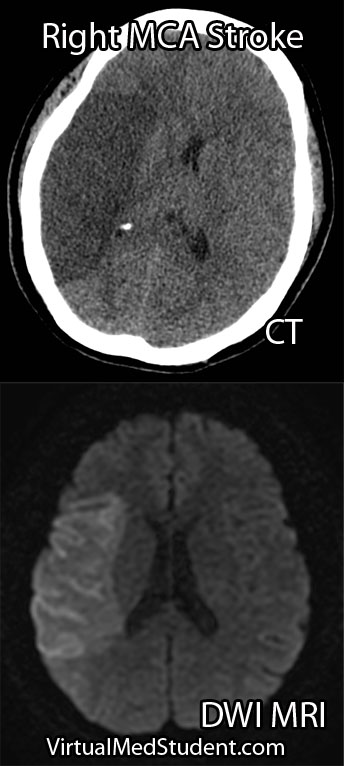
Right MCA stroke
Less "severe" cases, in which M2, M3 or M4 branches are affected can produce a variety of signs and symptoms depending on the specific branches involved.

Diagnosis

Diagnosis of MCA strokes are based on symptoms, CT scans, and MRI images. CT and MR angiograms frequently show the blocked blood vessel causing the stroke. CT perfusion scans are a newer technology that give information regarding the amount of blood flow to affected brain tissue.

Treatment

Treatment depends on the timing of the stroke. If the patient presents within 3 hours of symptom onset, and the head CT reveals no bleeding, than intravenous tissue plasminogen activator (tPA) may be given to help "break" up the blood clot causing the stroke.

Other treatments using catheter based approaches are frequently used in patients who are unable to receive tPA. Such treatments include mechanical clot removal with special catheter and wire devices. In addition, in patients more than 3 hours, but less than 6 hours out from symptom onset intra-arterial (not to be confused with intravenous) tPA may be used.

Less commonly, large "malignant" MCA strokes may cause significant swelling, which can put pressure on the brainstem. These patients sometimes undergo an open surgical procedure known as a "craniectomy", in which the bone overlying the affected brain tissue is removed. This surgery allows the edematous brain tissue to swell outwards preventing it from herniating downwards towards vital brainstem structures.

Overview

Middle cerebral artery strokes are most commonly caused by blood clots that break off from the heart or carotid artery. Symptoms of MCA strokes depend on the segment involved, as well as which MCA (right versus left) is involved. Diagnosis is made with a combination of MRI, CT, and symptomatology. Treatment consists of intravenous or intra-arterial tPA and/or mechanical clot removal depending on the time frame of the symptoms.

Related Articles

References and Resources

The Flaws of RVUs: Why They Fall Short in Measuring Physician Worth

Relative Value Units (RVUs) have been utilized in the United States healthcare system for several decades as a standardized method for quantifying the “value” of medical services. They have become an important component of determining physician reimbursement. While the RVU system was initially designed to bring uniformity and transparency to physician payments, it has been increasingly criticized for its shortcomings in accurately reflecting physician worth. This article will discuss the key reasons why RVUs are a flawed measure of physician worth and explore alternative methods of evaluation.

Focus on Quantity over Quality

One of the most significant criticisms of the RVU system is its emphasis on the volume of services provided rather than the quality of care. By assigning higher RVU values to more complex and time-consuming procedures, the system inadvertently incentivizes physicians to prioritize high-revenue-generating procedures over less profitable, but potentially necessary, patient care activities. This focus on quantity can compromise the quality of care and hinder a physician’s ability to provide patient-centered care.

Lack of Personalized Care

RVUs are based on standardized averages, which do not account for individual variations in patient needs, physician skill, and the complexity of cases. Consequently, the RVU system fails to recognize the nuances of personalized care that physicians provide. High-quality care often involves tailoring treatment plans to each patient’s unique circumstances, which may not align with the fixed values assigned by RVUs. For example, if a primary care physician spends an entire hour discussing a patient’s mental health issues in a caring and compassionate way they are reimbursed substantially less than an orthopedic surgeon who takes an hour to pin a fracture or a neurosurgeon who spends an hour doing a cervical discectomy and fusion.

Limited Scope of Measurement

The RVU system only captures a narrow scope of a physician’s worth by focusing solely on clinical procedures and services. It does not account for the many other essential aspects of healthcare, such as patient education, interdisciplinary collaboration, care coordination, and research contributions. By overlooking these non-clinical activities, the RVU system fails to provide a comprehensive evaluation of a physician’s value.

Inadequate Incentive for Preventative Care

Preventative care plays a critical role in promoting public health and reducing healthcare costs. However, the RVU system does not adequately incentivize physicians to engage in these activities, as they are generally assigned lower RVUs. This discrepancy may lead to an underemphasis on preventative care and a potential increase in long-term healthcare costs.

Perpetuation of Disparities

RVUs can contribute to healthcare disparities by allocating resources based on service volume rather than patient need. Physicians practicing in underserved areas may find it challenging to generate high RVU values due to lower patient volume or a greater focus on primary care. This imbalance in resource distribution may inadvertently widen the gap in healthcare access and quality.

Lack of Collegiality

RVUs also tend to foster a lack of collegiality amongst physicians. In my experience, especially doctors on an “eat what you kill” compensation model, meaning they are paid for the number of RVUs they produce, are much less likely to refer to colleagues who may have more expertise or skill in a particular area. In other words, the doctor may “hold on” to patients to generate more RVUs rather than getting them to a colleague who may be able to provide a higher level of care for their particular ailment.

While RVUs were initially intended to standardize physician reimbursement and provide a transparent measure of physician worth, their shortcomings have become increasingly apparent over time. By focusing on quantity over quality and failing to capture the full spectrum of a physician’s value, the RVU system has inadvertently compromised patient care and perpetuated healthcare disparities. It is essential for the healthcare industry to consider alternative methods of evaluation and reimbursement that better align with the goals of patient-centered care, quality improvement, and equitable resource distribution.

It can be a fine line between incentivizing physicians to “work harder” and “earn more” while maintaining a high level of care and maintaining a highly ethical medical practice. I don’t think RVUs are a great answer to this dilemma, but for the time being it is the best we have in the United States. Let’s here your thoughts below…

Letters of Intent to Trap Unwary Physicians

The following article is courtesy of Dennis Hursh, author of The Final Hurdle – A Physician’s Guide to Negotiating a Fair Employment Agreement. Dennis is an attorney who focuses on review and negotiation of physician employment agreements.

The contracting process for a physician employment agreement sometimes (but not always) begins with a letter of intent, so it makes sense for you to be aware of a common trap that many physicians find themselves in after they sign a letter of intent.

A letter of intent (“LOI”) is simply a very brief summary of the main terms of what the parties assume will be in a binding physician employment agreement. The purpose of an LOI is to make sure that the both parties are “on the same page” as far as the major terms of the agreement they hope to form.

For example, in a physician employment agreement, if you expect to be paid $300,000 a year but the employer is expecting to pay $200,000 a year, there probably isn’t any value in continuing negotiations. A letter of intent can save a lot of pain and aggravation (not to mention attorney’s fees) by avoiding negotiations that are not likely to lead to a signed deal.

Since the purpose of a LOI in physician employment agreements is just to determine if further negotiations are in order, the provisions in a LOI are generally not legally binding. Accordingly, it is very common for physicians to treat a LOI as an unimportant formality. Having been told that the document is not legally binding, they sign the LOI even though it contains some terms that they hope to negotiate.

You must remember that the purpose of a LOI is to make sure that you and your potential employer are on the same page with respect to the major terms of the physician employment agreement you hope to conclude. Although there will be many terms and conditions of the final agreement that the parties will negotiate, you should assume that anything you agree to in the LOI is “off the table”. Accordingly, do not sign a letter of intent unless you are completely comfortable with the compensation and other material terms set forth in the LOI. Legally binding or not, make sure you aren’t “agreeing” to something that you don’t want in the final agreement.

Footnotes, References, and Resources

(1) Some provisions of the LOI are typically legally binding. Specifically, the LOI will most likely provide that each party is responsible for its own attorney’s fees, that the negotiations will remain confidential, and that the physician will negotiate exclusively with this employer for some period of time. These provisions generally are legally binding. In other words, even if you do not sign a physician employment agreement, you are still bound to pay your own attorney’s fees and keep the terms of the negotiations confidential.

(2) For additional information about physician employment agreements, see www.TheFinalHurdle.com and The Physician Contract Blog.

The Frustrations of Dealing with a Demanding Patient

Dealing with a demanding patient can be extremely frustrating! But before we delve further into how to handle these pesky buggers, let’s take a moment to reflect on what constitutes a “demanding” patient.

Demanding patients exhibit a variety of behaviors and attitudes. Patients may be rude, down right nasty, non-compliant, litigious, passive-aggressive, or inconsiderate of your time. In essence, a "demanding" patient is one that requires a tremendous amount of investment on the doctor’s part.

It is important that being sick can make people nasty! I know I turn into a real asshole when I am sick (just ask my wife). Not to mention many patients are on medications (ie: steroids) or illicit drugs, which can alter their behavior. Therefore, it is extremely important to not take the threats of demanding patients personally. In fact, placating a demanding patient can be difficult, if not impossible!

But fret not! There are many ways to deal with a demanding patient! I recommend learning as many different ways as possible because each method is a "weapon" in your arsenal. One method may work on patient A, but not on patient B. Remember that no two patients are the same! Enough intro, let’s get into some real world approaches and solutions…

The first type of demanding patient is the "confused" patient. This patient is "demanding" because they just don’t seem to get it. They are lost in the massive sea that is healthcare and no one has bothered to explain why they are adrift! Ask the patient to tell you, in their own words, why they are in the hospital or clinic. If they cannot, a few minutes (literally minutes!) explaining what is happening to them can turn this difficult patient into a bucket of smiles!

Educating your patients about their disease is paramount to being a great physician! I cannot tell you how many patients have literally ZERO idea why they are taking such and such medication. Very few have actually seen their x-rays or CT scans. Show them and educate them; I guarantee you will develop instant rapport if you do this.

Sometimes, no matter how much time you spend, or how much you educate a patient there are some people who will continue to be a pain in the tookus! I call this patient the "asshole". These patients can be particularly tricky to deal with, and many of them have underlying personality disorders (remember you psych rotation!). However, many will respond to the "tough love" approach. These patients typically need firm, stern, and direct confrontation otherwise they will continue to walk all over doctors, nurses, and other members of the healthcare team. Remember that you didn’t go to medical school and spend countless hours studying to be told by the patient what tests should be done or what treatment should be administered (and just so you know this isn’t advice they will likely give you in medical school!). A little bit (and I do mean a little bit) of paternalism should still exist in medical care, in my honest opinion.

When all else fails and the patient continues to be overly demanding and unsatisfied with their care a simple, "you are free to get a second opinion somewhere else" will often free both the patient and the doctor from turning a nasty situation into a total crap storm. If they take you up on the offer you’re free of their painful tactics, if not, they may re-think their demanding ways and become a more amenable patient.

And perhaps my best piece of advice… Ask a nurse! Nurses are the front line and deal with ghastly behavior on the regular! When you learn some nursing skills as a medical student it makes you look like a rock star, and will endear you to the health care team you work with on a daily basis.

Overall, the doctor patient relationship is exactly what it sounds like, a relationship! The patient does not have a right to abuse their doctor. In my personal experience I have found that some patients respond extremely well to firm and direct discussion, others to education, and others need to be coddled a bit.

It is important to recognize that there are a thousand different ways to approach a demanding patient. In fact, there are books written about it! The best piece of advice I can give is to learn many different methods and practice them! Interacting with patients is part of the “art of medicine”… and with a little bit of practice (and perhaps luck) you wont get any poo thrown at you!

Related Content

References and Resources

What Do New Orleans and Canada Have in Common? Head CT Rules

Background

CT scans have become the sine-qua-non of assessing traumatic brain injury. CT scans of the head are fast and relatively inexpensive. In addition, they are able to pick up injuries that require emergent intervention.

CT scanners are so ubiquitous, at least in the United States, that it is easy to order a scan regardless of whether or not it is clinically indicated. The “scan everybody with a head bonk” mentality is dangerous for several reason. First, it exposes the patient to unnecessary radiation. Second, the term “relatively” inexpensive is exactly that, “relative”. CT scans are still costly by comparison, and every unnecessary scan only adds to the economic health care crisis.

So who should we scan? The literature on who to scan is based heavily on the Glasgow Coma Scale (GCS). This scale divides patients with head trauma into three categories: mildly injured, moderately injured, or severely injured.

The Glasgow Coma Score is based on three behavioral components: eye opening, verbal performance, and motor responsiveness. Scores range from 3 to 15, with 15 being a "normal" score and 3 being completely comatose (or even dead!). Patients with a score of 8 or less are considered severely injured. Those with a score of 9 to 12 are considered moderately injured, and those with a score of 13 to 15 are considered mildly injured.

Patients with moderate to severe GCS scores should always be scanned. These people are at high risk for clinically important brain injury.

That brings us to the next question… What should we do with all the mild head bonks? The mildly injured patient usually looks good clinically (ie: a normal neurological examination), but may still harbor intracranial nastiness! So how do we determine which mild injuries to scan and which ones to send home?

The New Orleans Criteria

New Orleans Criteria:
Scan if GCS 15 and
any of the following
are present…

– Headache
– Vomiting
– Age 60 or older
– Short term memory
problems
– Seizure
– Intoxicated
– Visible injury above
clavicles
The answer lies in two commonly used guidelines. The first set of guidelines is known as the "New Orleans Criteria for Minor Head Injury". The New Orleans Criteria state that anyone with a normal GCS should be scanned if any of the following criteria are present: headache, vomiting, 60 years of age or older, short term memory problems, seizure, intoxication (ie: alcohol or drugs), or visible injuries above the clavicles.

The Canadian Head CT Rules

The second set of guidelines is known as the "Canadian Head CT Rules in Minor Head Injury". This set of guidelines states that any patient with a mild GCS score (ie: a 13, 14 or 15) are at high risk for neurosurgical intervention if the following factors are present: GCS score of less than 15 for longer than 2 hours after the injury, open or depressed skull fracture, signs of basilar skull fracture on physical examination, greater than two episodes of vomiting, and age greater than 65. In addition, patients at risk for brain injury (although not necessarily requiring surgical intervention) include those with amnesia longer than 30 minutes from the injury, or those involved in a dangerous mechanism of injury.

Comparing the Two Criteria

Canadian Head CT Rules:
Scan if GCS 13, 14, or 15 and any
of the following are present…

– GCS < 15 at 2 hours
– Open/depressed skull fracture
– Vomiting > 2 times
– Signs of basilar skull fracture
– Age 65 or older
– Dangerous mechanism
– Antegrade amnesia > 30 minutes
The two sets of criteria for scanning are surprisingly different. I would argue that the New Orleans Criteria are more "loose" compared to the Canadian Rules. For example, most people have a "headache" after traumatic injury, and based on the New Orleans criteria this alone would be enough to warrant a scan. In a head-to-head comparison both criteria were very sensitive at picking up clinically important head injuries, but the Canadian Rules were more specific.

Overview

Deciding who to scan after mild head injuries has been studied extensively. Currently, two common sets of criteria are used to decide who gets a CT scan. Both sets of criteria are sensitive in picking up clinically significant head injury, but the Canadian Head CT Rules are more specific than the New Orleans Criteria and may help further reduce unnecessary scanning.

References and Resources

  • Washington CW, Grubb RL Jr. Are routine repeat imaging and intensive care unit admission necessary in mild traumatic brain injury? J Neurosurg. 2012 Mar;116(3):549-57.
  • Stiell IG, Clement CM, Rowe BH, et al. Comparison of the Canadian CT Head Rule and the New Orleans Criteria in patients with minor head injury. JAMA. 2005 Sep 28;294(12):1511-8.
  • Teasdale G, Jennett B. Assessment of coma and impaired consciousness. A practical scale. Lancet. 1974 Jul 13;2(7872):81-4. This is the original GCS paper.
  • Bouida W, Marghli S, Souissi S, et al. Prediction value of the Canadian CT head rule and the New Orleans criteria for positive head CT scan and acute neurosurgical procedures in minor head trauma: a multicenter external validation study. Ann Emerg Med. 2013 May;61(5):521-7.

Pitting Edema or Look How Swollen My Legs Are

Pitting edema is a physical examination finding that occurs when you press on a patient’s skin, usually the shins, ankles, or feet, and a “pit” forms at the site of pressure.

Pitting edema is graded on a scale from 1 to 4, which is based on both the depth the “pit” leaves and how long the pit remains. A patient with a score of 1 has edema that is slight (roughly 2mm in depth) and disappears rapidly. A score of 2 is deeper (4mm) and disappears within 15 seconds. A score of 3 is deeper yet (6mm), and can last longer than a minute; in stage 3 pitting edema the extremity also looks grossly swollen. Finally, stage 4 is the most severe with deep pitting (8mm or greater in depth) that may last more than 2 minutes.

Causes

Pitting edema is most commonly seen in patients with heart, liver, or kidney failure. These three conditions cause the body to hold onto excess sodium and water. It can also be seen in patients with rheumatological diseases such as rheumatoid arthritis and systemic lupus erythematosus. Pitting edema can also be seen in patients receiving excess intravenous fluids.

Regardless of the cause, the excess fluid leaks out of the capillaries and into the surrounding tissues. When it leaks into the subcutaneous tissues it is seen clinically as pitting edema. Patients with pitting edema can also suffer from pulmonary edema as well as ascites.

Overview

Pitting edema occurs when the examiner can make an indentation or “pit” in the extremity of a patient. This is caused by excessive fluid seeping out of the capillaries and into the subcutaneous tissues. It occurs most commonly in patients with heart, kidney, or liver failure, as well as in patients who have received too much intravenous fluid.

Related Articles

References and Resources

How To Systematically Interpret a Head CT: Blood Can Be Bad

Head CTs are a common, inexpensive, and fast way of evaluating intracranial pathology. Although they do not give the anatomical detail of an MRI, they are still extremely important in diagnosing “gross” pathology that needs emergent intervention.

CT scans are based on the Hounsfield unit (HU), which is an indirect way to measure density. Interestingly, Sir Godfrey Newbold Hounsfield won a Nobel prize for his work on developing the CT scanner, but I digress…

The importance of the Hounsfield unit is that things that are hyper-dense (very dense) appear bright; those things that are hypo-dense (not very dense) appear dark. The different tissues and fluids within the confines of the skull have varying densities. The most dense materials, like bone, have very high Hounsfield units; less dense materials such as air and cerebrospinal fluid have very low Hounsfield units.

It is important to approach head CTs in a systematic fashion so that subtle (and not so subtle) pathology is not missed. The easiest way I have found to read a head CT is to remember the following mnemonic:

Blood Can Be Very Bad

The first “B” in the mnemonic stands for, you guessed it, blood. There are five different pathological locations that blood can be located: epidural, subdural, subarachnoid, intraventricular, and intraparenchymal. Depending on the age of the blood, it may be hyper-dense (acute/active bleeding), isodense (roughly 3 to 7 days old), or hypo-dense (older than 7 days).

CT scans of Intracerebral Hemorrhages
The "C" in the mnemonic stands for "cisterns". Cisterns are enlarged subarachnoid spaces where cerebrospinal fluid pools. The most important cisterns are around the brainstem. They include the interpeduncular, suprasellar, ambient, quadrigeminal and pre-pontine cisterns. A healthy amount of cerebrospinal fluid should “bathe” the brainstem; if there is increased intracranial pressure cerebrospinal fluid will get pushed out of these cisterns as brain tissue starts to herniate into them. And that as they say is “no bueno”.

The second "B" stands for "brain". Although blatant pathology such as blood clots are usually readily apparent, more subtle pathology can also be obtained from a CT. For example, blurring of the gray-white junction may indicate evolving stroke. Any areas of hypodensity (ie: dark areas) within the brain may indicate edema associated with a tumor.

The "V" represents the ventricular system. The ventricular system consists of a pair of lateral ventricles, a third ventricle, and a fourth ventricle (don’t ask me what happened to the first and second ventricle!). The ventricles are in communication with one another via holes known as foramen. The paired foramen of Monroe connect the lateral ventricles to the third ventricle; the cerebral aqueduct of Sylvius connects the third ventricle to the fourth ventricle. The fourth ventricle drains into the subarachnoid space surrounding the spinal cord via the foramen of Magendie and Lushka.

The ventricular system is quite symmetric. Any obvious asymmetries may indicate a pathologic process "pushing" on a ventricle causing it to become distorted. In addition, if the ventricles are larger than normal it may indicate the presence of hydrocephalus, a condition in which cerebrospinal fluid is not reabsorbed appropriately.

The final "B" in the mnemonic stands for "bone". The skull should be assessed for fractures, especially in trauma patients. A common place for fractures is at the skull base. Time should be spent assessing this area to rule out fractures that extend across the canals and foramen that house the carotid arteries, jugular veins, and cranial nerves.

Reading a head CT is the first step in determining what additional imaging studies are necessary, or what treatment should be given. By using the above mnemonic it allows the interpreter of the scan to quickly and effectively assess if there is underlying pathology that needs further evaluation.

Overview

The mnemonic – blood can be very bad – can be used to systematically interpret a head CT. The first "B" stands for blood. The "C" stands for cisterns. The second "B" stands for brain. The "V" represents the ventricular system. And the last "B" stands for bone. By looking at these five components it is possible to assess all the important pathology that may require further imaging and/or treatment.

References and Resources