The ACOMM Aneurysm: Balloons and Blood

In order to understand the anterior communicating artery, we have to first appreciate the anatomy of the anterior cerebral arteries. The anterior cerebral arteries are one of the two terminal branches of each internal carotid artery (the other being the middle cerebral artery). Each anterior cerebral artery has several sections from A1 to A5.

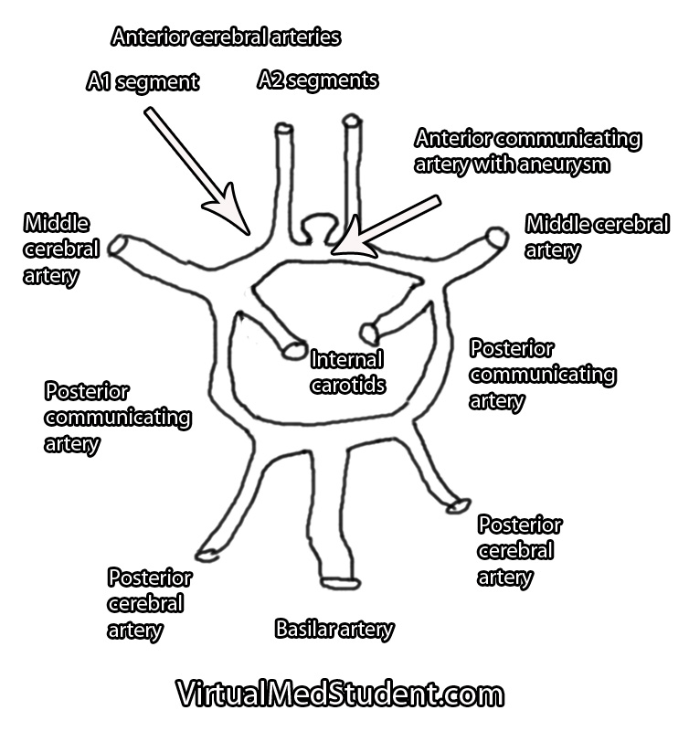
Each A1 segment branches into an A2 segment and into the anterior communicating artery. The anterior communicating artery connects each anterior cerebral arteries’ A1 segment together to form a circle (see schematic below).

The anterior communicating artery is one of the most common sites for intracranial aneurysm formation. Patients at risk for developing aneurysms include those with atherosclerosis, those with a family history of intracranial aneurysms, those with a history of hypertension or collagen vascular disease, and those with polycystic kidney disease. Smokers are also at a higher risk of developing aneurysms.

Basilar Tip Schematic Drawing
Anterior communicating artery aneurysms form when the lining of the vessel wall is thinned and the muscular layer of the blood vessel (tunica media) becomes weakened.

This thinning allows turbulent blood flow to form out-pouchings in the vessel wall. Typically these out-pouchings occur at points where blood vessels branch.

Signs and Symptoms

Anterior communicating artery aneurysms commonly present after a subarachnoid hemorrhage, which can cause a variety of signs and symptoms. The most common being a severe headache, although cranial nerve dysfunction, stroke, coma, and death can also occur.

Less commonly, aneurysms in this location can compress the optic chiasm or optic nerves leading to problems with vision.

How Do You Diagnose Aneurysms

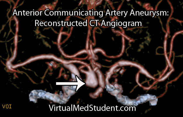
Anterior cerebral artery aneurysms are most commonly diagnosed after a subarachnoid hemorrhage when a patient presents with the "worst headache of their life". The best imaging methods for diagnosing these aneurysms are CT angiograms (see image below), MR angiograms, and formal cerebral angiograms.

Treatment

Like other intracranial aneurysms, anterior communicating artery aneurysms may be clipped or coiled. Clipping of an aneurysm involves an open surgical procedure where the surgeon dissects down to the aneurysm and places a clip across its neck. This excludes it from the circulation and prevents it from rupturing.

Anterior Communicating Artery Aneurysm CT Angiogram

Aneurysms may also be treated from inside the blood vessel. In this procedure a catheter is threaded from the femoral artery in the groin up towards the location of the aneurysm. Small metallic coils are placed within the dome of the aneurysm, which also excludes it from the normal circulation.

Regardless of how the aneurysm is treated – either with clipping or coiling – the end result is that the aneurysm is excluded from the normal circulation. This prevents it from rupturing.

The merits of clipping versus coiling are still under debate. Ultimately, the treatment depends on the size and location of the aneurysm, as well as other medical problems that the patient may have.

The Highlights…

Anterior communicating artery aneurysms are the most common intracranial aneurysm. They typically present after rupturing into the subarachnoid space and/or adjacent frontal lobes. They are diagnosed using CT angiograms or formal cerebral angiography. Treatment is with clipping and/or coiling.

Related Readings

Not Satisfied? More Reading on the Subject…

  • Bederson JB, Awad IA, Wiebers DO, et al. Recommendations for the management of patients with unruptured intracranial aneurysms. Stroke 2000;31:2742-2750.
  • li>Hunt WE, Hess RM. Surgical Risk as Related to Time of Intervention in the Repair of Intracranial Aneurysms. Journal of Neurosurgery 1968; 28:14-20.
  • Brisman JL, Song JK, Newell DW. Cerebral Aneurysms. NEJM 2006; 355:928-939.
  • Kumar V, Abbas AK, Fausto N. Robbins and Cotran Pathologic Basis of Disease. Seventh Edition. Philadelphia: Elsevier Saunders, 2004.
  • Frontera JA. Decision Making in Neurocritical Care. First Edition. New York: Thieme, 2009.
  • Greenberg MS. Handbook of Neurosurgery. Sixth Edition. New York: Thieme, 2006. Chapter 25.

Meningioma and the Arachnoid Cap Cell

A meningioma is a tumor that arises from the lining of the brain or spinal cord (ie: the “meninges”). They arise from cells known as “arachnoid cap cells”. Meningiomas are usually slow growing, “benign” tumors, which means that they are not usually considered cancerous in the strictest sense of the term.

When viewed under the light microscope, meningiomas can have many different appearances. The most common type is a dense sheet-like formation of cells interspersed with closely packed blood vessels. Sometimes the sheets of cells can be separated by connective tissue.

Frequently, meningiomas will have calcium deposits in them known as “psammoma bodies”. Uncommonly, meningioma cells may take on a malignant appearance characterized by increased cellular division (ie: “mitotic figures”) and invasion of the tumor cells into surrounding brain or bone.

Meningiomas test positive for epithelial membrane antigen (EMA) and vimentin (a marker of connective tissue). Ki-67 (a marker of proliferation) can be elevated in more aggressively behaving tumors.

The most common genetic abnormality seen in patients with meningiomas is found on chromosome 22. If present, a mutation in the NF2 gene on this chromosome causes type 2 neurofibromatosis. This disease predisposes individuals to developing multiple tumor types including meningiomas. Other less common genetic abnormalities can be seen on other chromosomes as well.

Signs and Symptoms

Due to their slow growing and benign nature, many meningiomas cause no symptoms. However, if they become too large or start to compress adjacent brain tissue they can cause headache, seizures, confusion, or visual problems. Spinal cord compression can result in myelopathy.

The most common location for a meningioma is in between the two hemispheres of the brain – the so called “parasagittal” location. Parasagittal meningiomas near the portion of the brain responsible for muscle movements may cause weakness of the opposite leg.

Diagnosis

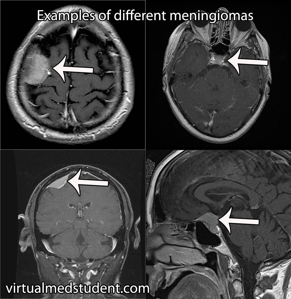
Diagnosis of meningioma can reliably be made on characteristic findings seen on CT or MRI scans. Interestingly, many meningiomas are found incidentally when a CT or MRI is done for other reasons.

Meningioma MRIs

However, like any other tumor, meningiomas can only be truly diagnosed once a specimen is sent to the pathology lab for analysis. Pathologists can reliably make the diagnosis based on typical histological features.

Meningiomas must be distinguished from a more malignant tumor known as a hemangiopericytoma. Hemangiopericytomas can look similar to meningiomas on imaging studies.

Dish Me Up Some Treatment Sir

Many meningiomas can be watched over time with repeat imaging studies; this is especially true if they are small and not causing neurological signs or symptoms.

On the other hand, large or symptomatic meningiomas require surgical resection. Many meningiomas can be removed completely. However, some meningiomas may be near vital structures such as the carotid artery, cranial nerves, or venous draining systems of the brain where complete surgical removal may be very difficult without causing significant neurologic impairment. In these cases the tumor is debulked as much as possible. The residual tumor can be followed or irradiated depending on the grade of meningioma.

Residual tumor after incomplete surgical resection, or meningiomas in difficult to access locations are candidates for radiation therapy. Many studies have shown long term growth control rates.

Overview

Meningiomas are considered “benign” tumors of the brain. They arise from arachnoid cap cells, which are located in a layer of the meninges (ie: the covering of the brain) known as the arachnoid. Symptoms include headache, weakness, vision problems, paresthesias (ie: abnormal sensations), amongst many other possible symptoms. Diagnosis can be made reliably from imaging studies such as CT or MRI. If symptomatic, or large, treatment is surgical resection. Small asymptomatic meningiomas can be managed with repeat imaging to assess for growth over time.

Related Articles

Curated References for Your Pleasure…

Cerebral Cavernous Malformations: Leaky Vessels

Cavernous malformations (aka: cavernomas or cavernous hemangiomas) can be thought of as vascular tumors. They are composed of a capillary-like network of endothelial cells (the cells that normally line blood vessels). However, unlike normal capillaries throughout the body, the capillaries of cavernomas can leak.

Interestingly, cavernous malformations do not have any brain tissue within them. This helps distinguish them from another related vascular abnormality known as an arteriovenous malformation.

The genetics of cavernous malformations have been elucidated by studying familial forms of the disorder. There are at least three known genetic defects that predispose patients to develop cavernomas. These genes appear to be important in the formation of blood vessels (a process known as "angiogenesis") and the blood brain barrier. Therefore, mutations in these genes can cause the abnormal growth of vascular tissue.

What Havock Do These Guys Cause?

Cavernomas can cause numerous signs and symptoms depending on their location within the brain. Seizures are the most common symptom. However, progressive neurological impairment such as worsening weakness can also occur.

Sometimes cavernomas can block the flow of cerebrospinal fluid leading to hydrocephalus. Hydrocephalus can cause increased intracranial pressure leading to headaches, nausea, and vomiting. However, it is important to realize that many patients with cavernomas have no symptoms at all!

If symptoms are present, they tend to progress over time. This is because cavernous malformations bleed and re-bleed resulting in an expansion of its size over time. As the malformation increases in size it can push on adjacent brain tissue causing worsening symptoms.

Unlike arteriovenous malformations, life threatening and severe hemorrhages are rare. Cavernomas bleed at an initial yearly rate of anywhere between 1% to 5%. After the first bleed, the risk of re-bleeding increases to as high as 10% per year. In other words, if a cavernoma bleeds, it is more likely to re-bleed at a later date.

How Do You Diagnose These Buggers?

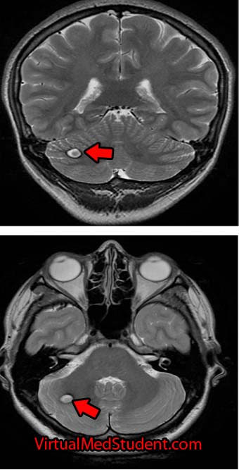
Cavernoma Marked

The diagnosis of cavernous malformations are made via imaging studies. They are usually detected via MRIs that are ordered for evaluation of neurological symptoms. Cavernomas are seen best on T2 and gradient echo MRI sequences. They typically look like a piece of popcorn.

Select patients undergo a more invasive imaging procedure known as angiography. Angiography is used to rule out another similar lesion known as an arteriovenous malformation. Since cavernomas are venous malformations they are not seen on angiograms.

How Are These Treated?

Depending on the location, most cavernous malformations in the brain or spinal cord are removed surgically. Some institutions offer radiation as a means of treatment, especially in difficult to access areas (ie: where the risk of surgical removal is very high).

Let’s Recap this MoFo

Cavernous malformations of the brain are abnormal vascular growths composed of capillary networks. They are likely the result of genetic mutations in genes responsible for blood vessel growth. Depending on their location they can cause numerous neurological symptoms such as seizures and weakness. MRI often shows the characteristic “popcorn” lesion. Treatment is usually with surgical resection, although some cavernomas may be radiated depending on their location.

Related Articles

Not Satisfied? More References and Resources…

Cerebral Ateriovenous Malformations: A Disease of Eloquence

A cerebral arteriovenous malformation is an abnormal tangle of blood vessels within the brain.

In order to understand these tangles we have to first understand normal blood flow. Blood flows from arteries to smaller arteries and then into capillary beds. In the capillary beds, gas, nutrients, and "wastes" are exchanged between the blood and adjacent body tissue. Once past the capillaries, the blood drains into successively larger veins where it eventually returns to the heart to be re-oxygenated.

In arteriovenous malformations there are no capillaries. Because of this, blood is shunted from the high pressure arterial system directly into the low pressure venous system. The "shunted" blood is unable to deliver its nutrients or oxygen to the nearby brain.

The risk of an arteriovenous malformation rupturing is relatively high because the pressure of arterial blood is "banging" into the walls of low pressure veins. The body tries to compensate for this by "arterializing" the blood vessels associated with the AVM.

The term "nidus" is often used to describe the center of the malformation. This is the point where the arterial feeding vessels meet the draining venous structures.

In addition, any brain tissue around, or within the AVM is usually gliotic (a term used to describe scarring within the brain). Macrophages are sometimes present and are usually there to "gobble up" hemosiderin (a breakdown product of blood).

Signs and Symptoms

The signs and symptoms of cerebral arteriovenous malformations are dependent on the location of the malformation.

Most patients discover they have an AVM after it bleeds into the surrounding brain tissue. Patients can present with everything from a mild headache to a severe neurological deficit depending on the location and size of the malformation.

In addition, AVMs may cause transient neurological symptoms. These transient symptoms are caused by blood being shunted away from the surrounding normal brain tissue. Again, the location of the AVM dictates what symptoms may develop (ie: weakness if near the motor strip, difficulty with speech if located near Wernicke’s or Broca’s area, balance problems if in the cerebellum, disturbances in sensation if in the parietal lobe, etc., etc.).

Patients may also present with seizures as a result of irritation of the surrounding cortex by hemosiderin (a breakdown product of blood). In fact, seizures are the second most common presenting symptom.

Interestingly, headache is an uncommon symptom of arteriovenous malformations.

Diagnosis and Classification

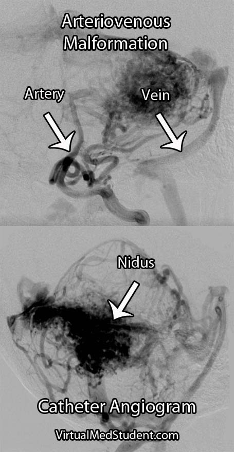
Cerebral Arteriovenous Malformation
Diagnosis is made with special imaging studies like CT angiography, MR angiography, and formal catheter angiography (formal angiography is the gold standard).

AVMs are characterized by an abnormal tangle of blood vessels. The tell tale sign of an AVM on an angiogram is that both arterial and venous structures are seen at the same time (normally the venous phase follows the arterial phase).

The Spetzler-Martin grading system helps guide treatment decisions. This system takes into account the size, location, and type of venous drainage (see the first reference below).

Treatment

Treatment is highly individualized. There are currently three accepted treatment strategies: surgery, radiation, and embolization.

Surgery is still the treatment of choice, especially for AVMs near the surface of the brain or in non-eloquent cortex. Surgery is also considered "definitive" therapy (ie: the AVM is removed all at once), which is ideal for lesions considered high risk for rupture. Patient’s with deep seated lesions (ie: basal ganglia, thalamus, etc.), or those located in very "eloquent" cortical areas may be better treated with radiation or embolization.

Radiation works by causing changes in the vessels of the AVM. Over the course of several months to years the vessels are "cooked" by the radiation. This effectively eliminates blood flow into the AVM. Since the effects of radiation take months to years to shut down the AVM, the patient remains at risk for rupture. In addition, side effects from radiation may be permanent in a small percentage of patients.

Embolization is usually used as an adjunct to surgical resection. During embolization, various substances are injected into the AVM. These substances deprive the AVM of its arterial blood flow. This can be very useful prior to surgery to help with intra-operative blood loss (especially for very large AVMs!). Embolization is less commonly used as a stand alone treatment.

Overview

Arteriovenous malformations are abnormal tangles of blood vessels within the brain tissue. They have no intervening capillary bed so arterial blood flows directly into dilated veins. The main risk of an arteriovenous malformation is when it ruptures and bleeds into the surrounding brain. They can cause numerous signs and symptoms depending on their location. They are diagnosed with CT angiograms, MR angiograms, or formal catheter angiograms. Treatment is with surgery, radiation, and/or embolization depending on the risk of rupture and the location of the lesion.

Other Interesting Neurovascular Diseases…

References and Resources

  • Spetzler RF, Martin NA. A proposed grading system for arteriovenous malformations. J Neurosurg. 1986 Oct;65(4):476-83.
  • Ding D, Yen CP, Xu Z, et al. Radiosurgery for patients with unruptured intracranial arteriovenous malformations. J Neurosurg. 2013 May;118(5):958-66
  • Fokas E, Henzel M, Wittig A, et al. Stereotactic radiosurgery of cerebral arteriovenous malformations: long-term follow-up in 164 patients of a single institution. J Neurol. 2013 May 28.
  • Albuquerque FC, Ducruet AF, Crowley RW, et al. Transvenous to arterial Onyx embolization. J Neurointerv Surg. 2013 Mar 6.
  • Nataraj A, Mohamed MB, Gholkar A, et al. Multimodality Treatment of Cerebral Arteriovenous Malformations. World Neurosurg. 2013 Feb 20.

Subarachnoid Hemorrhage: Aneurysms, Vasospasm, and Hyponatremia

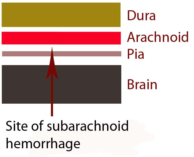
In order to understand subarachnoid hemorrhage we have to first appreciate the layers that make up the brain and its surrounding tissues. The brain itself has three protective layers: dura mater, the arachnoid, and the pia.

The dura is a thick layer of fibrous tissue immediately below the skull. Below the dura is the arachnoid, which is a layer of delicate web-like tissue (hence the name "arachnoid"). Finally, below the arachnoid is the pia mater. The pia is a very thin layer that is directly adjacent to brain tissue.

The subarachnoid space, or the region between the arachnoid tissue and the pia contains cerebrospinal fluid, which acts like a liquid shock absorber for the brain. Also contained within the subarachnoid space are blood vessels that penetrate down into the brain tissue. Sometimes these blood vessels "leak", which can cause a "sub-arachnoid" hemorrhage.

Brain Layers

The most common cause of subarachnoid hemorrhage is traumatic injury; the most common non-traumatic cause is a ruptured aneurysm.

An aneurysm is an abnormal ballooning out of a blood vessel’s wall. The balloon’s dome is much weaker than the rest of the vessel wall. These weak areas can rupture allowing blood to leak out of into the subarachnoid space.

Other causes of subarachnoid hemorrhage include idiopathic (ie: unknown) causes, arteriovenous malformations, vessel dissections, and very rarely tumors. Regardless of the cause, blood will pool in the subarachnoid space.

The remainder of this article will focus on the most common non-traumatic cause of subarachnoid hemorrhage – aneurysm rupture.

Signs and Symptoms

The classic symptom of a subarachnoid hemorrhage is a horrific headache described as the “worst headache of their life". Photophobia, nausea, vomiting, and nuchal rigidity are also common. Seizures may also occur. In addition, depending on how severe the subarachnoid hemorrhage is, patients may have decreased levels of consciousness; some patients become comatose, and many die before reaching medical attention.

The patient’s clinical status is graded according to the Hunt and Hess system. It only applies to patients in whom subarachnoid hemorrhage is caused by rupture of an aneurysm. This grading system was initially established to help determine mortality and clinical outcomes. In modern practice, these numbers are likely high given modern improvements in critical care and neurosurgical intervention since Hunt and Hess first developed their grading system.

Hunt and Hess Clinical Grading Scale
Grade Patient’s Clinical Status Associated Mortality
1 Mild headache and/or nuchal rigidity 1%
2 Cranial nerve dysfunction, moderate to severe headache and/or nuchal rigidity 5%
3 Mild focal neurological deficit, lethargic, confused 19%
4 Stuporous, moderate to severe hemiparesis, early decerebrate posturing 40%
5 Coma, decerebrate posturing 77%

The world federation of neurological surgeons also has a clinical score based on the Glasgow Coma Scale (GCS). It associates the patient’s GCS with the likelihood of death.

World Federation of Neurological Surgeons Grading System
  GCS Major focal deficit Mortality
1 15 No 5%
2 13-14 No 9%
3 13-14 Yes or No 20%
4 7-12 Yes or No 33%
5 3-6 Yes or No 77%

It is very important to think about the possibility of subarachnoid hemorrhage in patients presenting with these signs and symptoms. Prompt diagnosis and treatment is necessary in order to prevent devastating consequences!

Complications

Blood in the subarachnoid space is very irritating to the brain and cerebral blood vessels. Because of this, several complications can occur.

One of the most common complications is known as "vasospasm." Vasospasm occurs when the blood vessels of the brain spasm several days after the initial hemorrhage. When this occurs blood is no longer able to flow past the blockage; if this occurs for a long enough period of time a stroke can occur. The peak period for vasospasm occurs between 3 and 14 days after the initial bleed.

Another complication of subarachnoid hemorrhage is known as cerebral salt wasting. This occurs when a patient urinates excessive amounts of sodium causing the blood level of sodium to drop precipitously. Because of the excessive urination the patient also becomes dehydrated. Aggressive fluid and salt resuscitation must be given to prevent profound hyponatremia (ie: decreased sodium levels in the blood), which can cause seizures, coma, and death.

In addition, for unknown reasons, many patients with subarachnoid hemorrhage also shower their cerebral hemispheres with micro-thrombi (ie: clots), which can lead to many small strokes. The reason why patients with subarachnoid hemorrhage become coagulopathic is still an area of intense research.

Diagnosis

Subarachnoid hemorrhage is most commonly diagnosed by head CT. CT scans are fast and readily pick up the extravasated blood, which layers in the subarachnoid space (see image below). If there is a high clinical index of suspicion but CT of the head is negative than a lumbar puncture should be performed. If the spinal fluid has xanthochromima (a product of red blood cell breakdown) this is highly concerning for subarachnoid hemorrhage.
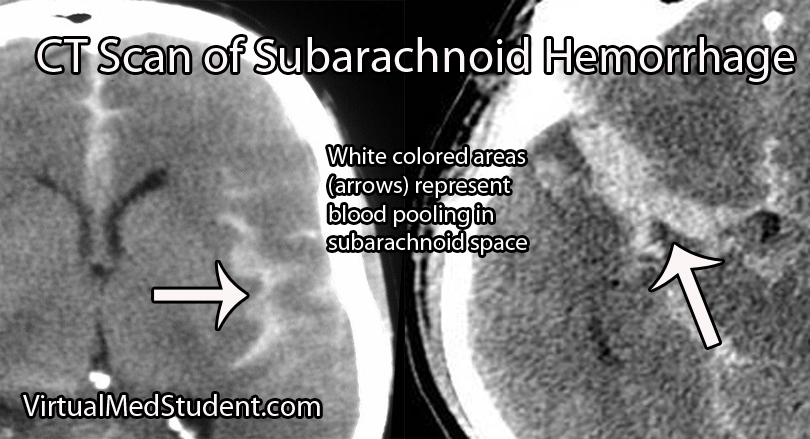
CT Scan of Subarachnoid Hemorrhage

Subarachnoid hemorrhage is not a diagnosis per say, but rather the result of some underlying pathology (ie: aneurysm, trauma, etc.). Many of these pathologies are treatable; therefore, it is important to figure out what caused the subarachnoid hemorrhage.

Since many are the result of ruptured aneurysms there are several other tests that are often done. The first test is a CT angiogram (CTA). In this test a radio-opaque material is injected into the blood vessels and a CT is performed.

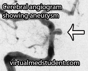
Cererbral Angiogram with Aneurysm
Dye in the dome of the aneurysm will appear as an abnormality helping to confirm the presence, and more importantly the location of the aneurysm.

A more invasive procedure known as a "cerebral angiogram" (image to the right) is also often performed.

In this test, a catheter is inserted into blood vessels in the groin and then threaded up into the blood vessels of the brain. Radio-opaque material is injected and x-rays can pick up abnormalities in the vessel.

The benefit of doing a cerebral angiogram is that it is diagnostic, and treatment can frequently be offered through the catheter itself.

Treatment

There are three main components of treating a subarachnoid hemorrhage: treating the underlying cause, preventing a "re-bleed", and preventing secondary complications.

Since many subarachnoid hemorrhages are caused by aneurysm rupture we’ll discuss the treatment for this common cause. Aneurysms are treated either “open” or “closed”.

“Open” refers to a surgical procedure in which part of the skull is removed. The surgeon then dissects down to the aneurysm. Once identified, a clip is placed around the neck of the aneurysm (ie: you can think of the clip as putting a knot in the neck of a balloon). This stops blood from flowing into the aneurysm, and therefore prevents re-rupture.

“Closed” treatment refers to endovascular technology in which a small micro-catheter is threaded from the blood vessels in the groin into the cerebral vasculature. The aneurysm is located via angiogram. Through a hole in the microcatheter tip the physician then fills the aneurysm dome with small metallic coils.

Once blood is in the subarachnoid space secondary complications often result. One of these complications is referred to as "vasospasm". Medications such as oral nimodipine and intra-arterial nifedipine are used to reduce the amount of vasospasm by inhibiting smooth muscle contraction in the wall of the blood vessel.

Overview

Subarachnoid hemorrhage occurs most commonly after an intracerebral aneurysm ruptures, although other causes exist. Regardless of the cause, blood spills out into the subarachnoid space. Symptoms include a horrible headache, focal neurological deficits (ie: weakness, difficulty speaking, etc.), and coma. If an aneurysm is the cause, it is secured with clipping or coiling. Preventing secondary complications such as vasospasm is also an important component of treatment.

References and Resources
Glioblastoma: A Real Beast of a Tumor

In order to understand what a glioblastoma is we have to first appreciate the different cell types that compose healthy brain tissue. Brain tissue has both neurons and glia. Neurons are the “action” cells of the brain. Glia are the “helper” cells of the brain. They ensure that neurons stay healthy.

A glioblastoma is a malignant brain tumor that arises from a specific type of glial cell known as an astrocyte. Glioblastomas are not only the most common astrocytic tumor, but they are also the most common primary brain tumor!

It is important to realize that there are less malignant tumors that arise from astrocytes (discussed in other articles). Many of these tumors have a much better prognosis, which is why it is important to distinguish glioblastoma from less malignant behaving tumors.

The first distinguishing characteristic is neovascularization. Neovascularization is a fancy medical term used to describe the proliferation of blood vessels within the tumor. As the tumor grows, it requires new vessels to feed it oxygen and nutrients; the process of neovascularization allows the tumor to obtain these essential factors so that it can continue to grow.

Interestingly, as the tumor expands, sections of it will get choked off from its own blood supply. The end result is that part(s) of the tumor actually dies. This is referred to as "necrosis", which is a common finding in glioblastoma.

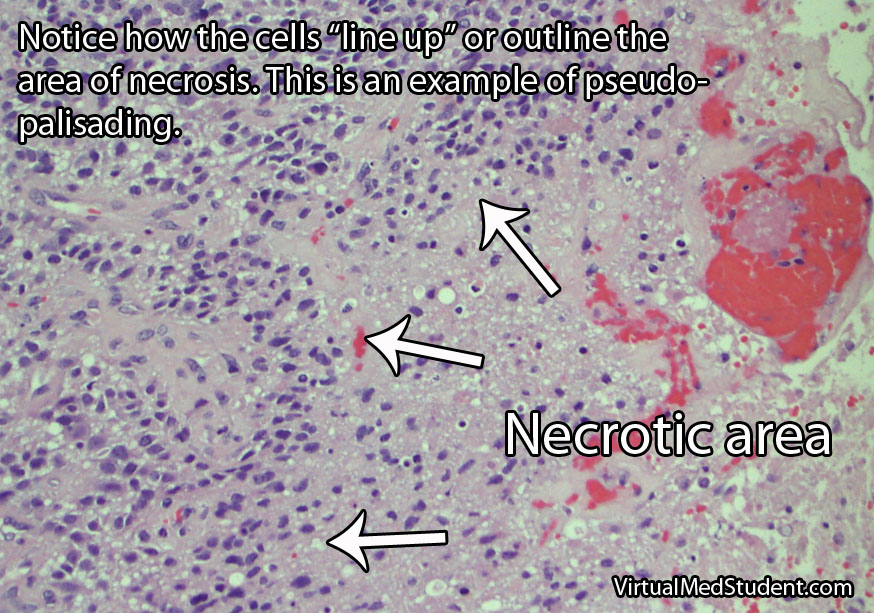
One of the most distinguishing features of glioblastomas is when cancerous astrocytes "line up" and outline areas of necrosis in a process known as pseudopalisading (see image below).

Given the malignant nature of glioblastoma it is common to see many mitotic figures. Mitotic figures are cells in various states of cell division; these figures indicate a relatively rapidly growing tumor type.

Glioblastoma Pathology - Pseudopalisading

Glibolastoma is therefore characterized by the following pathological characteristics: prominent microvascular proliferation (ie: development of new blood vessels within the tumor), mitosis (ie: an indicator of cell division/growth), and necrosis (ie: areas of dead tumor); pseudopalisading necrosis is a specific form of necrosis shown in the above image that is a hallmark of glioblastomas.

Signs and Symptoms

Glioblastomas may present with any number of signs and/or symptoms depending on their location within the brain. Lesions that are located on the left side of the brain may cause problems with speech if they involve the Broca or Wernicke areas. Tumors in the areas of the brain that control motor movement may cause weakness. Additionally, tumors that arise in the frontal lobes may cause odd behavioral changes. Some patients present with seizures, and others with only a dull headache.

Diagnosis

An official diagnosis of glioblastoma can only be made when a pathologist looks at a sample of the tumor under a microscope. These samples are typically obtained by a neurosurgeon who resects or biopsies the tumor.

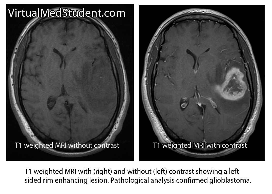
However, glioblastomas also have typical features seen on imaging studies such as MRI. For example, these tumors will “rim-enhance” when a contrast material such as gadolinium is infused into the patient during the scan. Rim enhancement is a result of the contrast material leaking out of all of the blood vessels present within the tumor. It is important to note that other diseases such as abscesses, lymphomas, and other infections can also cause rim-enhancement.

MRI of glioblastoma

Another useful study known as MR spectroscopy measures the relative amounts of different molecules present within the tumomr. In a glioblastoma the amount of lactate, choline, and lipid are all increased. Lactate is a marker of brain tissue that is not receiving enough oxygen, which is common in necrotic tumor areas. Choline is a molecule that is present in cell membranes. When neurons are rapidly dividing, which is what occurs in glioblastoma, the amount of choline present also increases. A different molecule known as N-acetyl aspartate (NAA) is present in mature cells. Therefore, unlike lactate and choline levels, NAA is decreased in glioblastoma because these cells are "immature" (ie: poorly differentiated).

Treatment

Treatment combines a mixture of surgery, radiation therapy, and different chemotherapeutic drugs, the most common being temozolomide (Temodar®). Surgery is only useful when a significant amount of the tumor can be removed. Despite optimal treatment the prognosis for patients with glioblastoma remains extremely poor.

It is also highly important to treat the edema that frequently surrounds the tumor. Steroids, most commonly dexamethasone (Decadron®), are used to decrease the amount of edema, which usually improves symptoms.

Patients are often started on an anti-seizure medication such as levetiracetam (Keppra®) or phenytoin (Dilantin®).

Overview

Glioblastoma is a malignant astrocytic tumor. It is the most common primary brain tumor. It has unique characteristics that distinguish it from more benign brain tumors that also arise from astrocytes. It is treated with a combination of surgery, radiation, and chemotherapy.

References and Resources