Hemangioblastoma of the Central Nervous System

Hemangioblastomas are benign tumors that occur in the central nervous system, most commonly in the cerebellum. The exact cell from which they arise is unknown; however, they are believed to be meningeal (ie: the cells that make up the covering of the brain) in origin.

Hemangioblastomas can be solid or cystic in appearance. However, all hemangioblastomas have capillary networks that are lined by endothelial cells. Interspersed between the capillaries are pericytes and stromal cells with a polygonal appearance.

The arrangement of these various cell types can occur in three different architectures: juvenile, clear cell, or transitional. Each architecture has a specific ratio of capillaries to stroma (connective tissue), with the juvenile type having the greatest amount of capillary tissue, and the clear cell type having the greatest amount of stromal tissue.

About a quarter of cases are related to a genetic disorder known as von Hippel-Lindau syndrome. von Hippel-Lindau syndrome is the result of a genetic mutation in the VHL gene on chromosome 3. The protein product of this gene is a tumor suppressor; when mutated it is unable to suppress the abnormal growth of tumor cells. As a result, patients with von Hippel-Lindau syndrome develop hemangioblastomas of the brain and retina, renal cell carcinoma, and other tumor types.

Hemangioblastomas are most commonly found in the cerebellum, but on occassion will affect the cervical spinal cord and brainstem. They are most commonly seen in males starting at around 20 years of age.

Signs and Symptoms

Patients with cerebellar hemangioblastomas present with numerous signs and symptoms. Many patients will complain of headache, nausea, and vomiting. This is often due to the tumor compressing the cerebral aqueduct, which causes cerebrospinal fluid to “back up” in the brain leading to hydrocephalus and increased pressure inside the head.

In addition, many people with hemangioblastomas will have evidence of cerebellar dysfunction on physical exam. These signs include ataxia (ie: wobbly gait), dysmetria (ie: uncoordinated movements of the limbs), and dysdiadochokinesia (ie: difficulty repeating rapid alternating movements).

If the tumor is present in the spinal cord, symptoms may include weakness, spasticity, numbness, or other sensory changes.

Interestingly, hemangioblastomas can secrete an analogue of the hormone erythropoietin. This hormone causes bone marrow to pump out more red blood cells. As a result, some patients may have an increased number of red blood cells; this is known as polycythemia.

Diagnosis
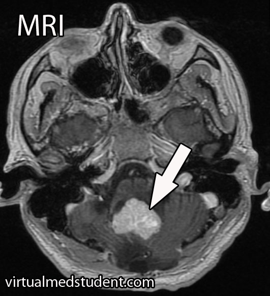
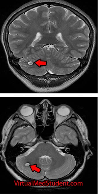
Hemangioblastoma MRI
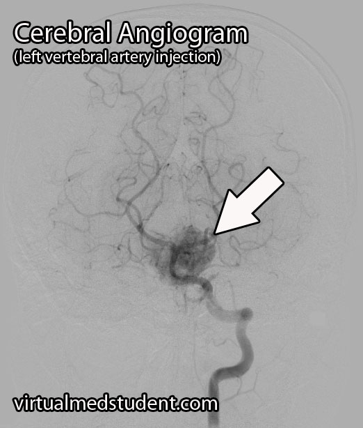
Hemangioblastoma Angiogram
A presumptive diagnosis can be made using epidemiology and imaging studies.

A tumor located in the cerebellum of an adult with certain characteristics on CT, MRI, and cerebral angiogram can make the diagnosis of hemangioblastoma likely.

However, the final diagnosis can only be made by looking at a sample of the tumor under the pathology microscope.

Treating These Bastards

Definitive treatment is surgical resection. However, hemangioblastomas can be highly vascular, which means they tend to bleed like stink! Therefore, preoperative embolization by an interventional neuro-radiologist can decrease the amount of bleeding that occurs during surgery.

Radiation therapy is also sometimes used as an adjunctive treatment. It may help slow the growth of the tumor, but will not cure it. Radiation therapy can be used in patients who are unable to undergo surgical resection, or if the tumor is inaccessible via traditional surgical means.

Recap It All…

Hemangioblastomas are highly vascular, but benign central nervous system tumors of undetermined origin. They are associated with von Hippel-Lindau syndrome. Signs and symptoms include headache, nausea, and vomiting, as well as cerebellar dysfunction. Diagnosis is based on imaging and pathological findings at the time of surgical resection. Surgery is the treatment of choice, although embolization and radiation therapy may also be used as an adjunct.

More Interesting Neurological Problems…

Some More Expert Ideas Below

Cerebral Cavernous Malformations: Leaky Vessels

Cavernous malformations (aka: cavernomas or cavernous hemangiomas) can be thought of as vascular tumors. They are composed of a capillary-like network of endothelial cells (the cells that normally line blood vessels). However, unlike normal capillaries throughout the body, the capillaries of cavernomas can leak.

Interestingly, cavernous malformations do not have any brain tissue within them. This helps distinguish them from another related vascular abnormality known as an arteriovenous malformation.

The genetics of cavernous malformations have been elucidated by studying familial forms of the disorder. There are at least three known genetic defects that predispose patients to develop cavernomas. These genes appear to be important in the formation of blood vessels (a process known as "angiogenesis") and the blood brain barrier. Therefore, mutations in these genes can cause the abnormal growth of vascular tissue.

What Havock Do These Guys Cause?

Cavernomas can cause numerous signs and symptoms depending on their location within the brain. Seizures are the most common symptom. However, progressive neurological impairment such as worsening weakness can also occur.

Sometimes cavernomas can block the flow of cerebrospinal fluid leading to hydrocephalus. Hydrocephalus can cause increased intracranial pressure leading to headaches, nausea, and vomiting. However, it is important to realize that many patients with cavernomas have no symptoms at all!

If symptoms are present, they tend to progress over time. This is because cavernous malformations bleed and re-bleed resulting in an expansion of its size over time. As the malformation increases in size it can push on adjacent brain tissue causing worsening symptoms.

Unlike arteriovenous malformations, life threatening and severe hemorrhages are rare. Cavernomas bleed at an initial yearly rate of anywhere between 1% to 5%. After the first bleed, the risk of re-bleeding increases to as high as 10% per year. In other words, if a cavernoma bleeds, it is more likely to re-bleed at a later date.

How Do You Diagnose These Buggers?

Cavernoma Marked

The diagnosis of cavernous malformations are made via imaging studies. They are usually detected via MRIs that are ordered for evaluation of neurological symptoms. Cavernomas are seen best on T2 and gradient echo MRI sequences. They typically look like a piece of popcorn.

Select patients undergo a more invasive imaging procedure known as angiography. Angiography is used to rule out another similar lesion known as an arteriovenous malformation. Since cavernomas are venous malformations they are not seen on angiograms.

How Are These Treated?

Depending on the location, most cavernous malformations in the brain or spinal cord are removed surgically. Some institutions offer radiation as a means of treatment, especially in difficult to access areas (ie: where the risk of surgical removal is very high).

Let’s Recap this MoFo

Cavernous malformations of the brain are abnormal vascular growths composed of capillary networks. They are likely the result of genetic mutations in genes responsible for blood vessel growth. Depending on their location they can cause numerous neurological symptoms such as seizures and weakness. MRI often shows the characteristic “popcorn” lesion. Treatment is usually with surgical resection, although some cavernomas may be radiated depending on their location.

Related Articles

Not Satisfied? More References and Resources…

Spinal Shock and Neurogenic Shock: The Battle Begins

Endless hours of studying, relentless exams, and the never-ending confusion between two perplexing phenomena: spinal shock and neurogenic shock. So, buckle up (so you don’t end up in spinal shock) and prepare for the journey through the world of spinal and neurogenic shock as we break down their differences.

The Tale of Two Shocks

Picture this: you’re in the ER, and a patient comes in with a recent spinal cord injury. You rack your brain to differentiate between spinal shock and neurogenic shock, but all you can remember is the cranial nerves mnemonic and your chief resident is about to pimp the you know what out of you… Let’s make it easy!

  • Spinal Shock: Think of it as your body’s initial reaction to “breaking up” with your spinal cord. It’s a temporary and unexpected “break-up shock” that leaves your reflexes, motor function, and sensation feeling lost and numb (physically, not emotionally) below the level of injury.
  • Neurogenic Shock: Now, imagine your body losing its balance between the sympathetic and parasympathetic nervous systems after a spinal cord injury. It’s like a tug-of-war, but the parasympathetic system wins, causing blood vessels to dilate and blood pressure to drop. Your heart, not knowing how to cope, slows down in response (bradycardia). Neurogenic shock occurs because the descending sympathetic fibers of the cord are injured, whereas the parasympathetic supply to the body provided by the vagus nerve (ie: “wandering nerve”) off of the brainstem is still intact and is now un-inhibited.

Diagnostic Dilemmas: The Hints are There You Just Have to Look

When you’re trying to diagnose spinal or neurogenic shock, look for these clues:

  • Spinal Shock: Your patient’s reflexes have gone on vacation, and they’re not telling you when they’ll be back. The muscles are flaccid, and sensations are playing hide-and-seek. But fear not, because those reflexes will eventually return. The delayed plantar response and bulbocavernosus reflex (don’t ask us how someone figured this one out!) are two of the earlier reflexes to come back from vacation. There is a lot of debate about how to define spinal shock and how long spinal shock lasts. However, you cannot prognosticate about the severity of cord injury until at least one reflex has returned.
  • Neurogenic Shock: Here, your patient’s blood pressure is lower than your motivation on a Monday morning, and their heart rate is slower than a sloth doing yoga. The skin may be warm and dry, resembling a cozy blanket you wish you were under instead of being in the ER.

Treatments: The Cures

Now that you’ve (hopefully) identified which shock you’re dealing with, it’s time to strategize and take action:

  • Spinal Shock: When faced with this shock, channel your inner superhero and protect the injured spinal cord at all costs! Immobilize the spine, maintain blood pressure, and ensure proper oxygenation to minimize further damage. Neurosurgical consultation is often indicated if the spinal column is unstable and requires surgical fixation.
  • Neurogenic Shock: Roll up your sleeves and get ready for some serious hemodynamic management. Rehydrate your patient with IV fluids, bring out the vasopressors to constrict those dilated blood vessels, and, if necessary, consider a temporary pacemaker to speed up the slow-motion heart rate.

Remember that both neurogenic and spinal shock are often occuring at the same time! Additionally, remember that neurogenic shock can also co-exist with other types of shock like hypovolemic shock in polytrauma patients.

As future health practitioners, you’ll face many confusing and challenging scenarios, like differentiating between spinal shock and neurogenic shock. But remember, amidst the stress, it’s essential to find some humor and light-heartedness. After all, laughter is the best medicine, and knowing the difference between these two conditions will not only help your patients, but also save you from those embarrassing moments during rounds. So, hold your head high, and step into the world of medicine with a smile on your face and the ability to distinguish between spinal and neurogenic shock in your ever-expanding medical knowledge toolbox.

More Fun Spinal Cord Pathology

References and Resources

The Internal Capsule: Some Pricey Brain Real Estate

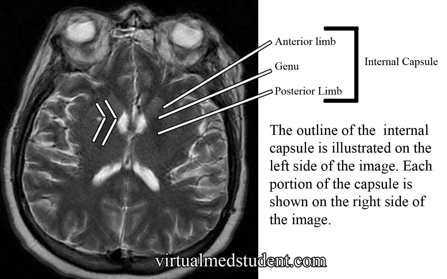
The internal capsule is one pricey piece of brain real estate! It contains all of the pathways that allow information to be transferred between the cerebral cortex and the spinal cord, brainstem, and subcortical structures (ie: thalamus, basal ganglia). It is divided into an anterior limb, posterior limb, and genu (ie: the area where the anterior and posterior limbs meet).

The anterior limb contains axons that send information between the thalamus and the cingulate gyrus and pre-frontal cortex. It also contains axons in the frontopontine pathway (ie: axons going from the frontal cortex to a portion of the brainstem known as the pons).

The genu contains the corticobulbar tract, which originate in the motor areas of the frontal lobes and extend to the cranial nerve nuclei in the brainstem. It also contains axons that connect the motor section of the thalamus (ie: VA and VL nuclei) with the motor areas of the frontal cortex.

The posterior limb contains the corticospinal tract, which are axons that come from the motor area of the frontal cortex and travel all the way to the anterior horns of the spinal cord where α-motor neurons are located. The posterior limb also contains sensory information coming from the body via the medial lemniscus and the anterolateral (aka: spinothalamic tract) systems.

Internal Capsule MRI

The blood supply to most of the internal capsule comes from the lenticulostriate arteries. These small arteries originate from the first portion of the middle cerebral artery. Two other important arteries also supply portions of the internal capsule: the anterior choroidal artery and the recurrent artery of Heubner. The anterior choroidal artery is a branch of the internal carotid. It supplies the inferior portion of the posterior limb. The recurrent artery of Heubner is a branch of the anterior cerebral artery. It supplies the inferior portions of the anterior limb and the genu.

Anatomy of the Internal Capsule
Division Major Communication Tracts Blood Supply
Anterior limb

– Tracts between the frontal lobe and pons (brainstem)

– Tracts between the thalamus and prefrontal cortex

– Tracts between the thalamus and cingulate gyrus

– Lenticulostriate arteries (branches of the middle cerebral artery)

– Recurrent artery of Heubner (branch of the anterior cerebral artery)

Genu – Tracts between the motor cortex in the frontal lobe and the cranial nerve nuclei in the brainstem (aka: corticobulbar tract)

– Lenticulostriate arteries (branches of the middle cerebral artery)

– Recurrent artery of Heubner (branch of the anterior cerebral artery)
Posterior limb

– Tracts between the motor cortex of frontal lobe and anterior horn of spinal cord (aka: corticospinal tract)

– Medial lemniscus tract (a continuation of the dorsal columns), which carries information about light touch, vibration, and pressure sensation from the body and spinal cord.

– Anterolateral (aka: spinothalamic) tract, which carries pain and temperature information

– Lenticulostriate arteries (branches of the middle cerebral artery)

– Anterior choroidal artery (branch of the internal carotid)

Importance in Disease

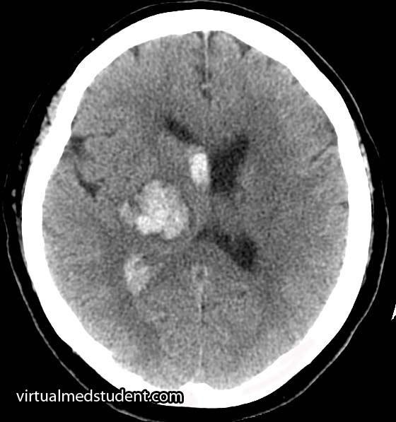
Thalamic Hemorrhage
Thalamic intracerebral hematoma
compressing the posterior limb
of the internal capsule

Damage to the internal capsule can be devastating neurologically because it contains so many vital tracts.

For example, a stroke of the anterior choroidal artery can lead to posterior limb damage. This can cause paralysis of the contralateral (ie: opposite) arm and leg secondary to interruption of the corticospinal tract.

Posterior limb disruption can also cause co-existent sensory deficits including an inability to feel light touch, pain, and temperature due to damage of the spinothalamic and medial lemniscal pathways.

Hypertensive hemorrhages in the thalamus or basal ganglia can compress the adjacent fibers of the internal capsule leading to similar clinical findings. The head CT to the right shows a thalamic hemorrhage secondary to severely elevated blood pressure. The patient had compression of the posterior limb of the internal capsule. As a result she was unable to move her left arm and leg, and could not feel pain or light touch on the left side of her body.

Overview

The internal capsule is a vital structure. It contains many communication pathways between the brain’s cortex, brainstem, spinal cord, and subcortical nuclei (ie: thalamus, basal ganglia). Its blood supply comes from branches of the middle cerebral artery (ie: lenticulostriates), anterior cerebral artery (ie: recurrent artery of Heubner), and the internal carotid (ie: anterior choroidal artery). Lesions in this area caused by strokes or hypertensive hemorrhages can have devastating clinical consequences.

Other Pertinent Articles…

References and Resources

  • Greenberg MS. Handbook of Neurosurgery. 9th Edition. New York: Thieme, 2006. Chapter 25.
  • Chowdhury F, Haque M, Sarkar M, et al. White fiber dissection of brain; the internal capsule: a cadaveric study. Turk Neurosurg. 2010 Jul;20(3):314-22. doi: 10.5137/1019-5149.JTN.3052-10.2.
  • Simon RP, Aminoff MJ, Greenberg DA. Clinical Neurology, Seventh Edition (LANGE Clinical Medicine). Seventh Edition. New York: McGraw Hill, 2009.
  • Nolte J. The Human Brain: An Introduction to its Functional Anatomy. Sixth Edition. Philadelphia: Mosby, 2008.
  • Bickley LS, Szilagyi PG. Bates’ Guide to Physical Examination and History Taking. Ninth Edition. New York: Lippincott Williams and Wilkins, 2007.