Glioblastoma: A Real Beast of a Tumor

In order to understand what a glioblastoma is we have to first appreciate the different cell types that compose healthy brain tissue. Brain tissue has both neurons and glia. Neurons are the “action” cells of the brain. Glia are the “helper” cells of the brain. They ensure that neurons stay healthy.

A glioblastoma is a malignant brain tumor that arises from a specific type of glial cell known as an astrocyte. Glioblastomas are not only the most common astrocytic tumor, but they are also the most common primary brain tumor!

It is important to realize that there are less malignant tumors that arise from astrocytes (discussed in other articles). Many of these tumors have a much better prognosis, which is why it is important to distinguish glioblastoma from less malignant behaving tumors.

The first distinguishing characteristic is neovascularization. Neovascularization is a fancy medical term used to describe the proliferation of blood vessels within the tumor. As the tumor grows, it requires new vessels to feed it oxygen and nutrients; the process of neovascularization allows the tumor to obtain these essential factors so that it can continue to grow.

Interestingly, as the tumor expands, sections of it will get choked off from its own blood supply. The end result is that part(s) of the tumor actually dies. This is referred to as "necrosis", which is a common finding in glioblastoma.

One of the most distinguishing features of glioblastomas is when cancerous astrocytes "line up" and outline areas of necrosis in a process known as pseudopalisading (see image below).

Given the malignant nature of glioblastoma it is common to see many mitotic figures. Mitotic figures are cells in various states of cell division; these figures indicate a relatively rapidly growing tumor type.

Glioblastoma Pathology - Pseudopalisading

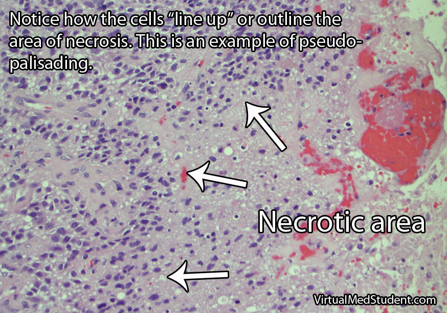
Glibolastoma is therefore characterized by the following pathological characteristics: prominent microvascular proliferation (ie: development of new blood vessels within the tumor), mitosis (ie: an indicator of cell division/growth), and necrosis (ie: areas of dead tumor); pseudopalisading necrosis is a specific form of necrosis shown in the above image that is a hallmark of glioblastomas.

Signs and Symptoms

Glioblastomas may present with any number of signs and/or symptoms depending on their location within the brain. Lesions that are located on the left side of the brain may cause problems with speech if they involve the Broca or Wernicke areas. Tumors in the areas of the brain that control motor movement may cause weakness. Additionally, tumors that arise in the frontal lobes may cause odd behavioral changes. Some patients present with seizures, and others with only a dull headache.

Diagnosis

An official diagnosis of glioblastoma can only be made when a pathologist looks at a sample of the tumor under a microscope. These samples are typically obtained by a neurosurgeon who resects or biopsies the tumor.

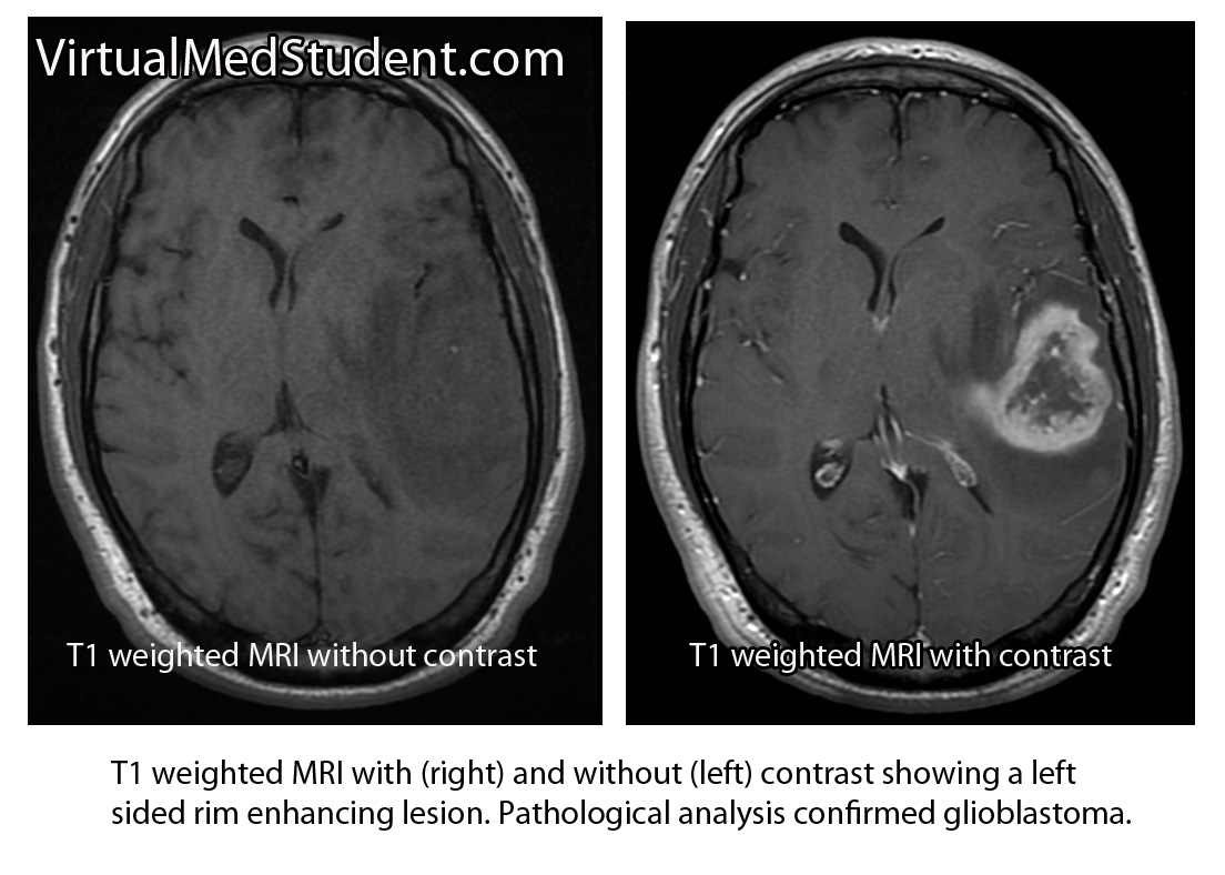
However, glioblastomas also have typical features seen on imaging studies such as MRI. For example, these tumors will “rim-enhance” when a contrast material such as gadolinium is infused into the patient during the scan. Rim enhancement is a result of the contrast material leaking out of all of the blood vessels present within the tumor. It is important to note that other diseases such as abscesses, lymphomas, and other infections can also cause rim-enhancement.

MRI of glioblastoma

Another useful study known as MR spectroscopy measures the relative amounts of different molecules present within the tumomr. In a glioblastoma the amount of lactate, choline, and lipid are all increased. Lactate is a marker of brain tissue that is not receiving enough oxygen, which is common in necrotic tumor areas. Choline is a molecule that is present in cell membranes. When neurons are rapidly dividing, which is what occurs in glioblastoma, the amount of choline present also increases. A different molecule known as N-acetyl aspartate (NAA) is present in mature cells. Therefore, unlike lactate and choline levels, NAA is decreased in glioblastoma because these cells are "immature" (ie: poorly differentiated).

Treatment

Treatment combines a mixture of surgery, radiation therapy, and different chemotherapeutic drugs, the most common being temozolomide (Temodar®). Surgery is only useful when a significant amount of the tumor can be removed. Despite optimal treatment the prognosis for patients with glioblastoma remains extremely poor.

It is also highly important to treat the edema that frequently surrounds the tumor. Steroids, most commonly dexamethasone (Decadron®), are used to decrease the amount of edema, which usually improves symptoms.

Patients are often started on an anti-seizure medication such as levetiracetam (Keppra®) or phenytoin (Dilantin®).

Overview

Glioblastoma is a malignant astrocytic tumor. It is the most common primary brain tumor. It has unique characteristics that distinguish it from more benign brain tumors that also arise from astrocytes. It is treated with a combination of surgery, radiation, and chemotherapy.

References and Resources

Latin for Toothlike: Fractures of the Odontoid Process

The odontoid process (also know as the "dens") is the finger of bone that sticks up from the second cervical vertebrae (ie: the axis).

It articulates via numerous ligaments to the anterior arch/ring of the first cervical vertebrae (ie: the atlas) to form a joint. This joint is what allows you to rotate your head from side to side as if you were nodding “no”.

Axis (C2)

Fractures of the odontoid typically occur after traumatic events. In younger, otherwise healthy individuals tremendous force is necessary to fracture the odontoid. Breaks are typically seen after car, motorcycle, or sporting accidents. In older, osteoporotic people simple ground level falls can result in a fracture. Less commonly, fractures of the odontoid may be caused by tumor chipping away at the underlying bone (a so called “pathologic” fracture).

Since the odontoid is a relatively long piece of bone it can fracture at one of several distinct sites. The most commonly used system (Anderson and D’Alonzo) categorizes fractures into one of three types:

  • Type 1 – a fracture at the tip of the odontoid.
  • Type 2 – a fracture at the base of the odontoid.
  • Type 3 – a fracture involving the body of the C2 vertebrae, which includes the odontoid within it.

Odontoid fractures

This grading system is important because it helps predict both stability of the C1-C2 (ie: atlanto-axial) joint and guides potential treatment options.

Signs and Symptoms

Roughly 80% of patients with odontoid fractures do not have any neurological injury to their spinal cord. The remaining patients can exhibit anything from quadriplegia to mild sensory disturbances. Patients with severe cervical spinal cord injury usually are unable to breath (secondary to diaphragm paralysis) and frequently die at the scene of the accident.

Many patients with odontoid fractures will have significant neck pain that radiates up into the scalp. This is usually caused by neck muscles spasming secondary to the injury.

Diagnosis

CT of odontoid fracture

CT scans, x-rays, and MRIs are all useful in diagnosing and properly treating odontoid fractures.

CT scans of the cervical spine provide excellent bony detail, and also help illustrate any additional fractures that may be present.

MRI of the cervical spine is useful for assessing any co-existent ligamentous injury. If ligamentous injury is present it drastically alters treatment decisions.

Treatment

Treatment of odontoid fractures is based on both bony and ligamentous injury. The goal of treatment is to stabilize the spine either by allowing the bone to heal on its own, or by fusing the spine artificially using rods, screws, and/or wires.

Placement of an odontoid screw is one method of fixing non-displaced type II fractures. However there are numerous contraindications for odontoid screw placement. For example severe angulation of the fractured segment precludes placement of a screw; barrel chested anatomy prevents an adequate angle for screw trajectory in the operating room. In addition, if the transverse ligament is disrupted, bony fixation with an odontoid screw alone will not stabilize the joint.

On the other hand, odontoid screws are beneficial because they typically stabilize the fracture with minimal restriction of neck motion.

Approaching the spine from behind is another option to stabilize odontoid fractures. Fusing the atlas (C1) to C2 or C3 is sometimes used if odontoid screw placement cannot be performed and the injury is deemed unstable. It is important to note that posterior approaches can restrict motion, especially in the high cervical spine.

Non-surgical options must ensure that the patient has minimal to no movement of the neck in order to give the bone an adequate chance to heal on its own. Rigid collars or halos are used to prevent neck motion.

Overview

Odontoid fractures come in three flavors depending on the location of the fracture. Symptoms can be anything from mild neck discomfort to quadriplegia, although neurological injury is surprisingly uncommon. Diagnosis is based on CT and MRI. Treatment is with cervical immobilization for an extended period of time, or surgical fusion. Treatment decisions are based on the degree of both bony and ligamentous injury, as well as the patient’s overall health status.

Related Articles

References and Resources

Cerebellum, Purkinje, Mossy, and All That Jazz!

Cellular Anatomy

The cerebellum is a large outgrowth on the backside of the brainstem that looks like a piece of cauliflower. It is the great "modulator" of movement. It compares the movement that the brain wants to do with what the body is actually doing. It then makes fine adjustments to ensure that the intended movement is smooth and fluid. It works in conjunction with the basal ganglia and motor cortex to modulate movement.

As can be expected, the cerebellum is highly complex with inputs and outputs going to various regions of the brain and spinal cord. Before we delve into the circuitry of the cerebellum, we need to first understand its cellular architecture…

Unlike most of the cerebral cortex, the cerebellar cortex has only three layers. They include – from most superficial to deep – the molecular layer, the Purkinje cell layer, and the granular layer.

The molecular layer is composed of connections between the dendrites (ie: information receiving processes) of Purkinje cells and the axons (ie: information sending processes) of granule cells. The molecular layer also contains stellate and basket cells, which help modulate the connections between Purkinje and granule neurons.

The Purkinje layer is, you guessed it, composed of Purkinje neurons. These cells send dendrites into the molecular layer where they receive information from granule cells. Purkinje cells also directly receive signals from other areas of the brain and spinal cord. Purkinje cells then send information along to the deep cerebellar nuclei, which are discrete collections of neurons within the cerebellum.

Finally, the granular layer is populated with granule and Golgi cells. They send axons into the molecular layer, which serve to pass on information to the dendrites of Purkinje cells in the molecular layer.

Confused yet???

Input Circuitry

In order for the cerebellum to modulate movement it must receive input from multiple sources. At this point, we have to introduce three new terms: climbing fibers, mossy fibers, and aminergic fibers.

Neurons in an area of the brainstem known as the "olive" (a part of the medulla oblongata) send climbing fibers into the cerebellum. Each climbing fiber forms direct and powerful excitatory connections with multiple Purkinje cells (interestingly, each Purkinje cell only receives input from one climbing fiber).

The second input, mossy fibers, originate from several different areas outside the cerebellum (see table below). These axons form connections with the granule cells. The granule cells, if you remember from our discussion above, send axons into the molecular layer where they form connections to Purkinje cell dendrites. Therefore, mossy fibers indirectly influence Purkinje cells through the "intermediary" granule cells.

Input Connections (ie: Afferent Fibers) to the Cerebellum

Source of Input Type of Fiber Target of Input
Olivary nuclei (brainstem) Climbing fibers Contralateral cerebellum
Pontine nuclei (brainstem) Mossy fibers Contralateral cerebellum
Reticular nuclei (brainstem) Mossy fibers Ipsilateral cerebellum
Ventral spinocerebellar tract Mossy fibers Ipsilateral cerebellum
Dorsal spinocerebellar tract Mossy fibers Ipsilateral cerebellum
Vestibular nuclei Mossy fibers Ipsilateral cerebellum
Locus ceruleus Norepinephrine Bilateral projections
Raphe nucleus Serotonin Bilateral projections

Aminergic fibers originate from the locus ceruleus in the pons, and the raphe nuclei of the midbrain, pons, and medulla. The locus ceruleus fibers "spit" norepinephrine and the raphe nuclei "spit" serotonin onto multiple areas within the cerebellum.

All of these inputs (as well as basket, stellate and Golgi cells, which are intrinsic to the cerebellum itself) are trying to influence the output of the Purkinje neurons. The Purkinje cells ultimately synthesize and pass along all of this competing information via an inhibitory message to the deep cerebellar nuclei.

And that folks brings us to our next section: the output circuitry…

Output from the cerebellum passes exclusively from the deep cerebellar nuclei. The deep nuclei are four discrete collections of neurons, which are given specific (and funky) names; they include the fastigial, globose, emboliform, and dentate nuclei.

Remember that the Purkinje cells inhibit the output of the deep cerebellar nuclei. Therefore, the more active the incoming messages (via mossy and climbing fibers) –> the more active the Purkinje cells –> the less active the output of the deep cerebellar nuclei.

The output of the deep nuclei goes to four major structures outside the cerebellum: the red nucleus, the vestibular nucleus, the reticular formation, and the thalamus. From these structures the information is passed along to the cerebral cortex and/or the spinal cord for additional processing.

Output Connections (ie: Efferent Fibers) from the Cerebellar Nuclei

Source of Output Target of Output Function
Globose nucleus Contralateral red nucleus
Contralateral thalamus
Influences tone of flexor
muscles
Emboliform nucleus Contralateral red nucleus
Contralateral thalamus
Influences tone of flexor
muscles
Dentate nucleus Contralateral thalamus Influences motor cortex
and coordination
Fastigial nucleus Bilateral vestibular nucleus
Bilateral reticular formation
Influences motor neurons in
spinal cord and helps
control tone of extensor
muscles

Ultimately, the output of the cerebellum influences not only coordination, but also the tone of flexor and extensor muscles. This allows movement to be smooth and coordinated (unless of course, you are me on the dance floor… in that case all bets are off!).

Overview

The cerebellum is an extremely complex part of the brain. It receives information about an intended movement from the cerebral cortex and compares that to sensory information coming back from the spinal cord. If the intended movement doesn’t match the actual movement the output of the cerebellum attempts to restore balance.

References and Resources

  • Ikai Y, Takada M, Shinonaga N, et al. Dopaminergic and non-dopaminergic neurons in the ventral tegmental area of the rat project, respectively, to the cerebellar cortex and deep cerebellar nuclei. Neuroscience, V51:3, p 719-28.
  • Asanuma C, Thach WT, Jones EG. Brainstem and spinal projections of the deep cerebellar nuclei in the monkey, with observations on the brainstem projections of the dorsal column nuclei. Brain Research Reviews. V5:3, May 1983. pp 299-322.
  • Huang CC, Sugino K, Shima Y, et al. Convergence of pontine and proprioceptive streams onto multimodal cerebellar granule cells. Elife. 2013;2:e00400. Epub 2013 Feb 26.
  • Baehr M, Frotscher M. Duus’ Topical Diagnosis in Neurology: Anatomy, Physiology, Signs, Symptoms. Fourth Edition. Stuttgart: Thieme, 2005.
  • Purves D, Augustine GJ, Fitzpatrick D, et al. Neuroscience. Fourth Edition. Sinauer Associates, Inc., 2007.

Vein of Galen Malformations: A Misnomer of Sorts

The vein of Galen is located deep within the brain. The internal cerebral veins, basal veins of Rosenthal, atrial veins, and precentral cerebellar veins join together to form the vein of Galen.

As expected, the vein of Galen drains blood from deeply located brain structures. The vein of Galen then connects with the inferior sagittal sinus to form the straight sinus; blood then drains from the straight sinus into the transverse and sigmoid sinuses, where it eventually finds its way into the internal jugular veins and back to the heart.

Interestingly, a "vein of Galen" malformation is not actually a malformation of the true vein of Galen; the term is a misnomer. It is actually a malformation of primitive fetal anatomical structures that normally regress during development. These primitive structures include a dilated venous structure, as well as "feeding" arteries. Therefore, vein of Galen malformations represent true arteriovenous fistulas; in other words, blood moves directly from an artery to a vein without an intermediary capillary bed.

Between the 3rd and 11th weeks of fetal development a large primitive vein known as the median prosencephalic vein of Markowitz drains the deepest parts of the brain. As the brain develops, the internal cerebral veins annex the territory normally drained by the anterior portion of this vein. As a result, this portion of the median prosencephalic vein regresses. The internal cerebral veins then plug in to the posterior portion of the median prosencephalic vein, which becomes the "true", or "normal", vein of Galen.

The most common arterial "feeders" of the malformation can also be explained by aberrant embryology. During early fetal brain development the distal branches of the anterior cerebral arteries (ie: the pericallosal branches) make connections with the posterior cerebral arteries. These connections usually regress to form the anterior and posterior circulations, which are connected to one another via the posterior communicating arteries.

So how does a vein of Galen malformation form? In some infants the median prosencephalic vein of Markowitz does not regress like it should. As a result, a large abnormal venous midline pouch remains. It also retains its primitive arterial blood supply from the distal branches of the anterior cerebral artery (ie: pericallosal branches), anterior choroidal arteries, posterior communicating arteries, and branches of the posterior cerebral arteries (ie: posterior choroidal arteries).

Vein of Galen malformations are also associated with other abnormalities in the venous structure of the brain. Not uncommonly, the straight sinus is absent or severely narrowed. As a result venous blood drains into a persistent falcine sinus, which is a structure that normally regresses in-utero.

To summarize, vein of Galen malformations are primitive direct arteriovenous fistulas. They are composed of a dilated venous pouch (ie: the median prosencephalic vein of Markowitz) with any combination of anterior and posterior circulation feeding arteries.

Signs and Symptoms

Signs and symptoms depend on the severity of the malformation. Severe malformations present in new borns with high output cardiac failure. This is because so much blood is being shunted into the malformation that the heart cannot keep pace!

Less significant malformations present later in infancy with a rapidly enlarging head circumference secondary to hydrocephalus, developmental delay, and seizures. The increase in venous blood pressure within the head can cause a "melting brain" syndrome in which the white matter of the brain fails to develop properly. This can lead to severe mental retardation later in life if left untreated.

Diagnosis

Vein of Galen Malformations
Diagnosis is made with a combination of MRI, CT, and diagnostic angiograms. MR venograms can show the dilated venous pouch, as well as associated venous anomalies.

CT angiograms can show associated arterial feeding vessels. Formal diagnostic angiograms are the gold standard test; they delineate both the spatial and temporal relationship of the arterial feeding vessels to the venous pouch.

Formal catheter angiograms are also necessary to distinguish true vein of Galen malformations from arteriovenous malformations of the adjacent brain tissue.

Treatment

Treatment is dependent on the age of the child as well as the severity of the malformation. The most commonly used grading system developed by Dr. Lasjaunias is known as the Bicetre score. It takes into account the child’s cardiac, pulmonary, hepatic (liver), and renal (kidney) function. Lower scores indicate more severe disease with poorer outcomes.

The Bicetre score also dictates the optimal time for treatment. If the score is very low then aggressive treatment, even in the neo-natal period, may be indicated to try and prevent death and severe disability. Higher scores are typically treated later in life; however, worse outcomes, in terms of mental retardation, have been illustrated if treatment is delayed.

Most of these lesions are treated endovascularly (ie: from inside the blood vessels). The arterial feeders are embolized with a glue like material, which ultimately shuts down the fistula in an attempt to restore normal venous pressures. The vein itself may also be filled with tiny metal coils to help reduce flow through the fistula; this is known as trans-venous endovascular therapy.

Surgical ligation of the arterial feeders has mostly become a treatment of the past. Radiation therapy with Gamma Knife has also been used in some cases; it is showing some promise as an alternative treatment modality in select cases.

Overview

Vein of Galen malformations are fetal abnormalities in the brain’s normal venous drainage. They represent true arteriovenous fistulas. They are composed of a dilated median prosencephalic vein of Markowitz and numerous arterial feeding vessels. Feeders may come from the anterior cerebral arteries, posterior cerebral arteries, or posterior communicating arteries. Symptoms are usually from high output heart failure in the neonatal period; older infants and children suffer from increasing head circumference, seizures, and developmental delay. Treatment is usually with endovascular techniques.

References and Resources

Dr. Bror Rexed and His Laminae (The Rexed Laminae)

The Rexed laminae are a system for organizing the neurons of the spinal cord (they were actually designed to torture medical students, I joke of course, sort of…). They are named after Dr. Bror Rexed who was a Swedish neuroscientist.

There are ten Rexed laminae and they follow a topographic organization, with the lower numbers being towards the back of the spinal cord (ie: posterior) and the higher numbers being towards the front of the spinal cord (ie: anterior).

The Rexed laminae are not strictly organized based on anatomical location, but are actually based on the types, and functions, of the neurons in each laminae. Let’s discuss each of the ten layers in more detail (let the fun begin!)…

Layer one contains neurons that receive pain and temperature information from the body and limbs via the axons coming from the dorsal root ganglia. The neurons in layer one then pass this information along to the brain via the spinothalamic tract on the opposite side of the cord.

Layer two, which is also known as the substantia gelatinosa, gets information from the spinothalamic tract as well as the dorsal columns. The spinothalamic tract relays information about painful stimuli and the dorsal columns relay information about non-painful stimuli.

Therefore, the neurons in layer two receive information about both painful and non-painful stimuli. These neurons then send information to Rexed laminae three and four. The neurons in these laminae then pass the sensory information to the brain where it is further interpreted. Interestingly, there are large amounts of opiate (ie: think morphine or heroin) responsive neurons in laminae two of the spinal cord.

The nucleus proprius, which constitutes layers three and four, receives information from the body about touch and proprioception (hence the name “proprius”). It then relays this information to numerous areas in the brainstem, brain, and other Rexed laminae for further processing.

Nobody knows exactly what the heck layer five does, but it receives information from a wide variety of sources including pain sensation from the bodies’ organs, as well as information about movement from the brain (via the corticospinal tracts) and brainstem (via the rubrospinal tracts).

The sixth layer can be divided into two sections: medial and lateral. The medial layer (ie: towards the middle of the spinal cord) gets input from muscle spindles. Muscle spindles (by way of type Ia fibers) tell the spinal cord how much a given muscle is being stretched. The neurons in the medial layer act as messengers for this information.

The lateral layer (ie: towards the periphery of the cord) gets information from the brain and brainstem via multiple descending tracts.

So what is the purpose of the sixth layer? The neurons in this layer send information to two places: the cerebellum (via the ventral spinocerebellar tracts) and motor neurons in the anterior horn of the spinal cord.

The cerebellum interprets the information and modulates movement and muscle tone accordingly. The direct communication between the neurons in Rexed laminae six and the motor neurons in the anterior horns are responsible for spinal reflexes.

Layer seven is most prominent between C8 and L2. This prominence is known as the dorsal nucleus of Clarke. The neurons in Clarke’s nucleus receive lower extremity position and sensory information and then pass that information to the cerebellum via the dorsal spinocerebellar tract.

Layer seven also receives and sends information from, and to, the bodies’ organs. The sympathetic and parasympathetic autonomic system have their pre-ganglionic neurons in Rexed laminae seven. These neurons are responsible for the fight or flight response (sympathetic) and rest and digest (parasympathetic) functions of the bodies’ organs.

Neurons in layer eight obtain information from the reticulospinal and vestibulospinal tracts. The reticulospinal tract is important in maintaing the tone of muscles that flex joints. The vestibulospinal system helps maintain the tone of muscles that are important in extending joints.

So what do the neurons in layer eight do? They take competing information from the reticulospinal and vestibulospinal tracts and help modulate movement and muscular tone. Pretty cool, huh?

Finally an easy layer! This layer contains the α, β, and γ motor neurons of the cord. Simply stated, these neurons send impulses to muscles leading to movement and its’ modulation. The more complex story is that motor neurons in layer nine are influenced by numerous inputs from other Rexed laminae, as well as by descending information coming from the brain.

The neurons in layer nine are topographically organized based on what type of muscle (flexor or extender) they control, as well as where in the body that muscle is located (axial or limb).

Neurons that are located towards the center of the cord control axial muscles and those located towards the periphery of the cord control the muscles of the limbs. In similar fashion, neurons that control muscles that extend joints are located towards the front of the layer (ie: anterior), and those that flex joints are located towards the back of layer nine (ie: posterior).

Like layer five, no one really knows what layer ten actually does. We do know that it is composed of neurons that surround the fluid filled central canal of the spinal cord like a donut. Hungry yet???

Overview

The Rexed laminae are layers of neurons within the spinal cord that perform specific functions. In general, neurons in the laminae towards the back of the cord (ie: laminaes one, two, three, four, and five) are predominately involved in interpreting and relaying sensory information from the body to the brain. On the other hand, neurons in the laminae towards the front of the cord (ie: laminaes seven, eight, and nine) are involved primarily in executing movement and controlling the functions of the bodies organs.

References and Resources

  • Kitahata L, Kosaka Y, Taub A, et al. Lamina-specific suppression of dorsal-horn unit activity by morphine sulfate. Anesthesiology, V41, issue 1, 1974.
  • Anamizu Y, Seichi A, Tsuzuki N, et al. Age-related changes in histogram pattern of anterior horn cells in human cervical spinal cord. Neuropathology. 2006 Dec;26(6):533-9.
  • Stephens B, Guiloff RJ, Navarrete R, et al. Widespread loss of neuronal populations in the spinal ventral horn in sporadic motor neuron disease. A morphometric study. J Neurol Sci. 2006 May 15;244(1-2):41-58.
  • Baehr M, Frotscher M. Duus’ Topical Diagnosis in Neurology: Anatomy, Physiology, Signs, Symptoms. Fourth Edition. Stuttgart: Thieme, 2005.
  • Neuroscience. Fourth Edition. Sinauer Associates, Inc., 2007.

Parkinson’s Disease: Pathology of Movement

Parkinson’s disease is caused by the death of neurons in a part of the brain known as the substantia nigra.

The substantia nigra is part of a system of connected neurons known collectively as the basal ganglia. This system is very important in the control of movement. The substantia nigra contains neurons that secrete a molecule known as dopamine. The loss of dopamine’s effect on the basal ganglia leads to the signs and symptoms of Parkinsonism.

Visible abnormalities are also seen in the neurons of patients with Parkinson’s disease. These abnormalities are called Lewy bodies. They are collections of abnormal protein (specifically, a protein known as α-synuclein) that clump together to form a redish-pink cytoplasmic inclusion in the substantia nigra neurons.

Lewy body
Interestingly, it is not known what causes most cases of Parkinson’s disease. However, there are some known causes, almost all of which involve insults to the substantia nigra.

For example, toxins such as carbon disulfide, manganese, and certain street drugs have been known to kill substantia nigra cells resulting in Parkinson’s.

Another toxin known as MPTP (1-methyl-4-phenyl-1,2,3,6-tetrahydropyridine, trying saying that four times fast!), which was produced accidentally by drug chemists trying to make a synthetic opiod, is extremely toxic to substantia nigra cells. It caused irreversible Parkinson’s disease in those who were unfortunate enough to ingest it!

Finally, the genetics of Parkinson’s disease are not well known. There appears to be multiple genetic causes of the disease. One such genetic mutation involves the gene for α-synuclein, the main component of Lewy bodies. Other genetic mutations may also play a role in Parkinson’s, but they appear to contribute to only a small percentage of cases.

Diagnosis

Parkinson’s disease is a clinical diagnosis. This means that there are currently no special laboratory tests that can reliably detect the disease. Instead, the disorder is diagnosed based on the clinical symptoms discussed below. A true pathological diagnosis can only be made at autopsy by looking at the neurons of the substantia nigra under a microscope.

Signs and Symptoms

Parkinson’s disease has a number of symptoms that commonly begin in the 6th and 7th decade’s of life. Most of these are related to difficulties with movement. The classic description of Parkinson’s involves several signs and symptoms: resting tremor (4-7 Hz frequency), postural instability, bradykinesia (slow movements), rigidity (of the cog-wheeling type), and masked facies.

In addition, late in the disease course other symptoms can become apparent. Roughly 10 to 15% of patients develop dementia. This is believed to be due to the development of Lewy bodies in the cerebral cortex. The dementia of Parkinson’s disease is likely on a continuum with another form of dementia known as Lewy body dementia.

This YouTube video illustrates the common signs and symptoms seen in Parkinson’s disease:

Treatment

The mainstay of treatment for Parkinson’s disease is replacing or augmenting dopamine levels in the brain. A molecule known as levodopa (L-dopa), which is a precursor of dopamine is commonly used. The reason L-dopa is given instead of dopamine is because it crosses the blood brain barrier more readily.

Once inside the brain, L-dopa is converted to dopamine by neuronal enzymes. Sinemet® is a common formulation of L-dopa. It also contains a molecule known as carbidopa. Carbidopa inhibits the breakdown of L-dopa in the body so that more of it can reach the brain. L-dopa is most effective at improving tremor and bradykinesia.

Other medications are designed to mimic the actions of dopamine in the brain. These medications are known as dopamine agonists. They bind to dopamine receptors and cause the same types of cellular reactions that dopamine normally would. The two common dopamine agonists in use today are ropinirole and pramipexole.

Treating Parkinson’s –
(1) Levodopa/carbidopa
(2) MAO-B inhibitors
(3) COMT inhibitors
(4) Dopamine agonists
(5) Anticholinergics
(6) Glutamate antagonists
(7) Deep brain stimulation
Other medications used to treat Parkinson’s disease attempt to indirectly increase the amount of dopamine.

One category of medications that does this is known as MAO-B inhibitors. MAO, or monoamine oxidase, is an enzyme that normally breaks down monoamines like dopamine. Therefore, inhibitors of this enzyme decrease the break down of dopamine allowing it to remain in the brain longer. Selegiline and rasagiline are example of MAO-B inhibitors.

Another class of medications that perform a similar function to the MAOIs are the COMT inhibitors. COMT (catechol-O-methyl transferase) is an enzyme that breaks down dopamine. Entacapone is a COMT inhibitor that may increase the amount of dopamine in brain tissue.

Medications that interfere with another neurotransmitter known as acetylcholine are also sometimes used. These anticholinergic medications, namely benztropine (Cogentin) and trihexylphenidyl (Artane), are most useful for tremor reduction. Since acetylcholine is present in many parts of the body, the anticholinergics tend to have many side effects (the mnemonic "dry as a bone (dry mouth), mad as a hatter (delirium), blind as a bat (pupil dilation), and hot as a hare (fever)" is commonly used for symptoms pertaining to anticholinergic medications).

Finally, medications that antagonize the neurotransmitter glutamate are less commonly used. Amantadine is the name of one medication in this category. It helps mostly with decreasing, or smoothing out, fluctuations in movement.

If medical therapy fails to control symptoms then surgical interventions may be used. In the past a procedure known as pallidotomy was used. This involved making a tiny incision in part of the basal ganglia. This procedure has been replaced by deep brain stimulation (DBS). During DBS surgery small electrodes are implanted in certain areas of the basal ganglia.

Deep Brain Stimulation

Overview

Parkinson’s disease is characterized by postural instability, resting tremor, slow movements, and rigidity. It’s caused by loss of dopaminergic neurons in the substantia nigra of the brain. Why these neurons die is not entirely understood. Diagnosis is based on clinical signs and symptoms. Treatment is by replacing or augmenting dopamine in the brain.

Related Articles

References and Resources

The Indirect Basal Ganglia Pathway

The basal ganglia represent a system of several discrete collections of neurons within the brain. These collections of neurons interact closely with the part of the cerebral cortex that initiates movement. The basal ganglia fine tune the starting and stopping of movements.

The term "basal ganglia" encompasses several separate, but interrelated neuron populations. The putamen, caudate, globus pallidus internus (GPi), globus pallidus externus (GPe), substantia nigra (SN), and subthalamic nucleus (STN) are all discrete neuron populations that, as a whole, compose the "basal ganglia". These named populations of neurons work together to achieve a common goal. The term "striatum" includes the caudate and putamen only, and the term "lentiform nuclei" includes the putamen and globus pallidus.

The basal ganglia modulate movement through a complex loop of both inhibitory and excitatory signals. When you decide to move, your frontal lobes send an excitatory signal via the neurotransmitter glutamate to the striatum (FYI: striatum = caudate and putamen).

In the indirect basal ganglia pathway the striatum then sends an inhibitory signal via the neurotransmitter GABA to the external segment of the globus pallidus. This is different from the direct pathway where the striatum sends a signal to the internal segment of the globus pallidus.

The external segment of the globus pallidus normally indirectly (more on this in the next few paragraphs) inhibits its internal counterpart. Thus, when the striatum inhibits the external segment, it is, in effect, releasing the internal segment from inhibition (that sure seems like a lot of double negatives!).

At this point, the internal segment of the globus pallidus is able to send its inhibitory signals to the thalamus, which causes thalamic neurons to stop sending excitatory signals to the motor cortex. The cortex is then unable to send an impulse down the spinal cord and, ta-da, the net result is a decrease in movement.

It would be easier to understand if the external segment of the globus pallidus “talked” directly to the internal segment, but that is not how it works. The message is relayed through another nucleus known as the subthalamic nucleus.

The subthalamic nucleus is usually inhibited by the external segment of the globus pallidus. Therefore, when the striatum inhibits the external globus pallidus, it causes the cells in the subthalamic nucleus to become more active (ie: the subthalamic nucleus is released from the inhibitory effects of the external globus pallidus).

The subthalamic nucleus, in turn, is able to send an excitatory signal to the neurons in the internal segment of the globus pallidus. The cells in GPi then become more active, which means that they suppress the activity of the thalamus more robustly. The thalamus is then unable to send its normal excitatory messages to the motor cortex. End result? Decreased movement!


Basal Ganglia Indirect Pathway Schematic


So how does dopamine act on the indirect pathway? Dopamine is secreted by the substantia nigra and binds to D2 receptors (these are different than the D1 receptors of the direct pathway) in the striatum. This causes striatal neurons to decrease their inhibitory message to the external segment of the globus pallidus. The external segment of the globus pallidus is then free to carry out its “normal” job and suppress the excitatory actions of the subthalamic nucleus on its internal counterpart. Less excitation going to the internal globus pallidus translates to less inhibition of the thalamus, and ultimately more excitation of the cortex!

Therefore, if you’ve managed to work through these complicated systems, you’ll realize that dopamine causes increased movement by activating the direct pathway and inhibiting the indirect pathway.

Overall, the indirect basal ganglia pathway has the exact opposite effect of the direct pathway. The indirect pathway serves as a negative modulator of movement and the direct pathway serves as a positive modulator of movement. Now that is some complicated sh**t!

Importance in Disease

Diseases of the basal ganglia cause unwanted movements, or a failure to initiate movement.

The classic basal ganglia disease is Parkinson’s disease, which has elements of both unwanted movement (resting tremor) and difficulty initiating movement (bradykinesia). Other diseases such as hemiballismus, in which the affected person violently flings an extremity, can occur when there is damage to the subthalamic nucleus.

Additionally, in Huntington’s disease the GABA and enkephalin projections from the caudate nucleus to the external globus pallidus are affected. This is believed to be responsible for many of the movement abnormalities seen in patients with this disease.

Overview

The indirect basal ganglia pathway fine tunes motor movements. It involves both excitatory and inhibitory signals through the striatum, globus pallidus, substantia nigra, thalamus, and motor cortex. Diseases such as Parkinson’s disease, hemiballismus, and Huntington’s disease may occur when there is damage to one of the components of the basal ganglia.

References and Resources

  • Baehr M, Frotscher M. Fourth Edition. Stuttgart: Thieme, 2005.
  • Bickley LS, Szilagyi PG. Bates’ Guide to Physical Examination and History Taking. Ninth Edition. New York: Lippincott Williams and Wilkins, 2007.
  • Nolte J. The Human Brain: An Introduction to its Functional Anatomy. Sixth Edition. Philadelphia: Mosby, 2008.
  • Purves D, Augustine GJ, Fitzpatrick D, et al. Neuroscience. Fourth Edition. Sinauer Associates, Inc., 2007.
  • Related Articles

The Direct Basal Ganglia Pathway

The basal ganglia are a complex system of several discrete collections of neurons within the brain. These collections of neurons interact closely with the part of the cerebral cortex that initiates movement. As such, the basal ganglia are important in fine-tuning the starting and stopping of movements.

The term "basal ganglia" encompasses several separate, but interrelated neuron populations. The putamen, caudate, globus pallidus internus (GPi), globus pallidus externus (GPe), substantia nigra (SN), and subthalamic nucleus (STN) are all discrete neuron populations that, as a whole, compose the "basal ganglia". These named populations of neurons work together to achieve a common goal. The term "striatum" includes the caudate and putamen only, and the term "lentiform nuclei" includes the putamen and globus pallidus.

The basal ganglia modulate movement through a complex loop of inhibitory and excitatory signals. When you decide to move, your frontal lobes send an excitatory signal via the neurotransmitter glutamate to the striatum (ie: caudate and putamen).

The neurons in the striatum then send an inhibitory signal to the globus pallidus internus (GPi) and the substantia nigra pars reticulata (SNpr). As a result, GPi and SNpr are no longer able to inhibit the thalamus, which is their normal resting function.

The thalamus now finds itself disinhibited and is able to send a message back to the cerebral cortex saying it is ok to allow the desired movement to occur. The motor cortex then sends a message down the spinal cord causing the desired movement.

So where does the neurotransmitter dopamine enter the picture? Dopamine is secreted by a different part of the substantia nigra known as the pars compacta. These neurons secrete dopamine onto specific cells in the striatum. The dopamine interacts with the D1 (dopamine-1) receptors on these cells causing them to become more active (ie: dopamine has a stimulatory effect via the D1 receptor).

Basal Ganglia Direct Pathway

The overall effect is that dopamine activates the striatum (ie: caudate and putamen), which, as we discussed above, inhibits the internal segment of the globus pallidus. The GPi is then unable to inhibit the thalamus, which in turn allows the thalamus to stimulate the cortex. Huh??? Simply stated, dopamine causes an increased propensity towards movement. Dopamine has a similar role, but via a different mechanism in the indirect pathway, which is discussed in detail in another article.

Importance in Disease

When the basal ganglia malfunction it causes unwanted movements or a failure to initiate movements. The classic basal ganglia disease is Parkinson’s disease, which has elements of both unwanted movements (resting tremors) and difficulty initiating movement (bradykinesia). Other diseases such as hemiballismus, in which the affected person violently flings an extremity, can occur when there is damage to the subthalamic nucleus (the subthalamic nucleus is discussed in more detail on the indirect pathway article).

Overview

The direct basal ganglia pathway fine-tunes motor movements. It involves both excitatory and inhibitory signals through the striatum, globus pallidus, substantia nigra, thalamus, and motor cortex. Diseases such as Parkinson’s disease and hemiballismus may occur when there is damage to one of the components of the basal ganglia.

Related Articles

References and Resources

  • Baehr M, Frotscher M. Duus’ Topical Diagnosis in Neurology: Anatomy, Physiology, Signs, Symptoms. Fourth Edition. Stuttgart: Thieme, 2005.
  • Bickley LS, Szilagyi PG. Bate’s Guide to Physical Examination and History Taking. Ninth Edition. New York: Lippincott Williams and Wilkins, 2007.
  • Nolte J. The Human Brain: An Introduction to its Functional Anatomy. Sixth Edition. Philadelphia: Mosby, 2008.
  • Purves D, Augustine GJ, Fitzpatrick D, et al. Neuroscience. Fourth Edition. Sinauer Associates, Inc., 2007.

When Brain Veins Go Bad: Cerebral Sinus Thrombosis

In order to understand cerebral sinus thrombosis, we need a quick overview of blood vessel anatomy and the normal direction of blood flow in the body. In the most general terms, blood flows from the heart to large arteries (ie: aorta) then smaller arteries (branches off the aorta) then even smaller vessels called capillaries. It is at this point where the different body tissues extract nutrients and oxygen from the blood. The blood then drains into progressively larger veins until it empties back into the heart where it is re-oxygenated.

The largest veins of the brain are referred to as cerebral venous sinuses. They include the superior sagittal sinus, inferior sagittal sinus, a pair of transverse sinuses, a pair of sigmoid sinuses, straight sinus, cavernous sinus, a pair of superior petrosal sinuses, a pair of inferior petrosal sinuses, and the occipital sinus. The role of these sinuses is to collect all the “used” blood from the brain and deliver it back to the heart.

Like other veins in the body, the cerebral sinuses can form blood clots in them (the technical term for a blood clot is actually a "thrombosis", hence the name “sinus thrombosis”). When this happens a back up of blood in the brain occurs leading to increased pressure and sometimes hemorrhage within the brain tissue itself.

The exact cause of a dural venous thrombosis is not always clear. However, there are numerous risk factors associated with their development. They include inherited defects in proteins responsible for blood clot formation. These defects are collectively known as “thrombophilias”, which in Latin means “thrombus loving”. People with these inherited issues are more prone to forming blood clots.

In addition, patients with a kidney condition known as nephrotic syndrome are at increased risk. In this condition patients urinate out proteins responsible for keeping the blood clotting system at bay. The resulting imbalance can cause blood clots to form where they normally would not.

Infections such as mastoiditis and meningitis can cause inflammation of the sinuses, which can result in blood clot formation. Trauma to the head can also cause clot formation. In addition, that beautiful parasitic infection known as pregnancy (a joke of course!) also increases the risk of developing blood clots. Many commonly used birth control pills, especially those containing estrogen, can also increase a person’s risk.

Signs and Symptoms

Depending on the severity of the clot, everything from a mild headache (the most common presenting symptom) to death is possible. Patients with severe headaches may also have associated nausea and vomiting secondary to elevated intracranial pressures. Decreased mental status is also sometimes observed.

It is also important to remember that not all sinuses are created equal. For example, thrombosis in the superior sagittal sinus can present with leg weakness secondary to edema (ie: swelling) of the adjacent motor cortex.

If a clot forms in the cavernous sinus it may cause dysfunction of the third, fourth, fifth, and/or sixth cranial nerves. In addition, when blood pools in the brain behind the clot it can result in a type of stroke known as venous infarction.

Diagnosis

With todays modern imaging studies, MRI, and more specifically, MR venography has become a crucial diagnostic aid.

An example of an MR venogram is shown in the picture below. Other commonly used tests include traditional angiograms, in which radio-opaque dye is injected directly into the sinuses through catheters inserted in the groin. Finally, CT scans are also commonly obtained, especially to evaluate for possible co-existent hemorrhage into the brain.

MRV of Cerebral Sinus Thrombosis

Treatment

Treatment is usually with a blood thinning medication known as heparin. It is delivered through an IV and helps prevent further clot formation. If a venous stroke is present the use of blood thinning medications must be weighed against the possibility of causing bleeding into the stroked brain tissue. Aggressive hydration with normal saline is also often advocated.

Treating other underlying co-problems such as seizures and increased intracranial pressure is also an important part of managing patients with cerebral sinus thrombosis. Patients with substantial increases in intracranial pressure may require removal of the skull (craniectomy).

Overview

Cerebral sinus thrombosis is a abnormal blood clot in one of the large venous draining systems of the brain. They are uncommon and can present with everything from a mild headache to coma and death. Diagnosis is made most commonly with MRI. Treatment is based on preventing further clot formation with heparin and aggressive hydration.

References and Resources

  • Xu H, Chen K, Lin D, et al. Cerebral venous sinus thrombosis in adult nephrotic syndrome. Clin Nephrol. 2010 Aug;74(2):144-9. Review.
  • Dlamini N, Billinghurst L, Kirkham FJ. Cerebral venous sinus (sinovenous) thrombosis in children. Neurosurg Clin N Am. 2010 Jul;21(3):511-27.
  • Ju YE, Schwedt TJ. Abrupt-onset severe headaches. Semin Neurol. 2010 Apr;30(2):192-200. Epub 2010 Mar 29.
  • Kamal AK. Thrombolytic therapy in cerebral venous sinus thrombosis. J Pak Med Assoc. 2006 Nov;56(11):538-40.
  • Greenberg MS. Handbook of Neurosurgery. Sixth Edition. New York: Thieme, 2006.