Subarachnoid Hemorrhage: Aneurysms, Vasospasm, and Hyponatremia

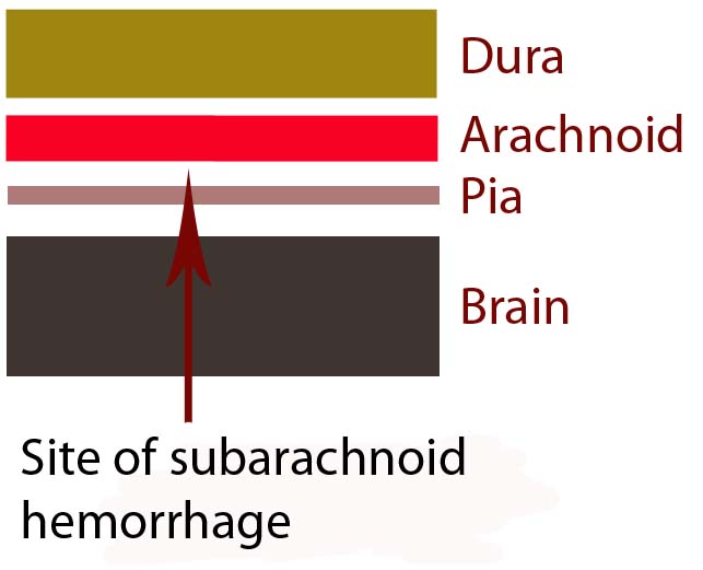
In order to understand subarachnoid hemorrhage we have to first appreciate the layers that make up the brain and its surrounding tissues. The brain itself has three protective layers: dura mater, the arachnoid, and the pia.

The dura is a thick layer of fibrous tissue immediately below the skull. Below the dura is the arachnoid, which is a layer of delicate web-like tissue (hence the name "arachnoid"). Finally, below the arachnoid is the pia mater. The pia is a very thin layer that is directly adjacent to brain tissue.

The subarachnoid space, or the region between the arachnoid tissue and the pia contains cerebrospinal fluid, which acts like a liquid shock absorber for the brain. Also contained within the subarachnoid space are blood vessels that penetrate down into the brain tissue. Sometimes these blood vessels "leak", which can cause a "sub-arachnoid" hemorrhage.

Brain Layers

The most common cause of subarachnoid hemorrhage is traumatic injury; the most common non-traumatic cause is a ruptured aneurysm.

An aneurysm is an abnormal ballooning out of a blood vessel’s wall. The balloon’s dome is much weaker than the rest of the vessel wall. These weak areas can rupture allowing blood to leak out of into the subarachnoid space.

Other causes of subarachnoid hemorrhage include idiopathic (ie: unknown) causes, arteriovenous malformations, vessel dissections, and very rarely tumors. Regardless of the cause, blood will pool in the subarachnoid space.

The remainder of this article will focus on the most common non-traumatic cause of subarachnoid hemorrhage – aneurysm rupture.

Signs and Symptoms

The classic symptom of a subarachnoid hemorrhage is a horrific headache described as the “worst headache of their life". Photophobia, nausea, vomiting, and nuchal rigidity are also common. Seizures may also occur. In addition, depending on how severe the subarachnoid hemorrhage is, patients may have decreased levels of consciousness; some patients become comatose, and many die before reaching medical attention.

The patient’s clinical status is graded according to the Hunt and Hess system. It only applies to patients in whom subarachnoid hemorrhage is caused by rupture of an aneurysm. This grading system was initially established to help determine mortality and clinical outcomes. In modern practice, these numbers are likely high given modern improvements in critical care and neurosurgical intervention since Hunt and Hess first developed their grading system.

Hunt and Hess Clinical Grading Scale
Grade Patient’s Clinical Status Associated Mortality
1 Mild headache and/or nuchal rigidity 1%
2 Cranial nerve dysfunction, moderate to severe headache and/or nuchal rigidity 5%
3 Mild focal neurological deficit, lethargic, confused 19%
4 Stuporous, moderate to severe hemiparesis, early decerebrate posturing 40%
5 Coma, decerebrate posturing 77%

The world federation of neurological surgeons also has a clinical score based on the Glasgow Coma Scale (GCS). It associates the patient’s GCS with the likelihood of death.

World Federation of Neurological Surgeons Grading System
  GCS Major focal deficit Mortality
1 15 No 5%
2 13-14 No 9%
3 13-14 Yes or No 20%
4 7-12 Yes or No 33%
5 3-6 Yes or No 77%

It is very important to think about the possibility of subarachnoid hemorrhage in patients presenting with these signs and symptoms. Prompt diagnosis and treatment is necessary in order to prevent devastating consequences!

Complications

Blood in the subarachnoid space is very irritating to the brain and cerebral blood vessels. Because of this, several complications can occur.

One of the most common complications is known as "vasospasm." Vasospasm occurs when the blood vessels of the brain spasm several days after the initial hemorrhage. When this occurs blood is no longer able to flow past the blockage; if this occurs for a long enough period of time a stroke can occur. The peak period for vasospasm occurs between 3 and 14 days after the initial bleed.

Another complication of subarachnoid hemorrhage is known as cerebral salt wasting. This occurs when a patient urinates excessive amounts of sodium causing the blood level of sodium to drop precipitously. Because of the excessive urination the patient also becomes dehydrated. Aggressive fluid and salt resuscitation must be given to prevent profound hyponatremia (ie: decreased sodium levels in the blood), which can cause seizures, coma, and death.

In addition, for unknown reasons, many patients with subarachnoid hemorrhage also shower their cerebral hemispheres with micro-thrombi (ie: clots), which can lead to many small strokes. The reason why patients with subarachnoid hemorrhage become coagulopathic is still an area of intense research.

Diagnosis

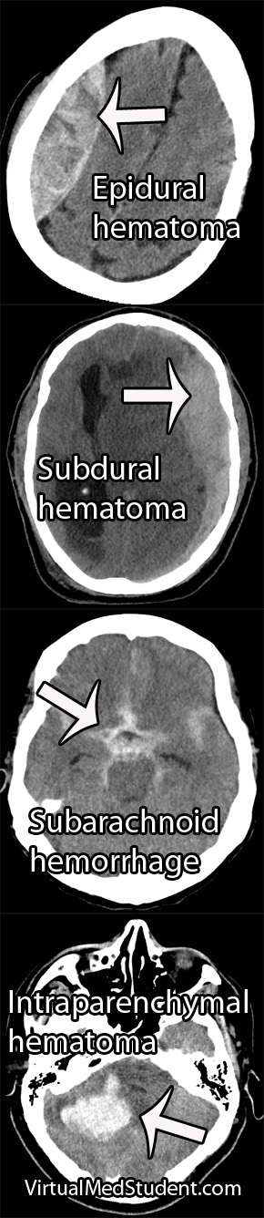
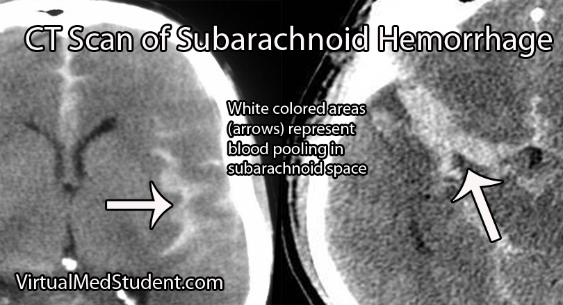
Subarachnoid hemorrhage is most commonly diagnosed by head CT. CT scans are fast and readily pick up the extravasated blood, which layers in the subarachnoid space (see image below). If there is a high clinical index of suspicion but CT of the head is negative than a lumbar puncture should be performed. If the spinal fluid has xanthochromima (a product of red blood cell breakdown) this is highly concerning for subarachnoid hemorrhage.

CT Scan of Subarachnoid Hemorrhage

Subarachnoid hemorrhage is not a diagnosis per say, but rather the result of some underlying pathology (ie: aneurysm, trauma, etc.). Many of these pathologies are treatable; therefore, it is important to figure out what caused the subarachnoid hemorrhage.

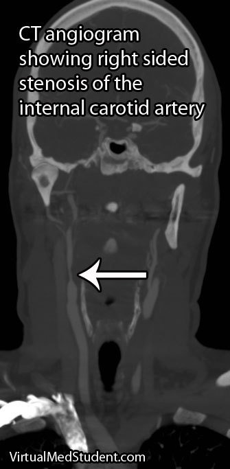
Since many are the result of ruptured aneurysms there are several other tests that are often done. The first test is a CT angiogram (CTA). In this test a radio-opaque material is injected into the blood vessels and a CT is performed.

Cererbral Angiogram with Aneurysm
Dye in the dome of the aneurysm will appear as an abnormality helping to confirm the presence, and more importantly the location of the aneurysm.

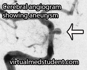
A more invasive procedure known as a "cerebral angiogram" (image to the right) is also often performed.

In this test, a catheter is inserted into blood vessels in the groin and then threaded up into the blood vessels of the brain. Radio-opaque material is injected and x-rays can pick up abnormalities in the vessel.

The benefit of doing a cerebral angiogram is that it is diagnostic, and treatment can frequently be offered through the catheter itself.

Treatment

There are three main components of treating a subarachnoid hemorrhage: treating the underlying cause, preventing a "re-bleed", and preventing secondary complications.

Since many subarachnoid hemorrhages are caused by aneurysm rupture we’ll discuss the treatment for this common cause. Aneurysms are treated either “open” or “closed”.

“Open” refers to a surgical procedure in which part of the skull is removed. The surgeon then dissects down to the aneurysm. Once identified, a clip is placed around the neck of the aneurysm (ie: you can think of the clip as putting a knot in the neck of a balloon). This stops blood from flowing into the aneurysm, and therefore prevents re-rupture.

“Closed” treatment refers to endovascular technology in which a small micro-catheter is threaded from the blood vessels in the groin into the cerebral vasculature. The aneurysm is located via angiogram. Through a hole in the microcatheter tip the physician then fills the aneurysm dome with small metallic coils.

Once blood is in the subarachnoid space secondary complications often result. One of these complications is referred to as "vasospasm". Medications such as oral nimodipine and intra-arterial nifedipine are used to reduce the amount of vasospasm by inhibiting smooth muscle contraction in the wall of the blood vessel.

Overview

Subarachnoid hemorrhage occurs most commonly after an intracerebral aneurysm ruptures, although other causes exist. Regardless of the cause, blood spills out into the subarachnoid space. Symptoms include a horrible headache, focal neurological deficits (ie: weakness, difficulty speaking, etc.), and coma. If an aneurysm is the cause, it is secured with clipping or coiling. Preventing secondary complications such as vasospasm is also an important component of treatment.

References and Resources

Carotid Stenosis: TIAs, Strokes, and Amaurosis Fugax

Carotid stenosis is narrowing of the carotid arteries. This is usually due to atherosclerotic disease present in the internal carotid arteries.

The internal carotid arteries supply a significant portion of the brain with blood, nutrients, and oxygen. If carotid stenosis is present it can result in transient ischemic attacks and possibly stroke.

Signs and Symptoms

Carotid stenosis may be either symptomatic or asymptomatic. Symptomatic carotid stenosis presents in one of three ways: transient ischemic attacks, stroke, or amaurosis fugax.

Transient ischemic attacks (TIA) occur when blood to the brain is blocked temporarily. TIAs cause the same symptoms as a stroke, but the symptoms resolve over 24 hours and there is no permanent brain injury.

A sub-category of TIA is a symptom known as amaurosis fugax, which is a temporary loss in vision, usually in one eye. It is described by patients as a curtain being drawn over the affected eye. It is caused by a small blood clot that breaks off from an atherosclerotic plaque present in the internal carotid artery. This small clot enters the retinal artery and occludes blood flow causing temporary blindness.

Stroke is by far the scariest problem associated with carotid stenosis. When a stroke occurs it can cause severe and irreversible brain injury. Strokes can cause life changing symptoms such as paralysis, aphasia (inability to speak or understand language), and even death!

Carotid Stenosis

Diagnosis

Diagnosis of carotid stenosis is based on imaging studies. The most commonly employed studies are carotid ultrasound, CT angiogram, MR angiogram, and formal angiography.

Treatment

Treatment is dependent on the location and degree of stenosis. Lesions that are near the origin of the internal carotid artery are frequently fixed with surgery in a procedure known as an endarterectomy. In a carotid endarterectomy the artery is surgically opened and the atherosclerotic material is "scooped" out.

The benefit of carotid endarterectomy on stroke risk for symptomatic patients is dependent on the degree of narrowing in the vessel. A landmark study in 1991 showed that carotid endarterectomy was very beneficial in preventing stroke in already symptomatic patients when the degree of narrowing was high (defined as 70 to 99%). A different study looked at lesser degrees of stenosis (50 to 69%) and found a benefit, albeit less robust. There is no clear benefit to having surgery in patients who have narrowing that is 50% or less. Surgery in asymptomatic patients with higher degrees of stenosis follows a more complicated algorithm.

If the diseased segment is higher up on the internal carotid, and surgical access is anatomically difficult, then procedures like carotid stenting or angioplasty can be performed. Stenting is a procedure in which a tiny metal tube-like device is threaded up through the femoral artery in the groin and then opened to “re-expand” the diseased arterial segment.

Patients who may not tolerate a procedure are often managed medically with antiplatelet medications such as aspirin, aspirin/dipyridamole combo (Aggrenox®), ticlopidine (Ticlid®) or clopidogrel (Plavix®). Currently there is no "best" medication to prescribe; the choice is highly dependent on the individual patient and physician.

Overview

Carotid stenosis refers to narrowing of the internal carotid artery. It is usually due to atherosclerotic disease. It can cause transient ischemic attacks, stroke, and amaurosis fugax. Diagnosis is with CT angiogram, carotid ultrasound, and/or formal invasive angiography. Treatment is highly variable, but usually involves a combination of surgery (endarterectomy), stenting, angioplasty, and antiplatelet medications like aspirin.

References and Resources

  • Barnett HJ, Taylor DW, Eliasziw M, et al. Benefit of carotid endarterectomy in patients with symptomatic moderate or severe stenosis. North American Symptomatic Carotid Endarterectomy Trial Collaborators. N Engl J Med. 1998 Nov 12;339(20):1415-25.
  • Chambers BR, Donnan GA. Carotid endarterectomy for asymptomatic carotid stenosis. Cochrane Database Syst Rev. 2005 Oct 19;(4):CD001923.
  • [No authors listed]. Beneficial effect of carotid endarterectomy in symptomatic patients wth high-grade carotid stenosis. North American symptomatic carotid endarterectomy trial collaborators. N Eng J Med. 1991 Aug 15;325(7):445-53.
  • Biller J, Feinberg WM, Castaldo JE, et al. Guidelines for carotid endarterectomy: a statement for healthcare professionals from a Special Writing Group of the Stroke Council, American Heart Association. Circulation. 1998 Feb 10;97(5):501-9.

Ossified Posterior Longitudinal Ligament: Its Clinical Significance

The posterior longitudinal ligament is a long ligament that runs from the base of the skull to the sacrum. It provides a significant amount of mechanical integrity to the spinal column. The ligament runs down the back of the vertebral bodies just in front of the spinal cord itself.

In some individuals this ligament becomes ossified, which means that it takes on a bone like quality. As a result, its overall size and hardness increase. Given the ligament’s location adjacent to the spinal cord, any increase in the girth or flexibility of the ligament can cause injury to the cord.

Ossification of the posterior longitudinal ligament can occur at multiple spots along the spinal column. The most common spot is in the cervical spine (usually C2 to C6), but ossification can also occur in the thoracic (usually T4 to T7) and lumbar spine as well.

Exactly why the longitudinal ligament ossifies in some people is unknown. Current thinking is that a combination of genetic and lifestyle factors play a role in its pathology. For example, family studies have shown increased rates of OPLL in first degree relatives of people known to have the disorder. OPLL is also more common in people of Japanese and Korean descent.

Both familial and racial linkage usually indicate a genetic component to the disease. In fact, patients with OPLL have higher rates of dysfunctional collagen gene regulation (specifically, type XI and VI collagens). Linkage to specific human leukocyte antigen haplotypes on chromosome 6 have also been implemented.

Lifestyle factors such as diet have also been shown to increase risk. Patient’s who are diabetic or pre-diabetic have higher rates of OPLL compared to the rest of the population. High protein diets seems to decrease the risk, whereas high salt diets seem to increase risk.

Overall, the reasons why the posterior longitudinal ligament ossifies in some people, but not others remains an area of ongoing debate and research.

Signs and Symptoms

When an ossified posterior longitudinal ligament pushes on the spinal cord it causes numerous signs and symptoms. The constellation of clinical findings seen in patient’s with symptomatic ossified posterior longitudinal ligaments is known as myelopathy.

Myelopathic patients present with a combination of weakness, clumsiness (ie: decreased ability to hold objects), bowel or bladder dysfunction, spasticity – which is manifested as increased reflexes, as well as changes in sensation (ie: numbness, tingling, etc.).

Ossified Posterior Longitudinal Ligament
Myelopathy can be subtle at first, but can become debilitating depending on how much compression of the cord is present.

Diagnosis and Classification

Diagnosis of an ossified posterior longitudinal ligament is usually made when a patient presents with myelopathic features, or after neurological injury from a traumatic event.

Imaging studies such as xrays and CT scans illustrate the bony quality of the ligament. MRIs are frequently ordered to assess how "squashed" the spinal cord is. CT myelograms also provide excellent detail of cord compression when MRI is not feasible.

The ossification is classified according to its anatomic location and continuity. There are four distinct patterns. They include a continuous pattern, in which there is ossification behind both the vertebral bodies and the disc spaces. The second pattern is known as segmental; in this type the ossification is only present behind the vertebral bodies and does not span the disc spaces. The third pattern is localized, which means that the ossified ligament is present and localized behind only one vertebral body. Finally, there is a mixed type, which is a combination of continuous and segmental.

Treatment

The problem with ossification of the posterior longitudinal ligament is that it progresses over time. Slowly, the ligament will compress the spinal cord. Therefore, surgical treatment is typically offered at the first sign of spinal cord compression (ie: myelopathy).

The best surgical treatment is controversial. Approaching the spine from the front (ie: anterior approach) allows direct removal of the ossified ligament. However, it is important to note that the ligament is often stuck to the dura mater overlying the spinal cord which can make dissection extremely difficult. A cerebrospinal fluid leak is not uncommon when an anterior approach is taken, and there is an increased risk of inadvertent injury to the spinal cord. That being said, when successful, surgery from the front offers several distinct advantages. The first is that myelopathic symptoms seem to respond better to this type of approach; in addition, removal of the ossified ligament can retard further progression of the disease.

The second surgical option is to approach the spine from the back (ie: posterior approach). Removing the bone behind the spinal cord allows the cord to "drift" backwards away from the ossified ligament. This approach is considered more safe because there is no direct removal of the ossified ligament, and therefore there is a decreased risk of inadvertent cord injury. That being said, progression of the ossification can still occur and myelopathic symptoms do not respond as well to this type of surgery.

Ultimately, the type of surgery offered – anterior versus posterior – is dictated by the severity of the ossification and symptoms present, as well as the preferred approach of the surgeon.

Overview

Ossification of the posterior longitudinal ligament is seen more commonly in patients of Japanese descent. There are genetic factors that appear to increase risk. Symptoms are related to compression of the spinal cord by the ossified ligament. Ossification is progressive in nature. Treatment consists of surgery from either an anterior, posterior, or combined approach.

Related Articles

References and Resources

Tuberculous Meningitis: Basal Cisterns, Strokes, Hydrocephalus

Tuberculosis is one of the most common infectious diseases in the world. It is caused by a bacteria of the genus mycobacterium. Tuberculosis usually infects the lungs, but may also infect the lymph nodes, vertebral bodies, kidneys, gastrointestinal system, or central nervous system.

Central nervous system disease comes in two flavors: a focal abscess-like lesion known as a tuberculoma or tuberculosis meningitis. The remainder of this article will focus on tuberculosis meningitis, which is an uncommon (although not rare!) form of extra-pulmonary tuberculosis.

Let’s start by discussing how the bacteria get into the central nervous system. After being inhaled the bacteria infect cells known as macrophages. The infected macrophages move towards lymph nodes, and eventually end up in the blood stream. Once in the blood stream, the mycobacterium-infected macrophages can travel anywhere in the body!

One spot the bacterium hitch a ride to is the lining of the brain (aka: the meninges). Collections of mycobacterium-laden macrophages (“Rich foci”) can rupture into the subarachnoid space causing an inflammatory reaction (ie: a "meningitis", or inflammation of the meninges).

For unclear reasons, the inflammation seen in tuberculosis meningitis preferentially affects the basal cisterns and base of the brain. Autopsy specimens show a gelatinous material coating the undersurface of the brain. The inflammatory reaction is what causes the signs and symptoms of tuberculosis meningitis.

Signs and Symptoms

Tuberculosis meningitis can present in a number of different ways. Many patients present with days to months of non-specific symptoms such as headache, lethargy, nausea, and vomiting.

Cranial neuropathies are commonly seen, especially since tuberculosis meningitis affects the basal cisterns and base of the brain, which is where many of the cranial nerves run.

Additionally, up to 40% of patients may present with stroke. Strokes occur because the inflammation can "eat up" the linings of small blood vessels. The basal ganglia, internal capsule, and thalamus are the most common locations where strokes occur in tuberculosis meningitis.

Diagnosis

The clinical history is extremely important. Tuberculosis meningitis may affect both immunocompetent and immunocompromised (ie: think HIV/AIDs, diabetics, people on immunosuppressives, etc.) people. If the clinical history and physical exam findings are concerning for meningitis, than confirmatory studies should be performed.

TB Meningitis MRI
TB Meningitis CT
Cerebrospinal fluid (CSF) analysis shows elevated opening pressures, increased cellularity with inflammatory cells like neutrophils (earlier) and lymphocytes (later), an increased amount of protein, and a decreased amount of glucose. An acid-fast stain of the CSF to look for bacteria is sometimes positive. Culturing the CSF for mycobacterium is routinely done, but results can take weeks to months, and therefore is not useful in deciding whether or not to start treatment. Polymerase chain reactions (PCR) to look for mycobacterium DNA are also commonly used, and are much quicker than culturing.

Appropriate imaging studies include CT or MRI scans with contrast. The basal cisterns and base of the brain will "light up" with contrast because of inflammation. Imaging may also show strokes and hydrocephalus. Under the right clinical scenario imaging studies can help support the diagnosis, but are not specific.

Treatment

Treatment should consist of antibiotics that target mycobacterium tuberculosis. Commonly used antibiotics include rifampin, isoniazid, streptomycin, and pyrazinamide. Other antibiotics may be necessary if the strain of bacteria is resistant to these drugs.

Steroids are also frequently given to help reduce inflammation. Dexamethasone, a commonly used steroid, has been shown to improve survival rates (although it may not affect outcome in those that survive).

The intense inflammatory reaction seen in tuberculosis meningitis may gunk up the re-absorption of cerebrospinal fluid and cause a communicating hydrocephalus. Hydrocephalus occurs in 70% of cases and may require surgically inserted shunts to fix.

Overview

Tuberculosis meningitis is a devastating manifestation of a common infectious disease. It can cause cranial neuropathies, strokes, and hydrocephalus. Prompt diagnosis is mandatory and is made from the clinical history, CSF analysis, and imaging studies. Treatment is with anti-TB medications and steroids. Many survivors have long term disabilities despite appropriate treatment.

Related Articles

References and Resources

Monro-Kellie and Their Doctrine: Blood, Brain, Spinal Fluid

The Monro-Kellie doctrine states that three things exist within the fixed dimensions of the skull: blood, cerebrospinal fluid, and brain. An increase in any one component must necessarily lead to a decrease in one (or both) of the other components, otherwise intracranial pressure will increase.

Increases in one of the three components can take many different shapes and sizes. For example, abnormal bleeding within the cranium such as in epidural and subdural hematomas are common examples, which typically occur after traumatic events. Bleeding within the brain tissue itself – known as an intraparenchymal or intracerebral hematoma – can also occur, especially in patients with untreated high blood pressure. Brain tumors of any type effectively increase the amount of brain tissue. And last, but not least, the cerebrospinal fluid can back up in a condition known as hydrocephalus.

Regardless of the cause, the end result is an abnormal increase in either blood, brain, or cerebrospinal fluid within the confines of the skull.

So what’s the big deal? If the abnormality becomes large enough, the pressure within the skull can increase rapidly. Eventually the pressure can become so great that the brain gets squished, and will pop over rigid boundaries and out the small holes within the skull.

This is known as “herniating” the brain tissue. It can occur in numerous places depending on where the pressure is greatest. However, the most important herniation clinically occurs at the base of the skull where a hole known as the foramen magnum exists.

When the brain herniates here it really pisses off a vital structure known as the brainstem. The brainstem is responsible for all the stuff we don’t consciously think about (heart rate, breathing, etc.), which ultimately keeps us alive. When herniation of the brainstem through the foramen magnum occurs it stretches all the “wires” that allow our brainstem to function properly. If severe enough, all those autopilot functions (ie: breathing, beating of the heart, etc.) stop working and brain death occurs.

Overview

The skull contains three components within it: blood, brain, and cerebrospinal fluid. An abnormal increase in any one of these components causes an increase in pressure, which if severe enough, can cause herniation of brain tissue out of the skull. This can lead to coma and brain death.

References and Resources

Cauda Equina Syndrome: Pee, Poop, and Leg Problems

The cauda equina is an anatomical term given to all of the nerves that dangle off the end of the spinal cord. In Latin, the term cauda equina means "horse’s tail". The dangling nerves are housed in the thecal sac, and are normally bathed in a sea of cerebral spinal fluid. Each of the nerves in the cauda equina eventually branches from the thecal sac. From there they exit the spinal column and travel to their respective target (ie: muscle, skin, organ, etc.).

Cauda equina syndrome occurs when the nerves in the "horse’s tail" get squished by some pathologic process. The most common pathology is a large herniated intervertebral disc that pushes back on the thecal sac. Other causes include tumors, blood clots (ie: hematomas), traumatic injuries such as burst fractures or fracture dislocations, severe lumbar stenosis, as well as abscesses.

Regardless of the cause, when the nerves get impinged they are unable to perform their functions. This leads to the classic signs and symptoms of cauda equina discussed below.

Signs and Symptoms

The classic teaching is that cauda equina syndrome presents with the acute onset of lower extremity weakness, poop incontinence (ie: fecal incontinence if you want to be "scientific" about it), urinary retention (ie: patient cannot pee), loss of leg reflexes, low back pain, sexual dysfunction, and loss of sensation, especially in the "saddle" and peri-anal region. Please note urinary RETENTION is a key factor and one that is often mixed up.

It is important to note that patients with cauda equina syndrome rarely present with all of these symptoms. The most worrisome symptoms are sudden weakness and bowel or bladder issues.

Additionally, radicular type symptoms such as numbness and tingling, or sharp pains down the legs can also be present.

Classic Signs and Symptoms
of Cauda Equina Syndrome:

– Lower extremity weakness
– Bowel incontinence
– Urinary retention

– Loss of reflexes
– Saddle anesthesia
– Sexual dysfunction

Overall, there is no single definition of cauda equina syndrome. there are numerous definitions in the scientific literature based on different permutations of the above signs and symptoms.

It is also important to distinguish between acute and chronic forms of the syndrome. Acute symptoms occur with rapid onset and require emergent evaluation. However, many patients have evidence of chronic dysfunction of the nerves that compose the cauda equina; their symptoms have been slowly evolving over months or years (ie: think older people with lumbar stenosis).

The time frame in which cauda equina symptoms develop is important for determining optimal treatment, and providing patients with a realistic prognosis for recovery.

Diagnosis


Diagnosis is based on the combination of appropriate symptoms and the cause is confirmed usually with an MRI or CT myelogram that shows compression of the cauda equina.

In addition to a thorough neurological examination, patients with suspected cauda equina syndrome should also have a post void residual (PVR) measurement. PVRs help diagnose urinary retention, which is a worrisome finding if present.

Treatment

"True" acute cauda equina syndrome caused by a large mass pushing on the nerve roots is managed with urgent surgical decompression. Typically, a laminectomy (ie: a procedure in which part of the bone in the back of the spinal column is removed) is performed to relieve pressure on the thecal sac.

Other therapies including antibiotics for infections, chemotherapy for tumors, and steroids for inflammatory causes are also used.

Overview

Cauda equina syndrome is caused by compression of the nerves in the thecal sac. Classic symptoms include weakness, saddle anesthesia, fecal incontinence, and urinary retention. Furthermore, both acute and chronic forms of cauda equina syndrome exist based on the rapidity of symptom onset. Diagnosis is based on clinical signs and symptoms in conjunction with MRI or CT myelogram imaging showing compression of the nerves.

References and Resources

  • Korse NS, Jacobs WC, Elzevier HW, et al. Complaints of micturition, defecation and sexual function in cauda equina syndrome due to lumbar disk herniation: a systematic review. Eur Spine J. 2012 Dec 13.
  • Fraser S, Roberts L, Murphy E. Cauda equina syndrome: a literature review of its definition and clinical presentation. Arch Phys Med Rehabil. 2009 Nov;90(11):1964-8.
  • Mauffrey C, Randhawa K, Lewis C, et al. Cauda equina syndrome: an anatomically driven review. Br J Hosp Med (Lond). 2008 Jun;69(6):344-7.
  • Shi J, Jia L, Yuan W, et al. Clinical classification of cauda equina syndrome for proper treatment. Acta Orthop. 2010 Jun;81(3):391-5.
  • Gitelman A, Hishmeh S, Morelli BN, et al. Cauda equina syndrome: a comprehensive review. Am J Orthop (Belle Mead NJ). 2008 Nov;37(11):556-62.
  • Baehr M, Frotscher M. Duus’ Topical Diagnosis in Neurology: Anatomy, Physiology, Signs, Symptoms. Fourth Edition. Stuttgart: Thieme, 2005.

How To Systematically Interpret a Head CT: Blood Can Be Bad

Head CTs are a common, inexpensive, and fast way of evaluating intracranial pathology. Although they do not give the anatomical detail of an MRI, they are still extremely important in diagnosing “gross” pathology that needs emergent intervention.

CT scans are based on the Hounsfield unit (HU), which is an indirect way to measure density. Interestingly, Sir Godfrey Newbold Hounsfield won a Nobel prize for his work on developing the CT scanner, but I digress…

The importance of the Hounsfield unit is that things that are hyper-dense (very dense) appear bright; those things that are hypo-dense (not very dense) appear dark. The different tissues and fluids within the confines of the skull have varying densities. The most dense materials, like bone, have very high Hounsfield units; less dense materials such as air and cerebrospinal fluid have very low Hounsfield units.

It is important to approach head CTs in a systematic fashion so that subtle (and not so subtle) pathology is not missed. The easiest way I have found to read a head CT is to remember the following mnemonic:

Blood Can Be Very Bad

The first “B” in the mnemonic stands for, you guessed it, blood. There are five different pathological locations that blood can be located: epidural, subdural, subarachnoid, intraventricular, and intraparenchymal. Depending on the age of the blood, it may be hyper-dense (acute/active bleeding), isodense (roughly 3 to 7 days old), or hypo-dense (older than 7 days).

CT scans of Intracerebral Hemorrhages
The "C" in the mnemonic stands for "cisterns". Cisterns are enlarged subarachnoid spaces where cerebrospinal fluid pools. The most important cisterns are around the brainstem. They include the interpeduncular, suprasellar, ambient, quadrigeminal and pre-pontine cisterns. A healthy amount of cerebrospinal fluid should “bathe” the brainstem; if there is increased intracranial pressure cerebrospinal fluid will get pushed out of these cisterns as brain tissue starts to herniate into them. And that as they say is “no bueno”.

The second "B" stands for "brain". Although blatant pathology such as blood clots are usually readily apparent, more subtle pathology can also be obtained from a CT. For example, blurring of the gray-white junction may indicate evolving stroke. Any areas of hypodensity (ie: dark areas) within the brain may indicate edema associated with a tumor.

The "V" represents the ventricular system. The ventricular system consists of a pair of lateral ventricles, a third ventricle, and a fourth ventricle (don’t ask me what happened to the first and second ventricle!). The ventricles are in communication with one another via holes known as foramen. The paired foramen of Monroe connect the lateral ventricles to the third ventricle; the cerebral aqueduct of Sylvius connects the third ventricle to the fourth ventricle. The fourth ventricle drains into the subarachnoid space surrounding the spinal cord via the foramen of Magendie and Lushka.

The ventricular system is quite symmetric. Any obvious asymmetries may indicate a pathologic process "pushing" on a ventricle causing it to become distorted. In addition, if the ventricles are larger than normal it may indicate the presence of hydrocephalus, a condition in which cerebrospinal fluid is not reabsorbed appropriately.

The final "B" in the mnemonic stands for "bone". The skull should be assessed for fractures, especially in trauma patients. A common place for fractures is at the skull base. Time should be spent assessing this area to rule out fractures that extend across the canals and foramen that house the carotid arteries, jugular veins, and cranial nerves.

Reading a head CT is the first step in determining what additional imaging studies are necessary, or what treatment should be given. By using the above mnemonic it allows the interpreter of the scan to quickly and effectively assess if there is underlying pathology that needs further evaluation.

Overview

The mnemonic – blood can be very bad – can be used to systematically interpret a head CT. The first "B" stands for blood. The "C" stands for cisterns. The second "B" stands for brain. The "V" represents the ventricular system. And the last "B" stands for bone. By looking at these five components it is possible to assess all the important pathology that may require further imaging and/or treatment.

References and Resources

Chiari Malformation: Type1, Tonsils and Syrinx

The Chiari malformations are a group of disorders characterized, at least in part, by herniation of hindbrain structures through the foramen magnum at the base of the skull. They are categorized as type 1, type 2, and type 3 depending on clinical and radiographic findings.

This article will focus on type 1 Chiari malformations. The definition of this malformation has been debated, but most agree that the combination of herniated cerebellar tonsils (usually defined as greater than 5mm below the foramen magnum), with or without a syrinx, in the setting of referable symptoms is sufficient to make the diagnosis.

So why do the cerebellar tonsils herniate? Nobody knows for sure! We do know that type 1 malformations can be congenital or acquired. One theory is that tonsillar herniation is a result of an abnormally small posterior fossa (ie: the bones that compose the base of the skull). A small posterior fossa may be caused by under-development of the occipital somites in-utero (the fetal precursors that form bone and connective tissues), premature fusion of the cranial bones (ie: craniosynostosis), or medical conditions that promote abnormal bony growth.

Other experts advocate that abnormal cerebrospinal fluid pressures between the brain and spine may cause the tonsils to herniate downwards.

Regardless of how you slice it, we can say with certainty that there are multiple potential etiologies for type 1 Chiari malformations.

Type 1 Chiari malformations are commonly associated with a finding known as a “syrinx”. A syrinx is an abnormal fluid filled cavity that is seen in the cervical and/or thoracic spinal cord. It may represent an enlargement and extension of the central canal of the cord, in which case it is termed hydromyelia; it may also represent a complex glial lined cavity, which is referred to as syringomyelia. Regardless, syrinxes are found in 30% to 70% of type 1 Chiari malformations.

For unclear reasons, type 1 Chiari malformations with a syrinx are associated with scoliotic curves of the spine (especially left sided curves). It is believed that the syrinx puts pressure on the motor pathways of the spinal cord. This results in weakness of the paraspinal muscles causing the vertebral column to curve.

Signs and Symptoms

The most common presenting symptom of a type 1 Chiari malformation is pain. The pain is usually located at the back of the head and upper neck. Additionally, a cape-like sensation loss, as well as problems with vision and/or hearing may also be present.

Myelopathic signs or symptoms may be present if there is a syrinx. Myelopathic patients present with a combination of gait dysfunction, hand clumsiness, weakness, abnormally brisk reflexes, spasticity, and Lhermitte’s sign.

Diagnosis

Type 1 Chiari Malformation
Diagnosis of a type 1 Chiari malformation is made when an MRI shows abnormal herniation of the cerebellar tonsils, with or without an associated syrinx, in the context of appropriate signs and/or symptoms.

Treatment

The treatment of Chiari malformation is with surgical decompression. Most commonly this involves "shaving" off part of the occipital bone and removing the C1 lamina. This effectively decompresses the spinal cord and cerebellar tonsils.

If an associated syrinx is present, many neurosurgeons will open the dura (ie: the lining of the spinal cord) and perform a "duraplasty"; during the duraplasty a patch is sewn into place to give the spinal cord and cerebellar tonsils more room. Duraplasty generally improves the size and severity of the syrinx over time, but adds risk and complications to the procedure.

Some neurosurgeons will surgically shrink the cerebellar tonsils after opening the dura. This is done with bipolar electrocautery and serves to further decompress the area.

Overview

Type 1 Chiari malformations are hindbrain abnormalities characterized by herniation of the cerebellar tonsils below the foramen magnum. They are associated with cervicothoracic syrinxes as well as neuromuscular scoliosis. Symptoms can range from pain to neurological deficits. Treatment is with surgical decompression, although the exact type of decompression has been the subject of intense research.

References and Resources

Burst Fractures: Axial Loading Leading to Ouch!

Burst fractures are a specific type of spine fracture in which the body of a given vertebrae “bursts” into pieces. By definition a burst fracture involves the entire vertebral body. The image below is an example of a normal lumbar spine with the vertebral bodies outlined.

Burst fractures most commonly occur at the junction between the thoracic and lumbar spine. This junction is an area where the rigid thoracic spine transitions to the more mobile lumbar spine, and hence is an intrinsic point of weakness. This is why most burst fractures occur between the T10 through L2 vertebrae.

CT vertebral body

Axial loading of the spine is what causes burst fractures. They typically occur after a traumatic events like car accidents or falls from significant heights. Elderly individuals, and those with poor bone quality, may suffer burst fractures after minor trauma such as falling from a chair.

Signs and Symptoms

Burst fractures invariably present with back pain at the site of the fracture. Depending on the exact location signs and symptoms of nerve root compression or lower spinal cord injury may occur.

If the nerves that dangle in the lumbar spine (aka: the cauda equina) get compressed by the fragments of bone then weakness, numbness, tingling, and even bowel and bladder problems may occur.

Burst fractures between T10 and L1 can cause damage to the end of the spinal cord (the spinal cord ends at L1 or L2 in most individuals), which can lead to lower extremity weakness, or even paralysis, as well as bowel and bladder dysfunction.

Diagnosis

Diagnosis of a burst fracture is made using a combination of x-rays, CT scans, and MRIs. These three imaging modalities serve different functions when evaluating the severity of a burst fracture.

X-rays are usually the first imaging ordered in patients with suspected spine fractures. If the plain x-rays show a burst fracture then CT scanning is usually done to further assess the degree of bony injury (see image below for an example of an L2 burst fracture).

MRI is used to detect ligamentous injury. The degree of ligamentous injury indicates a higher degree of instability; information about ligament integrity helps determine treatment options.

Burst Fracture Lumbar Spine

Treatment

Treatment of burst fractures is highly dependent on the severity of the burst fracture. Treatment is either conservative with immobilization in a brace (ie: a "TLSO" or thoracolumbar sacral orthotic brace) or surgical fixation.

Burst fracture after instrumentation
As a rough rule of thumb patients with any of the following criteria should be strongly considered for surgical correction:

  • Greater than 50% vertebral body height loss.
  • Greater than 25 to 40 degrees of kyphosis.
  • Greater than 50% spinal canal compromise.
  • Significant posterior ligamentous injury.
  • Any neurological signs or symptoms referable to the injury.
  • If the patient fails conservative therapy with a brace.

Surgical correction can be achieved in a variety of ways and is often related to surgeon preference. Some surgeons will remove a significant portion of the fractured vertebral body and place a “cage” in the area, a procedure known as a “corpectomy”. This, combined with rods and screws from posteriorly provides the greatest stability, but has a higher risk of nerve injury. Not uncommonly, the fractured vertebral body is left alone and rods and screws are placed from behind only. This is especially true if the fractured level shows minimal spinal canal compromise.

Overview

Burst fractures of the thoracolumbar spine typically occur after high impact axial loading. They usually occur between T10 and L2, but can be seen anywhere in the spine. Patients will almost invariably have pain at the fracture site and may or may not have neurological signs and symptoms depending on the severity of the fracture. Diagnosis is made with CT, plain x-rays, and MRI. Treatment is highly dependent on the individual fracture and ranges from bracing to surgical fixation.

Related Articles

References and Resources

Atlas Fractures: The Weight of the World On Its Shoulders

The atlas, or the first cervical vertebra (C1), is a ring shaped structure. It forms joints with the base of the skull above and the axis (ie: the second cervical vertebrae) below. It also has two foramen transversarium, which are holes that allow the passage of the vertebral arteries on either side of the spinal cord.

Fractures of the atlas occur most commonly with forceful axial loading of the head (ie: a downward force applied to the top of the head). Pressure on the top of the head causes the skull to push down on the atlas, which results in a break(s) of its ring-like structure. Specific fracture types such as a break in the front of the ring, the back of the ring, or one side of the ring versus the other, are dependent on additional force vectors at the time of loading (ie: flexion, extension, lateral bending, etc.).

Fractures of the atlas must also include a discussion of biomechanical stability, which is usually determined by the integrity of the transverse ligament. The transverse ligament attaches the dens (odontoid) of the axis to the anterior ring of the atlas.

Fractures of the atlas with co-existent rupture of the transverse ligament lead to instability of the joint between C1 and C2. In other words, the ring of C1 may be able to move forward relative to the dens of C2. Transverse ligament injury is more common when axial loading is combined with extension of the head.

Not surprisingly, fractures of the atlas often co-exist with fractures of other cervical spine vertebrae. The most common combination is with a fracture of the axis, occurring in up to 40% of cases.

Signs and Symptoms

Patient’s with isolated atlas fractures usually have neck pain and muscle spasms. Frequently they have no injury to the spinal cord because the ring splays outwards as it fractures.

It is important to rule out injuries to the vertebral arteries, which run in bony holes (ie: foramen transversarium) on the sides of the atlas. When injured, the vertebral arteries can cause strokes in the brainstem and cerebellum, which can be life threatening.

Since the atlas is so close to the brainstem, patients may have co-existent injury to the lower cranial nerves. Specifically, injury to the 12th nerve can cause problems with tongue movements, injury to the 11th nerve can cause weakness with shoulder shrug and the ability to turn the head to the side, and injuries to the 9th and 10th cranial nerves can cause problems with swallowing and paralysis of the larynx leading to difficulty with speech.

Co-existent head and brain trauma, which can cause a constellation of different signs and symptoms depending on severity can also occcur.

Diagnosis

Diagnosis of an atlas fracture is made using x-rays, CT scans, and MRIs. X-rays should include anterior-posterior views, open mouth odontoid views, and lateral views of the cervical spine. If there is no evidence of neurological injury, flexion-extension x-rays may also be obtained to assess for stability of the C1-C2 joint.

The bony injury associated with atlas fractures is categorized according to the Jefferson or Landell and Van Peteghem classification systems. The Landells classification has three types, whereas the Jefferson classification has four types:

Landell and Van Peteghem Classification
Type 1 Fracture of either the anterior or posterior ring, but not both (posterior ring fractures are most common type)
Type 2 Fractures of both the anterior and posterior ring
Type 3 Fracture of the lateral mass(es)

Jefferson Classification
Type 1 Fracture of the posterior ring only
Type 2 Fracture of the anterior ring only
Type 3 Fracture of the anterior and posterior rings on both sides; this is the classic "burst", or traditional “Jefferson” fracture
Type 4 Fracture of the lateral mass(es)

Atlas fracture

An important part of diagnosing atlas fractures involves assessing the integrity of the transverse ligament, which is best done using MRI. However, if an MRI cannot be performed then open mouth odontoid, flexion-extension x-rays, and CT scans can provide some information regarding transverse ligament injury.

The rule of Spence is one way of assessing the integrity of the transverse ligament on an open mouth odontoid x-ray. The rule states that if the right and left lateral masses of C1 overhang the lateral masses of C2 by greater than a total distance of 6.9mm than the likelihood of co-existent transverse ligament injury is high. The rule of Spence is not fool proof and should be supplemented with MRI and/or flexion-extension films whenever possible.


Atlantodental Interval
Another method for assessing transverse ligament injury is using the "atlantodental" interval (see image to the left). This is the distance between the anterior arch of C1 and the odontoid process (aka: dens) of C2.

This interval is usually quite small, typically less than 3mm in adults and 5mm in children. If the interval is greater than this, then co-existent transverse ligament injury should be suspected.

Treatment

Treatment of isolated atlas fractures is usually with cervical immobilization. This may be with a halo or with a rigid cervical collar such as a cervical-occipital-mandibular-immobilizer (SOMI).

Atlas fractures that have co-existent transverse ligament rupture often require an operation to stablize the bones of the spine. This is usually in the form of fusing the atlas or occiput (back of the head) to the second cervical vertebrae.

If other injuries (ie: fractures of C2) are present and/or there is significant ligamentous injury then open surgical fusion of the bones may be necessary to re-create stability of the craniocervical junction.

Overview

Atlas fractures occur in response to vertical compression of the head on the upper cervical spine. Fractures of the anterior, posterior, or both rings of C1 may be present. Biomechanical stability is typically determined by assessing the integrity of the transverse ligament. Patients with isolated C1 fractures usually complain of neck pain, and rarely have injury to the spinal cord. Diagnosis is based on CT, x-ray, and MRI findings. Treatment is with rigid external immobilization or operative spinal fusion.

Related Articles

References and Resources