The Persistent Carotid-Vertebrobasilar Anastomoses

The post-natal blood vessels of the brain are broadly broken up into the anterior (carotid) and posterior (vertebro-basilar) circulations. After birth, these circulations are, at least in theory, connected together via a circle of blood vessels at the base of the brain known as the Circle of Willis. The vessels that complete this circle are the paired posterior communicating arteries.

Prior to the development of the posterior communicating arteries in-utero, there are a series of blood vessels that appear and disappear as the arterial tree of the brain develops into its post-natal form. These vessels form transient connections between the anterior (carotid) and posterior (vertebro-basilar) circulations.

In some individuals these transient blood vessels do not regress and remain patent. These embryonic connections are collectively known as the "persistent carotid-vertebrobasilar anastomoses".

There are four known anastomoses. They are the persistent trigeminal artery, otic (acoustic) artery, hypoglossal artery, and proatlantal intersegmental artery. Some authors classify fetal posterior communicating arteries as a fifth anastomoses, but given how common these are we have dedicated an entire article to this variant. We will only discuss the remaining four.

The Anastomoses

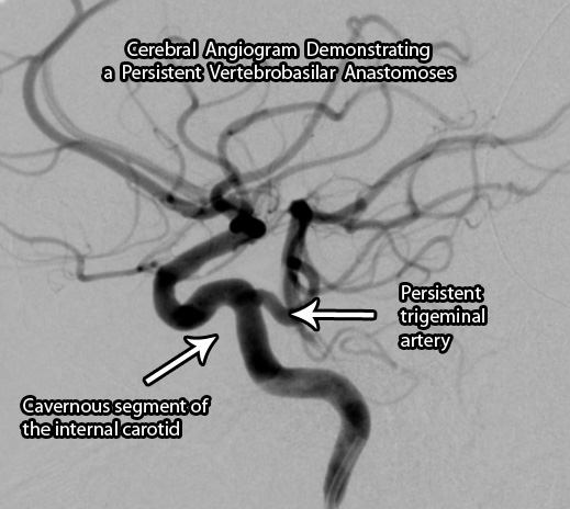
The most common of the persistent anastomoses is the trigeminal artery. This artery connects the cavernous segment of the internal carotid artery directly to one of the vertebral arteries. Saltzman further categorized these anastomoses into two types. A type I vessel has an absent ipsilateral (ie: same side) posterior communicating artery; whereas a type II has an ipsilateral fetal posterior communicating artery present. Individuals with trigeminal arteries are also at increased risk of developing aneurysms throughout their cerebral vasculature.

The least commonly seen anastomoses is the otic (acoustic) artery. This vessel arises from the petrous internal carotid artery and plugs into the basilar artery. This is the first vessel to regress in-utero and is rare to see in post-natal life.
Trigeminal Artery

The proatlantal intersegmental artery is a link between the cervical carotid artery and the vertebral artery. Like the trigeminal artery, it is further sub-divided into two different types. In a type I intersegmental artery the vessel arises from the internal carotid artery; whereas in a type II intersegemental artery the vessel arises from the external carotid artery.

The hypoglossal artery arises from the cervical internal carotid artery and plugs in to the basilar artery.

Persistent Carotid Vertebrobasilar Anastomoses
Name Vessels Connected
Trigeminal artery Cavernous internal carotid to vertebral artery
Otic (acoustic) artery Petrous internal carotid to basilar artery
Proatlantal intersegmental artery

Type I – cervical internal carotid artery to vertebral artery

Type II – external carotid artery to vertebral artery

Hypoglossal artery Cervical internal carotid artery to basilar artery

Overview

The persistent vertebrobasilar anastomoses are connections between the anterior and posterior cerebral circulations that fail to regress in-utero. Certain types are associated with increased risk of aneurysm formation. They are normally clinically silent and found incidentally during the work-up for other symptoms.

Related Articles

References and Resources

Enterobius Vermicularis and Itchy Butts

Enterobius vermicularis, better known as pinworm, is a nematode or roundworm. Like other worms it has a unique lifecycle that is quite interesting, albeit somewhat disgusting! It begins when an egg is ingested by a human. Eggs are usually ingested because of poor hygiene (ie: not washing your hands after doing number 2) and can sometimes be found in contaminated food. Once ingested the eggs hatch in the small intestine. From there the worms migrate to the large intestine where they mate. For unknown reasons the pregnant females head towards the anus at night where they lay their eggs.

The eggs are then shed in the feces and potentially picked up by another unlucky host. It is the most common worm related infection in the United States.

Signs and Symptoms

The eggs in the perianal area are extremely pruritic (ie: itchy). Scratching of the anus secondary to the intense pruritis can lead to skin breakdown and potential bacterial super infection. The symptoms are generally most severe at night because this is when the females migrate to the anus to lay their eggs. Systemic signs are generally not present although some patients can have malaise (ie: feeling "crappy").

Diagnosis

The traditional way of diagnosing is the "scotch tape" test. The clinician takes a piece of scotch tape and applies it to the patient’s perianal region. Eggs can be seen on the tape once its removed. The best time to perform this test is early in the morning before bathing or at night.

Treatment

Two anti-helminth medications are used to treat pinworm. The first is albendazole. Generally a single dose is enough to kill all worms, but since eggs may be present on clothing, bedding, etc. a second dose is often given two weeks later. Mebendazole is another medication that is also given as a single dose repeated two weeks later. These medications work by inhibiting microtubule polymerization in the cytoplasm of the worm’s cells.

Overview

Enterobius vermicularis (pinworm) is the most common worm infection in the United States. It causes an intense itch in the perianal area that is worst at night. It is diagnosed by the "scotch tape" test. Treatment is with albendazole or mebendazole.

Related Articles

References and Resources

Colloid Cysts of the Third Ventricle

Colloid cysts of the third ventricle are slow-growing, benign cranial tumors. They are believed to be composed of an epithelial wall with either mucous or protein like material trapped inside a spherical structure. However, their exact etiology is still under debate.

They are typically found in the anterior portion of the third ventricle near the foramen of Monroe (the channels that connect the lateral ventricles to the third ventricle). The third ventricle is one of the spinal fluid filled cavities of the brain.

Colloid cysts are “benign” because they are not cancerous (ie: don’t invade other parts of the body); however, they have the potential to block the flow of cerebrospinal fluid, which can lead to acute hydrocephalus and brain herniation. Therefore, in this regards they are certainly not “benign” tumors!

Signs and Symptoms

The most common presenting symptom of a colloid cyst is headache and difficulty walking. Acute hydrocephalus (dilation of the ventricular system secondary to blocked cerebrospinal fluid) can occur if the cyst blocks the flow of cerebrospinal fluid; this can cause nausea, vomiting, headache, and lethargy. Changes in mental status may also be seen in patients with these lesions.

There are numerous reports of patients dying suddenly from colloid cysts of the third ventricle. This is believed to be due to rapid obstruction of cerebrospinal fluid at the foramen of Monroe. The fluid builds up behind the blockage which puts pressure on the brain. Too much pressure can cause the brain to herniate through the base of the skull (see the Monro-Kellie doctrine).

Diagnosis

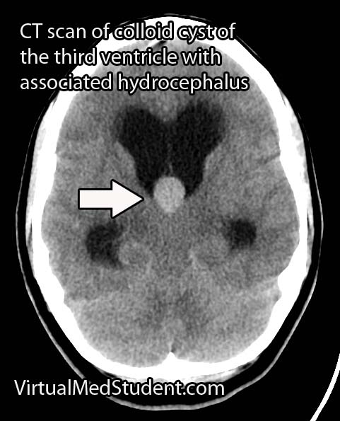
Colloid Cyst of the 3rd Ventricle CT
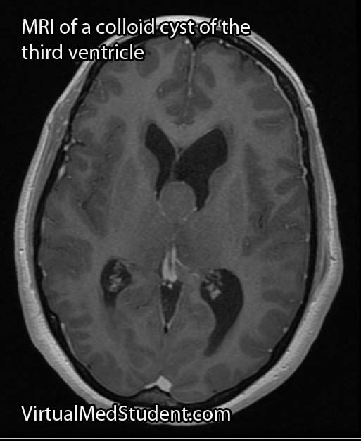
MRI of colloid cyst of the third ventricle
Diagnosis can be made with MRI or CT scan. Head CT scans will reveal a hyperdense (ie: bright or white colored) lesion. MRI is beneficial because it provides a superior picture of the regional anatomy around the cyst. Lumbar puncture should never be performed in a patient with a colloid cyst due to the risk of brain herniation.

Treatment

Treatment of colloid cysts is surgical. There are numerous approaches including the use of an endoscope, or the use of stereotactic guidance systems. In patients with contraindications to surgery bilateral cerebrospinal fluid shunts can be placed to prevent acute hydrocephalus from developing.

Overview

Colloid cysts of the third ventricle are "benign" tumors. They have the potential to block the flow of cerebrospinal fluid leading to acute hydrocephalus. The most common symptom is headache followed by gait instability. Diagnosis is made with CT and MRI imaging. Treatment is surgical resection.

Related Articles

References and Resources

Medulloblastoma: Sonic Hedgehog, Wingless, and Prognosis

Medulloblastomas are highly malignant brain tumors. They are the most common primary malignant brain tumor, and the second most common overall brain tumor in children. They are uncommonly seen in adults. Medulloblastomas are believed to arise from the granular cell layer of the cerebellum and are part of a broader category of tumors known as primitive neuroectodermal tumors (PNETs, coolloquially called "peanuts").

The term medulloblastoma is somewhat of a misnomer because it actually comprises several distinct pathologic types. These types include classic medulloblastoma, desmoplastic/nodular medulloblastoma, large cell medulloblastoma, anaplastic medulloblastoma, and medulloblastoma with extensive nodularity.

In addition to their pathologic appearance, medulloblastomas vary in their molecular make-up. There are currently four molecular categories. They include those that belong to the sonic hedgehog gene group (SHH), the wingless gene group (WNT), and two less well understood groups known as "group three" and "group four".

The SHH group contains roughly a third of all medulloblastomas. Aberrant activation of the SHH gene is responsible for the development of all pathologic types of medulloblastomas, but is most commonly seen in anaplastic, desmoplastic, and large cell types.

The least common molecular group is the WNT group. The wingless gene signaling pathway is extremely complicated and outside the scope of this article. Suffice it to say that aberrant activation of the WNT gene can cause medulloblastoma formation, most commonly of the classic variety.

The molecular nature of group three and four is still poorly understood.

As you can see, the classification of medulloblastomas is quite complex! Medulloblastomas can be categorized both molecularly and pathologically. The table below attempts to organize the complex nature of this heterogeneous group of tumors:

Pathologic Type Molecular Type Clinical Features Outcome
Classic SHH, WNT, group 3 and group 4 Midline location, mostly in children < 10 years old, second peak in 20 to 40 year olds Better prognosis
Large cell Group 3, group 4, SHH Uncommon, similar to anaplastic Worse prognosis
Anaplastic Group 3, group 4, SHH Midline with cysts, necrosis, and bleeding within tumor Worse prognosis
Desmoplastic SHH Located in the midline in children and off midline in adults Better prognosis
Extensive nodularity SHH Off midline and nodular architecture Better prognosis

Given the malignant nature of these tumors it is not uncommon for medulloblastomas to seed other areas of the central nervous system. Tumor frequently "coats" the spinal cord. These lesions are known as "drop" metastasis and are seen in 10% to 40% of patients at the time of diagnosis.

Signs and Symptoms

Patients with medulloblastoma can present with a variety of signs and symptoms. Headaches with nausea and vomiting secondary to obstructive hydrocephalus is frequently observed. In addition, signs of brainstem dysfunction including dizziness and trouble with eye movements may occur. Cerebellar signs like ataxia and dysdiadochokinesia are also commonly seen.

Diagnosis

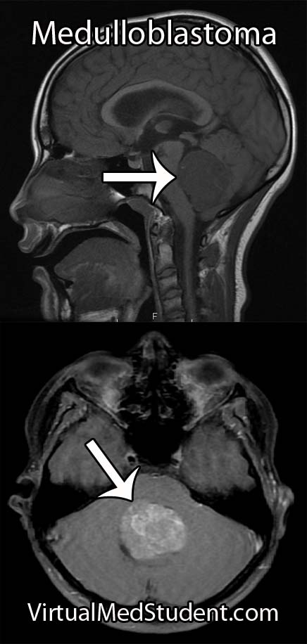
Medulloblastoma MRI
Characteristic imaging findings on MRI and CT scans, especially in the right age groups, can support the diagnosis. However, definitive diagnosis can only be made at the time of surgical resection by an experienced pathologist.

Treatment

Treatment is composed of surgical removal of the tumor, chemotherapy, and radiation. Surgery is always the first treatment because it decreases the disease "burden" so that radiation and chemotherapy can effectively treat any remaining tumor cells.

After surgery patients are classified as either “standard risk patients” or “poor risk patients”. Standard risk patients have complete surgical removal of their tumors and no dissemination of the disease to other areas of the central nervous system (ie: no “drop mets”). Poor risk patients have more than 1.5 cm2 of tumor left after surgery and evidence of dissemination in the cerebrospinal fluid.

Numerous chemotherapeutic medications including carboplatin, etoposide, cisplatin, cyclophosphamide, and vincristine have helped improve survival in poor risk patients. In addition, radiation therapy to the entire cranio-spinal axis has been shown to reduce recurrence rates.

Overview

Medulloblastoma is considered a malignant primitive neuroectodermal tumor. They are the second most common brain tumor in children, and the most common malignant brain tumor in children. They are rare in adults. There are several pathologic and molecular "sub-categories" of medulloblastoma; each category has different clinical features and outcome. Diagnosis is made with characteristic imaging findings in conjunction with pathologic analysis made at time of surgical resection. Treatment consists of surgery, radiation, and chemotherapy.

Related Articles

References and Resources

  • Northcott PA, Hielscher T, Dubuc A, et al. Pediatric and adult sonic hedgehog medulloblastomas are clinically and molecularly distinct. Acta Neuropathol. 2011 Aug;122(2):231-40.
  • Jones DT, Jäger N, Kool M, et al. Dissecting the genomic complexity underlying medulloblastoma. Nature. 2012 Aug 2;488(7409):100-5.
  • Northcott PA, Jones DT, Kool M, et al. Medulloblastomics: the end of the beginning. Nat Rev Cancer. 2012 Dec;12(12):818-34.
  • Byrd T, Grossman RG, Ahmed N. Medulloblastoma-biology and microenvironment: a review. Pediatr Hematol Oncol. 2012 Sep;29(6):495-506.
  • Robertson PL, Muraszko KM, Holmes EJ, et al. Incidence and severity of postoperative cerebellar mutism syndrome in children with medulloblastoma: a prospective study by the Children’s Oncology Group. J Neurosurg. 2006 Dec;105(6 Suppl):444-51.
  • Allen J, Donahue B, Mehta M, et al. A phase II study of preradiotherapy chemotherapy followed by hyperfractionated radiotherapy for newly diagnosed high-risk medulloblastoma/primitive neuroectodermal tumor: a report from the Children’s Oncology Group (CCG 9931). Int J Radiat Oncol Biol Phys. 2009 Jul 15;74(4):1006-11.
  • Packer RJ, Gajjar A, Vezina G, et al. Phase III study of craniospinal radiation therapy followed by adjuvant chemotherapy for newly diagnosed average-risk medulloblastoma. J Clin Oncol. 2006 Sep 1;24(25):4202-8.

Os Odontoideum: Floating Bone of the Axis

In order to understand what an os odontoideum is, we have to first appreciate the anatomy of the first two cervical vertebrae.

The first cervical vertebrae is known as the "atlas". It forms joints with the base of the skull and the second cervical vertebrae, which is also known as the axis. It has a an elongated structure on its ventral aspect called the “odontoid”. The odontoid of the axis connects to the atlas via numerous ligaments. This joint provides most of the flexibility that allows you to move your head in various directions.

An os odontoideum is a failure of the tip of the odontoid (ie: the part closest to the atlas) to fuse with its base on the axis.

Exactly why this occurs is still debated. The first theory is that it represents a congenital failure of the odontoid to fuse properly with the axis. The second, and more supported theory is that it may be caused by a previous fracture in early childhood that failed to heal properly. Regardless of the cause, the end result is a floating mass of bone that represents the superior (ie: top) most portion of the odontoid process.

This mass of bone may be fused to the base of the skull. If this is the case, the term "dystopic" os odontoideum is used. Or it may articulate and move with the atlas; if this is the case, the term "orthotopic" os odontoideum is used.

Signs and Symptoms

Many patients with os odontoideum are asymptomatic. However, because the tip of the odontoid is not technically connected to the base of the axis the patient may have an unstable neck. If the instability is severe, damage to the spinal cord can result causing myelopathy.

Myelopathy can manifest with several symptoms. Patients may have numbness and tingling in the upper and lower extremities. If damage to the nervous tissue responsible for motor movements occurs, patients may complain of weakness (and possibly even paralysis in extreme cases!).

On examination, patients may have both upper and lower motor neuron signs. Upper motor neuron signs refer to exaggerated reflexes – Babinski and Hoffmann signs, and clonus are all examples of this. These findings tend to be seen below the level of the actual spinal cord injury. Lower motor neuron findings typically occur at the level of the spinal cord damage, and consist of flaccid weakness with decreased reflexes.

Diagnosis

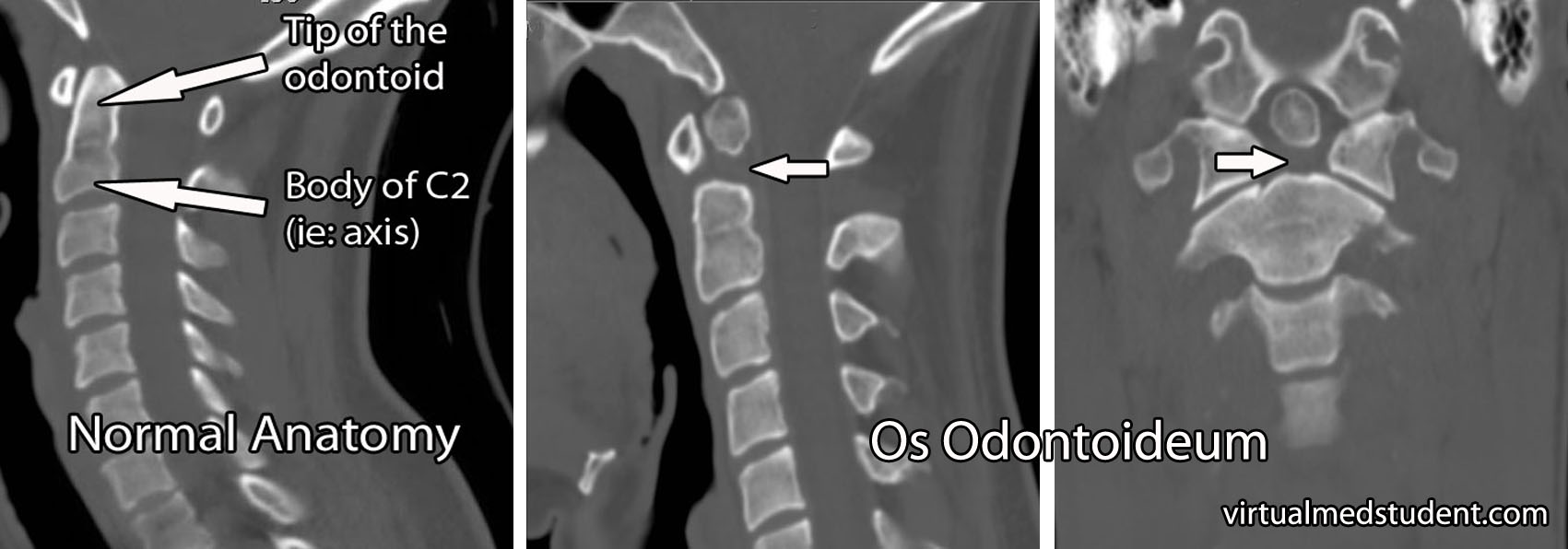
Diagnosis of os odontoideum is made by x-rays or CT of the cervical spine. To assess the degree of instability in the joint, some doctors will get flexion and extension x-rays as well.

The image to the right is a CT of the cervical spine that illustrates the missing portion of the odontoid process (marked by arrows in the image). A normal CT of the cervical spine is shown to the left for comparison.

Os Ondontoideum

Some patients may also get an MRI to assess for spinal cord and ligamentous injury, especially when symptoms or physical examination findings are present.

Treatment

Treatment depends on whether or not symptoms are present, and whether or not the cervical spine is unstable. Many patients without symptoms may be followed with serial X-rays or CT scans to assess for progression of instability.

If significant instability exists, or the patient has signs and symptoms consistent with spinal cord injury, then surgical stabilization is performed. There are numerous ways to achieve stabilization in this region surgically, which are outside the scope of this article. Regardless of which method is used, the end result is stabilization of the joint between the first and second cervical vertebrae.

Overview

Os odontoideum is an absence of part of the odontoid process. It may be due to a congenital malformation, or an early childhood fracture that fails to heal properly. Symptoms, when present, are due to spinal cord injury (ie: myelopathy) and consist of weakness, numbness, tingling, and other signs of spinal cord dysfunction. Imaging with x-rays or CT scan can show the bony defect. MRI is occasionally used to assess the spinal cord itself. Treatment depends on whether or not symptoms or significant instability is present. The best treatment options are surgical stabilization of the joint between C1 and C2 using one of several potential methods.

Related Articles

References and Resources

Basics of Pregnancy: GxPy, Naegele’s Rule, and that Bundle of Joy

This article will discuss the basics of pregnancy. In order to understand the basics we have to understand two important obstetric terms: gravity and parity.

Gravity refers to the number of times a woman has been pregnant. Parity refers to how many times the woman has given birth to an infant older than 20 weeks gestational age, or weighing greater than 500 grams.

On a medical chart a woman’s obstetric history is commonly written as “G x P y”. “G” refers to the gravity, or the number of times, “x”, a woman has been pregnant. The “P” stands for the parity, or the number of times, “y”, a woman has given birth to an infant older than 20 weeks gestation, or weighing greater than 500 grams. In other words the “y” equals the number of pregnancies that made it past 20 weeks gestation.

However, a woman’s obstetric history may also contain miscarriages, abortions, preterm births, and still births. In addition, some of her children may have died early in infancy. To account for these facts the “Gx Py” system is sometimes written as “G x P y, a, b, c, d”. The “a” refers to the number of term pregnancies; the "b" refers to the number of preterm pregnancies. The "c" refers to the number of abortions; abortions includes both spontaneous and induced, as well as ectopic pregnancies. And finally, the "d" refers to the number of living children the woman currently has.

So, as an example, a woman with two living children and one previous spontaneous abortion at twelve weeks gestation, who is currently pregnant for the third time would have an obstetric chart that reads "G 3 P 2, 2, 0, 1, 2."

Fetal Age nd Due Date

Pregnancy Wheel
There are different measures of fetal age. They include the following:

(1) Developmental age
(2) Gestational age

The developmental age is the age of the fetus measured from the time of fertilization. Since it is often difficult to determine exactly when fertilization occurred, a surrogate marker is used.

The surrogate marker is the gestational age. The gestational age is measured from the first day of the last menstrual period. Typically, the gestational age overestimates the developmental age by approximately two weeks.

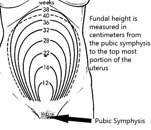
If the last menstrual period is not known there are other ways of determining the gestational age. One method is to use fundal height; fundal height is the location of the fundus (ie: the top most portion of the uterus) as felt by palpating the maternal abdomen. By 20 weeks of gestation the fundal height should be at the mother’s umbilicus (belly button). From there on, the height roughly correlates with gestational age (ie: at 28 weeks the fundal height is 28cm above the pubic symphysis).

The second method, albeit an even more imprecise one, is to use quickening. Quickening is the detection of fetal movements by the mother. It usually corresponds to a gestational age of 17 to 18 weeks. Ultrasound can also be used to determine the gestational age, and is the most reliable way, assuming the last menstrual period is not known.

In order to calculate the delivery date a simple mathematics trick known as Nagele’s rule is used. It states that the estimated delivery date is determined by adding nine months plus seven days to the first day of the last menstrual period.

Naegele’s rule -> Last menstrual period + 9 months + 7 days = expected due date

Pregnancy Tests

How exactly do you determine if someone is pregnant? The most common way is to use a urine pregnancy test. These tests detect a molecule known as beta-HCG (ie: beta human chorionic gonadotropin). Beta-HCG is a molecule secreted by the placenta. Its level doubles roughly ever 48 hours during the first few days of pregnancy. It peaks at about 10 weeks of gestation. Urine tests tend to have a high false negative rate. This means that they miss pregnancies, especially if the pregnancy has just begun.

A more sensitive test is the serum (ie: blood) pregnancy test. This test measures exactly the same molecule, beta-HCG, but it does so from a blood, rather than a urine sample. This test can pick up pregnancies earlier than a urine test can.

Related Articles

References and Resources

Bun in the Oven: Fetus Surveillance Techniques

Fetal growth is most commonly monitored by ultrasound. Most “routine” pregnancies begin with one fetal ultrasound between 18 and 20 weeks of gestation. This ultrasound can pick up important defects that may not be apparent at later gestational dates. If the 18-20 week ultrasound is normal, and the pregnancy is otherwise uncomplicated, repeat ultrasounds are not routinely performed.

Fetus

Fetal growth can also be monitored by measuring the "fundal height". The fundal height is the distance between the fundus (ie: top of the uterus), which is felt via palpation of the abdomen, and the pubic symphysis (see image to left).

A rule of the thumb is that the top of the uterus should be felt at the level of the umbilicus (ie: belly button) at approximately 20 weeks gestation. It then increases proportionally until about 36 weeks gestation. At this point the height may actually decrease as the fetal head enters the pelvis.

Fetal Maturity

There are three different ways to assess fetal maturity. They are used to determine if the fetus has adequate lung maturity to survive outside the uterus. These tests measure molecules that are present in the amniotic fluid. Therefore an invasive test known as an amniocentesis is necessary. The different methods are:

(1) Lecithin to sphingomyelin ratio
(2) Phosphatidylglycerol test
(3) Foam stability index

The first way of determining fetal lung maturity is to measure the lecithin to sphingomyelin ratio. Sphingomyelin is a specialized phospholipid that is produced in constant amounts by the fetus throughout pregnancy. Lecithin is a component of surfactant (ie: molecules that help keep the alveoli of the lung inflated) and its production increases as fetal lung maturity increases. An L:S ratio of greater than two indicates that the fetal lungs are mature enough to work outside the uterus.

The second test is known as the phosphatidylglycerol (PG) test. The presence of this molecule in the amniotic fluid indicates fetal lung maturity. PG is a component of pulmonary surfactant, which helps keep the gas exchange portions of the lung (ie: alveoli) from deflating at the end of exhalation.

The third test is the "foam stability index" test. This test measures the stability of bubbles when amniotic fluid is shaken with variable percent alcohol solutions. If the bubbles are stable this indicates fetal lung maturity.

Fetal Well-Being

Fetal well-being is assessed using several different methods. They include:

(1) Non-stress test
(2) Stress test
(3) Biophysical profile

Then on-stress test measures fetal well-being by looking at changes in the fetal heart rate during movement. A normal (aka: "reactive") non-stress test occurs when the fetus’ heart rate increases by 15 beats per minute over 15 seconds following fetal movement. If this increase in heart rate occurs twice within 20 minutes of monitoring the test is normal or “reactive”. Note: if the fetus is less than 32 weeks gestation then only a 10 beat per minute increase is needed to satisfy the non-stress test.

The stress test (aka: "contraction stress test", or "oxytocin stress test" depending on how contractions are elicited) measures the fetus’ ability to handle the "stress" of a uterine contraction. When the uterus contracts blood flow from the placenta is momentarily decreased. A healthy fetus will have no discernible change in their heart rate; however, an at risk fetus will have heart rate decelerations. Stress tests, especially oxytocin induced stress tests, commonly result in false-positives (ie: indicate fetal jeopardy when the fetus is normal); therefore, an abnormal stress test is followed by a non-stress test. If the non-stress test is normal/reactive then the stress test is often times considered a false-positive.

The biophysical profile uses five parameters for assessing fetal well-being. They include: fetal breathing movements, gross body movements, fetal muscle tone, fetal heart rate as measured by non-stress test, and qualitative amniotic fluid levels. Each of these parameters is given a score of 0 if absent/abnormal, or 2 if normal. A score of 8-10 is reassuring and indicates no problems with the fetus.

Related Articles

References and Resources

Vitamin B12: Function and Disease

Vitamin B12 and its derivatives serve many roles in the body. It functions primarily as a co-factor to help enzymes perform their molecular reactions. It is composed of the metal cobalt, which is coordinated to a large corrin ring structure composed of carbon and nitrogen atoms. Several important biochemical reactions in which vitamin B12 is a crucial component include:

(1) The conversion of odd chain fatty acids (specifically propionate) into succinate.
(2) The conversion of homocysteine into methionine via methyl group donation.

In the first reaction above B12 serves as a cofactor for the enzyme methylmalonyl-CoA mutase, which converts the odd chain fatty acid L-methylmalonyl-CoA into succinyl-CoA. Succinyl-CoA can then enter the citric acid cycle (Krebs cycle).

In the second reaction vitamin B12 serves as a methyl donor (ie: a "-CH3" unit) allowing the conversion of homocysteine into methionine. Interestingly, this reaction also requires a form of vitamin B9 (aka: folate or folic acid) known as N-methyltetrahydrofolate. The reason this reaction is important is because methionine will eventually go on to form S-adenosylmethionine, which is an important contributor of single carbon fragments to various other molecules.

Additionally, vitamin B12 is needed to convert N-methyltetrahydrofolate back into tetrahydrofolate, which serves as a cofactor in a slew of other important biochemical reactions.

Role in Disease

Patients typically become deficient in vitamin B12 in one of three ways: strict vegan diets, an autoimmune condition known as pernicious anemia, or impaired absorption via the gut.

Vitamin B12 is found mainly in animal products and vegans may become deficient if they fail to supplement. Patients with pernicious anemia have antibodies that attack the cells in the stomach that secrete a molecule (intrinsic factor), which helps the intestine absorb B12 from the diet. Any damage to the gut lining (ie: inflammatory bowel disease, celiac disease, etc.) can also cause impair absorption of B12.

Deficiencies of any cause usually take several years to develop because excess vitamin B12 is stored in the liver. These stores can last several years before becoming depleted.

When a person’s vitamin B12 level is low it can cause the accumulation of odd chain fatty acids. These are thought to "gunk-up" cellular membranes. This is most noticeable in the nervous system, and can result in neurological signs and symptoms including numbness and tingling, loss of proprioception (ie: the ability to "feel" where your limbs are in space), and difficulty with coordination. In its most severe form a condition known as subacute combined degeneration of the spinal cord can occur.

Vitamin B12 deficiency can also cause a megaloblastic anemia. This occurs because rapidly dividing cells in the bone marrow require a constant supply of tetrahydrofolate for methylation reactions in order to produce enough DNA. When B12 is deficient, N-methyltetrahydrofolate cannot be converted back to tetrahydrofolate. When this occurs all of the tetrahydrofolate gets trapped in the N-methyltetrahydrofolate form, which is not used as a methyl donor in nucleotide synthesis. The resulting DNA deficiency causes red blood cells to exit the bone marrow deformed, dysfunctional, and larger than normal (hence the term "megalo"-blastic anemia).

There is no evidence that excess vitamin B12 causes any significant health problems. It is well excreted in the urine.

Diagnosis

Deficiencies of vitamin B12 are diagnosed by measuring the amount of B12 in the blood. However, "normal" B12 levels do not necessarily rule out B12 deficiency, making this test less than useful.

Other blood tests that are often sent include methylmalonyl-CoA and homocysteine levels. Both of these will be elevated in B12 deficiency since the vitamin is necessary for the enzymatic conversion of these molecules to succinyl-CoA and methionine, respectively.

Although rarely used today, another test called the Schilling test can be performed. It involves several steps in which radiolabelled B12 is given orally. If the patient is having difficulty absorbing B12 because of either intrinsic factor deficiencies or intestinal problems the Schilling test will be able to differentiate the cause.

Treatment

Treatment of vitamin B12 deficiency is, you guessed it, giving the patient B12. Patients with pernicious anemia or intestinal absorption problems are given the vitamin intramuscularly. Other patients can supplement with oral forms.

Overview

Vitamin B12, also known as cobalamin, is important in several cellular reactions including the conversion of odd chain fatty acids into succinate and the replenishing of tetrahydrofolate (a form of vitamin B9 or folate). Deficiencies may occur in those who are vegan, have pernicious anemia, or have intestinal absorption problems. Diagnosis is made by measuring blood B12, homocysteine, and methylmalonyl-CoA levels. Treatment is with supplementation, either orally or intramuscularly.

Top

Related Articles

References and Resources

Thiamine (Vitamin B1): Wernicke-Korsakoff and Beriberi

Vitamin B1 (thiamine) and its derivatives serve many roles in the body. It functions primarily as a co-factor to help enzymes perform their molecular reactions. Several important biochemical reactions in which thiamine plays a crucial role include:

(1) Conversion of pyruvate to acetyl-CoA.
(2) Conversion of α-ketoglutarate to succinyl-CoA.
(3) Conversion of glyceraldehyde-3-phosphate to ribose-5-phosphate.
(4) It is used by the enzyme branched-chain α-keto acid dehydrogenase.

All of these reactions are crucial to human biochemistry. The conversion of pyruvate to acetyl-CoA feeds important molecules into the Krebs cycle, which allows the cell to produce energy.

The conversion of α-ketoglutarate to succinyl-CoA is necessary for the Krebs cycle to continue “spinning”. Without vitamin B1 (thiamine) the cycle would slow resulting in decreased energy production.

Thiamine is also important in the production of NADPH via the pentose phosphate pathway. NADPH plays a crucial role in biochemistry because it donates its electron pairs to numerous anabolic reactions.

The breakdown of branched chain amino acids such as valine, isoleucine, and leucine are also dependent on thiamine. The product that results from the breakdown of these amino acids are α-keto acids. They can also be fed into the Krebs cycle to replete molecules in the pathway that may have been siphoned off for other purposes. Maple syrup urine disease is a disorder of branched chain amino acid catabolism.

Since thiamine is involved in many energy producing pathways it is found abundantly in tissues that produce and use lots of energy. These include, but are certainly not limited to, the brain, muscle, and liver.

Role in Disease

Thiamine is a water soluble vitamin that is not actively stored in the body. It therefore must be obtained in the diet. Deficiencies can cause numerous disorders.

One such disorder is Wernicke-Korsakoff’s syndrome. It is seen in chronic alcoholics who may not be obtaining adequate nutrition. In Wernicke’s encephalopathy patients present with nystagmus (ie: jerky eye movements), ophthalmoplegia (ie: inability to move the eyes), and ataxia (ie: a wobbly unsteadiness).

These patients are often very confused. If Wernicke’s encephalopathy is left untreated it can progress to Wernicke-Korsakoff’s syndrome, in which the patient also develops short term memory problems and confabulations (ie: making up stories that aren’t true).

Another disease of thiamine deficiency is known as beriberi. There are numerous forms of this disease. It can affect infants who are being breast fed by thiamine deficient mothers, or who are receiving formulas with inadequate thiamine. In its worst case, infantile beriberi can manifest as severe heart problems.

Beriberi can also affect adults. The adult form exists in two types: wet and dry. Dry beriberi refers to the development of symmetric neuropathies (ie: damage to the peripheral nerves). Both sensory and motor nerves can be involved, and it tends to affect the parts of the nerve furthest from the spinal cord (ie: near the hands and feet). Wet beriberi is the combination of neuropathy plus heart problems, which can range in severity from cardiomegaly (ie: an enlarged heart) to congestive heart failure.

There is no evidence that excess thiamine (vitamin B1) causes any significant health problems. It is well excreted by the kidneys.

Diagnosis

Deficiencies of vitamin B1 (thiamine) are diagnosed by adding the vitamin to a preparation of the patient’s red blood cells. An increase in the activity of the transketolase enzyme is diagnostic of deficient levels.

Treatment

Treatment of thiamine deficiency is, you guessed it, giving the patient thiamine. If the patient has significant disease such as Wernicke’s encephalopathy thiamine can be given IV. Otherwise, oral supplementation and/or "prescribing" a diet rich in thiamine can be used to replenish any deficiencies.

Overview

Vitamin B1, also known as thiamine, is a vital component of numerous cellular reactions, especially those involving carbohydrate and amino acid catabolism (ie: breakdown). When deficiencies exist, several different diseases can occur. They include, but are not limited to, Wernicke-Korsakoff’s syndrome and beriberi. There are no known problems with excessive levels of thiamine since excess vitamin is efficiently excreted in the urine. Diagnosis of deficiencies are made by adding the vitamin to a preparation of the patient’s red blood cells. An increase in the activity of transketolase, an enzyme that relies on thiamine as a co-factor, is considered diagnostic of deficiency.

Related Articles

References and Resources

Pyogenic Liver Abscesses: Pus, Needle Drainage, and Antibiotics

Pyogenic liver abscesses are localized collections of pus and bacteria. The initial infection occurs when bacteria travel through the portal vein. This most commonly occurs after bowel contents leak into the peritoneal cavity from the gut. Other causes of liver abscesses include direct spread of infection from the bile duct system, or from other bacterial infections of the blood. Rarely, penetrating injuries (gunshots, stab wounds, surgery) may directly introduce infection. There are numerous “bugs” that can cause pyogenic liver abscesses. They include, but are not limited to, streptococcus species, klebsiella pneumoniae, and staphylococcus species.

Signs and Symptoms

Similar to other infections, liver abscesses can cause fevers, chills, decreased appetite, abdominal pain, and a generalized sense of not feeling well (ie: malaise). Interestingly, hiccups may also be present if the abscesses are causing adjacent inflammation/irritation of the diaphragm.

Diagnosis

Pyogenic Liver Abscesses
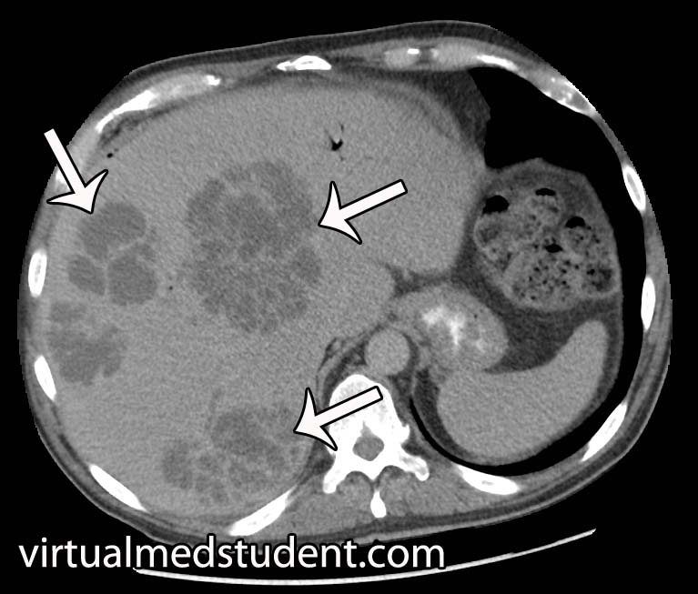
The diagnosis of pyogenic liver abscesses is made with imaging. The most commonly used method is a CT scan of the abdomen with and without contrast. If present, a liver abscess will look like a collection of fluid, with or without septated dividers.

Another commonly employed imaging modality is the use of ultrasound to detect the fluid filled pockets within the liver.

However, it is important to note that imaging studies alone cannot distinguish between the different types of liver abscesses. Imaging can also not tell you what bacteria is responsible for the abscess.

Treatment

Pyogenic abscesses must be drained and treated with antibiotics. Drainage is usually done under image guidance using a needle placed through the skin into the abscess. Although occasionally direct surgical evacuation of the abscess is necessary.

Draining the abscess is important for two reasons. First, it decreases the size of the abscess allowing antibiotic therapy to work more effectively. And secondly, it provides abscess fluid that can be sent to the lab for bacterial culture and gram stain.

The results of the culture help guide subsequent antibiotic therapy. Commonly used antibiotics include piperacillin-tazobactam (Zosyn®), vancomycin, metronidazole (Flagyl®), and ceftriaxone; once cultures confirm the causative bug antibiotic therapy can be narrowed.

Overview

Pyogenic liver abscesses are collections of pus and bacteria. They occur most commonly after the spilling of gut bacteria into the peritoneal space (ie: peritonitis). There are numerous causative bacteria. Symptoms include fever, chills, decreased appetite, and abdominal discomfort. Treatment is with drainage and antibiotics.

References and Resources

  • Hasper D, Schefold JC, Baumgart DC. Management of severe abdominal infections. Recent Pat Antiinfect Drug Discov. 2009 Jan;4(1):57-65.
  • Benedetti NJ, Desser TS, Jeffrey RB. Imaging of hepatic infections. Ultrasound Q. 2008 Dec;24(4):267-78.
  • Mortelé KJ, Segatto E, Ros PR. The infected liver: radiologic-pathologic correlation. Radiographics. 2004 Jul-Aug;24(4):937-55.
  • Kurland JE, Brann OS. Pyogenic and amebic liver abscesses. Curr Gastroenterol Rep. 2004 Aug;6(4):273-9.