Hodgkin’s Lymphoma: B-Cells, Pel-Ebstein, and EBV

Lymphoma is a cancer that develops from cells in the body known as lymphocytes. Lymphocytes are a subcategory of white blood cells, which are the cells that help ward off infection. There are two different types of lymphocytes: B-cells and T-cells. The majority of lymphomas, including Hodgkin’s disease, stem from B-cells.

In Hodgkin’s disease a B-cell, for unknown reasons, becomes cancerous. The cell then makes many clones of itself. These cells bundle together to form a solid tumor known as a lymphoma.

Why B-cells in Hodgkin’s lymphoma become cancerous is not entirely known. One belief is that infection with Epstein-Barr virus (the same virus that causes infectious mononucleosis) can cause the cells to turn cancerous in genetically susceptible people. Other theories are that certain genetic translocations may be the underlying factor. As of yet, no particular theory has significant supporting data to call it the true cause. It is likely that the development of Hodgkin’s disease is multi-factorial.

Types

Reed-Sternberg Cell

There are different types of Hodgkin’s lymphoma. They are based on unique histopathological (ie: what it looks like under a microscope) characteristics, and are important in determining prognosis.

The histopathological features the pathologist looks for are the number of Reed-Sternberg cells, as well as the number of lymphocytes present in the biopsy specimen. A Reed-Sternberg cell is a funny shaped cell with two nuclei that looks like an "owl’s eyes" (see image to the right). They are believed to form when two cells merge together under the influence of certain proteins produced by the Epstein-Barr virus.

The first category, and most common type, is nodular sclerosing Hodgkin’s lymphoma. In this type, there are very few Reed-Sternberg cells with a moderate number of lymphocytes. It commonly occurs in younger individuals, and with treatment, the prognosis is excellent.

The second category is mixed cellularity Hodgkin’s lymphoma. This type has many Reed-Sternberg cells, and a moderate number of lymphocytes when viewed under the microscope. It has an intermediate prognosis.

The third category is lymphocyte predominant Hodgkin’s. It has very few Reed-Sternberg cells and many lymphocytes. It occurs most commonly in males less than 35 years of age. It is also one of the few types that is not associated with Epstein-Barr virus infection.

The last category is lymphocyte depleted. It is the rarest form of Hodgkin’s lymphoma. It typically affects older males.
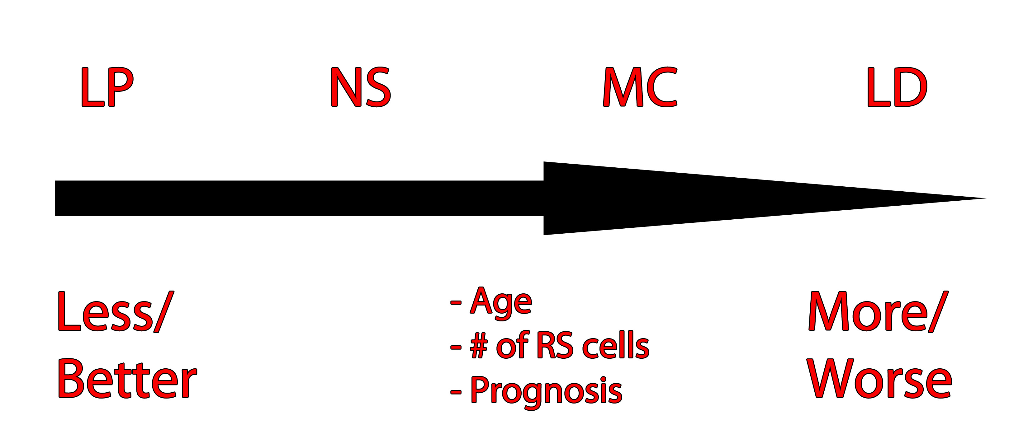
Hodgkin's Types
Unfortunately it has the worst prognosis of the four types types.

The image to the left is one way of organizing the different Hodgkin’s types and their prognosis based on age, number of RS cells, and prognosis. LP = lymphocyte predominant, NS = nodular sclerosing, MC = mixed cellularity, LD = lymphocyte depleted.

Signs and Symptoms

The classic presentation of Hodgkin’s lymphoma is painless enlargement of the lymph nodes. This is similar to non-Hodgkin’s lymphoma, and the only way to differentiate the two is through biopsy.

Systemic manifestations may occur and include night sweats, fever, and weight loss. However, these are more common in patients with disseminated (ie: metastatic) disease. Interestingly, a pathognomonic (ie: seen exclusively in Hodgkin’s lymphoma) feature that occurs in some cases is pain of the involved nodes after drinking alcohol. Finally, a symptom known as Pel-Ebstein fevers are also specific for the disease. A Pel-Ebstein fever is a cyclical fever that occurs for several weeks at a time followed by a fever free period.

Other signs related to the immune system can be seen in patients with Hodgkin’s lymphoma. A condition known as cutaneous anergy can occur. Anergy refers to a lack of response by the cell mediated immune system. For example, in patients with tuberculosis a reaction will occur underneath the skin when they get a TB test. This reaction is the result of their cell mediated immunity reacting to the tuberculosis components injected underneath the skin. However, in anergic patients no reaction is seen, even if they have tuberculosis! This can also occur in patients with Hodgkin’s disease.

Diagnosis
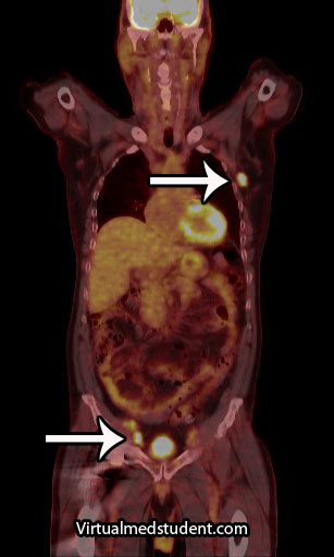
Hodgkin's Disease PET-CT
Diagnosis of Hodgkin’s lymphoma is made by looking at a biopsy specimen underneath the microscope. The most common way of obtaining a specimen is through biopsy of a lymph node. Differentiating Hodgkin’s from other types of lymphomas is important because it determines the best treatment options.

Additional studies are often performed to determine the number and location of involved lymph nodes. One such study is a positron emission tomograph (PET) combined with a CT scan. Any lymph nodes involved "light up" on the scan. An example, with the arrows pointing to involved nodes, is shown to the right.

Staging

Staging of Hodgkin’s lymphoma was traditionally based on a system known as the Ann Arbor Classification. It is divided into four stages. In stage 1 disease a single lymph node, or single organ is involved. In stage 2 disease involvement of multiple (two or more) lymph node regions on the same side of the diaphragm is present. In stage 3 disease involvement of nodes on both sides of the diaphragm is present; the spleen or other limited organ involvement may also be present. In stage 4 disease multiple organs are involved; interestingly, lymph node involvement is not necessary for a stage 4 diagnosis, although it is commonly present. Finally, each stage is further divided into “A” and “B” depending on whether or not symptoms are present. If symptoms are present, the stage is upgraded to a “B”.

Ann Arbor Classification (simplified)
Stage 1 Single lymph node or organ
Stage 2 Multiple lymph nodes on same side of diaphragm
Stage 3 Lymph nodes on both sides of diaphragm
Stage 4 Multiple organs involved

However, more recently the Lugano staging classification has become the preferred method. It divides disease into limited and advanced. Limited disease includes stage 1 and stage 2. Stage 1 involves a single lymph node or nearby nodes. Stage 2 involves two or more nodal groups. Advanced disease includes stages 3 and 4. In stage 3 disease nodes on both sides of the diaphragm are involved as is the spleen. Stage 4 disease is disseminated disease into organs. There are sub-categories in the Lugano model as well.

Treatment

Like most cancers treatment is highly dependent on the stage of the disease. In most cases chemotherapy and radiation are used. Radiation is directed at involved lymph nodes, as well as lymph nodes that are uninvolved, but nearby. Common chemotherapeutic agents used include: adriamycin, bleomycin, vinblastine, vincristine, prednisone, procarbazine, and mechlorethamine.

Overview

Hodgkin’s lymphoma is a cancer of a type of white blood cell known as a B-cell. There are numerous categories depending on its histopathological characteristics. Patients often have painless enlarged lymph nodes. Some patients have fever, weight loss, and other non-specific symptoms. Staging is based on the Ann-Arbor model. Treatment usually involves a combination of chemotherapy and radiation.

Related Articles

References and Resources

SOCRATES: Thinking About Pain

When I was in medical school one of the most useful mnemonics I came across was "SOCRATES". The mnemonic is designed to figure out the characteristics of someone’s pain. The characteristics of pain help the clinician develop a differential diagnosis from which testing can be ordered, and then hopefully, treatment can be given.

So what does each letter in the mnemonic SOCRATES stand for??? Let’s go letter by letter…

S   O   C   R   A   T   E   S

The first “S” stands for “site”. What body part or parts are involved? Is the pain in the leg? Is it in the abdomen? Is it a general sense of overall discomfort? The site of pain helps you fine tune your subsequent physical exam and diagnostic decision making.

The next letter is “O”, which stands for “onset”. When did the pain start? Asking about the onset of the pain is extremely important! For example, if someone has had chronic low back pain for 10 years that invokes a much lower sense of urgency than someone complaining of the sudden onset of severe belly pain or headache.

S O C R A T E S 


S – Site
O – Onset
C – Characteristics
R – Radiation
A – Associated
T – Timing
E – Exacerbating/
      Alleviating
S – Severity

“C” stands for “characteristics”. What are the characteristics of the pain? You want the patient to describe the pain in their own terms without influencing them too much. The pain may be sharp, dull, heavy, burning, etc, or a combination of descriptors.

The next letter is “R”, which represents “radiation”. I typically ask if the pain stays at the site or if it travels somewhere else in the body. For example, someone with chest pain radiating to the left arm might be experiencing a heart attack. Back pain that is associated with radiation down the leg might indicate a herniated lumbar disc that may require surgery. Back pain radiating to the abdomen could be intraabdominal pathology. Radiation of the pain is an important component to help guide your decision making.

“A” stands for associated symptoms. What other symptoms are present with the pain? For example, if the patient is complaining of belly pain do they also have nausea or vomiting? If they have a headache do they also complain of double vision or photophobia? Associated symptoms can provide a wealth of information to help you hone your differential diagnosis even more.

“T” stands for timing. When does the pain occur? Does it happen at specific times of the day, or is it constant? Does it happen during a certain movement? All of these can give you an idea of the origin for the pain.

The letter “E” represents “exacerbating” factors; grouped within this is also alleviating factors. The patient should be probed as to what makes their pain better or worse. Certain physical positions, medications, etc. may make the pain better or more unbearable. These factors can all provide historical clues about the root cause.

The final “S” stands for “severity”. In most hospitals this is formulated on a 1 to 10 scale with 10 being the most severe pain they’ve ever experienced. This can be a tricky one to gauge because many patients will describe 10 out of 10 pain when they are lying comfortably in bed; therefore, it is often necessary to ask more pointed questions and place pain in a context.

Overall, the answers obtained when using the mnemonic SOCRATES can provide a solid framework from which to order new testing and treatments.

More Good Stuff…

Maroteaux-Lamy Syndrome: GAGs and Dermatan Sulfate

Maroteaux-Lamy syndrome falls into a family of conditions called the “mucopolysaccharidoses”. This fancy term describes any disorder where patients are unable to metabolize long chains of repeating sugar units.

Another term for mucopolysaccharide is glycosaminoglycan, or “GAG”. GAGs are composed of long, repeating, un-branched sugar chains. Each repeating unit has a six carbon sugar (ie: the general term for a simple six carbon sugar is a “hexose”; glucose, mannose, galactose, etc. are specific examples) attached to another six carbon sugar that has a nitrogen group sticking off of it; the general name for a six carbon sugar with an attached nitrogen group is a “hexosamine”.

These two-sugar units, a hexose plus a hexosamine, connect to one another over and over again to form a very long chain called a glycosaminoglycan.

GAGs and mucopolysaccharides are important in the human body because they make up a significant proportion of connective tissue. Connective tissues provide support and structure to many of the organ systems. As such, the body is constantly forming, breaking down, and re-molding GAGs to provide the structural framework so that the rest of our cells can do their jobs well.

So what do GAGs have to do with Maroteaux-Lamy disease? A specific GAG known as dermatan sulfate is not broken down appropriately in patient’s suffering from this condition.

The reason these individuals are unable to break down dermatan sulfate is because they have a genetic mutation in the enzyme arylsulfatase B. In a normal person, arylsulfatase B degrades excess and unwanted dermatan sulfate; however, when the enzyme is defective it causes dermatan sulfate to build up to abnormally high levels within cells.

This excess dermatan sulfate eventually “gunks” up various organ systems and results in the characteristic signs and symptoms associated with the disease.

Signs and Symptoms

It is important to realize that there are numerous genetic mutations that can cause Maroteaux-Lamy syndrome and not all mutations are created equal! Therefore, there is a wide spectrum of signs and symptoms that may occur quickly, or slowly, depending on which particular mutation the patient has.

Most patients are short and have progressive changes in their facial features. Patients often develop early onset vision problems because their corneas become clouded by excess dermatan sulfate. Joint stiffness and skeletal abnormalities are also very common, as are heart and lung complications.

The lining of the brain and spinal cord can become excessively thick which can cause weakness secondary to spinal cord and nerve compression.

Patient’s also often have enlargement of the spleen – a condition called “splenomegaly”. It is also not uncommon for patients to have various types of hernias.

Unlike patients who suffer from some of the other mucopolysaccharidoses, people with Maroteaux-Lamy are of normal intelligence.

Diagnosis

Overall, Maroteaux-Lamy disease is more appropriately a “syndrome”, or constellation of signs and symptoms. By themselves, each sign or symptom is not diagnostic of the disorder, but when present together can support the diagnosis.

Genetic testing for mutations of the arylsulfatase B gene are done once the syndrome is suspected on clinical grounds. Additionally, urine samples contain elevated amounts of dermatan sulfate.

Treatment

Treatment consists of using a replacement enzyme known as Naglazyme® (aka: galsulfase). This enzyme performs the function of the deficient arylsulfatase B and helps in the degradation of dermatan sulfate.

Stem cell transplants can also provide the missing enzyme, but at significant risk. Unfortunately, few patients are candidates for stem cell transplant given difficulty with donor matching.

A very important component of treatment is monitoring for the development or progression of symptoms. This will allow specialists to step in and perform palliative procedures that can prevent worsening disability. For example, corneal transplants can be done to restore vision loss.

Overview

Maroteaux-Lamy is one of the mucopolysaccharidoses. It is caused by a genetic deficiency in the enzyme arylsulfatase-B, which prevents the degradation of dermatan sulfate. It presents with numerous signs and symptoms, which may be rapidly or slowly progressive depending on the specific genetic mutation. Treatment is with enzyme replacement therapies and palliative management of symptoms.

Related Articles

References and Resources

  • Calleja Gero ML, González Gutiérrez-Solana L, López Marín L, et al. Neuro-imaging findings in patient series with mucopolysaccharidosis. Neurologia. 2011 Dec 15.
  • Mtar A, Charfeddine B, Braham I, et al. Maroteaux-Lamy syndrome: a case report. Ann Biol Clin (Paris). 2011 Dec 1;69(6):693-697.
  • Golda A, Jurecka A, Tylki-Szymanska A. Cardiovascular manifestations of mucopolysaccharidosis type VI (Maroteaux-Lamy syndrome). Int J Cardiol. 2011 Jul 5.
  • Valayannopoulos V, Wijburg FA. Therapy for the mucopolysaccharidoses. Rheumatology (Oxford). 2011 Dec;50 Suppl 5:v49-v59.

Glioblastoma: A Real Beast of a Tumor

In order to understand what a glioblastoma is we have to first appreciate the different cell types that compose healthy brain tissue. Brain tissue has both neurons and glia. Neurons are the “action” cells of the brain. Glia are the “helper” cells of the brain. They ensure that neurons stay healthy.

A glioblastoma is a malignant brain tumor that arises from a specific type of glial cell known as an astrocyte. Glioblastomas are not only the most common astrocytic tumor, but they are also the most common primary brain tumor!

It is important to realize that there are less malignant tumors that arise from astrocytes (discussed in other articles). Many of these tumors have a much better prognosis, which is why it is important to distinguish glioblastoma from less malignant behaving tumors.

The first distinguishing characteristic is neovascularization. Neovascularization is a fancy medical term used to describe the proliferation of blood vessels within the tumor. As the tumor grows, it requires new vessels to feed it oxygen and nutrients; the process of neovascularization allows the tumor to obtain these essential factors so that it can continue to grow.

Interestingly, as the tumor expands, sections of it will get choked off from its own blood supply. The end result is that part(s) of the tumor actually dies. This is referred to as "necrosis", which is a common finding in glioblastoma.

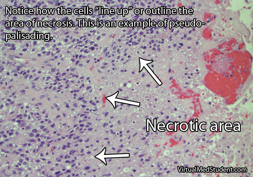
One of the most distinguishing features of glioblastomas is when cancerous astrocytes "line up" and outline areas of necrosis in a process known as pseudopalisading (see image below).

Given the malignant nature of glioblastoma it is common to see many mitotic figures. Mitotic figures are cells in various states of cell division; these figures indicate a relatively rapidly growing tumor type.

Glioblastoma Pathology - Pseudopalisading

Glibolastoma is therefore characterized by the following pathological characteristics: prominent microvascular proliferation (ie: development of new blood vessels within the tumor), mitosis (ie: an indicator of cell division/growth), and necrosis (ie: areas of dead tumor); pseudopalisading necrosis is a specific form of necrosis shown in the above image that is a hallmark of glioblastomas.

Signs and Symptoms

Glioblastomas may present with any number of signs and/or symptoms depending on their location within the brain. Lesions that are located on the left side of the brain may cause problems with speech if they involve the Broca or Wernicke areas. Tumors in the areas of the brain that control motor movement may cause weakness. Additionally, tumors that arise in the frontal lobes may cause odd behavioral changes. Some patients present with seizures, and others with only a dull headache.

Diagnosis

An official diagnosis of glioblastoma can only be made when a pathologist looks at a sample of the tumor under a microscope. These samples are typically obtained by a neurosurgeon who resects or biopsies the tumor.

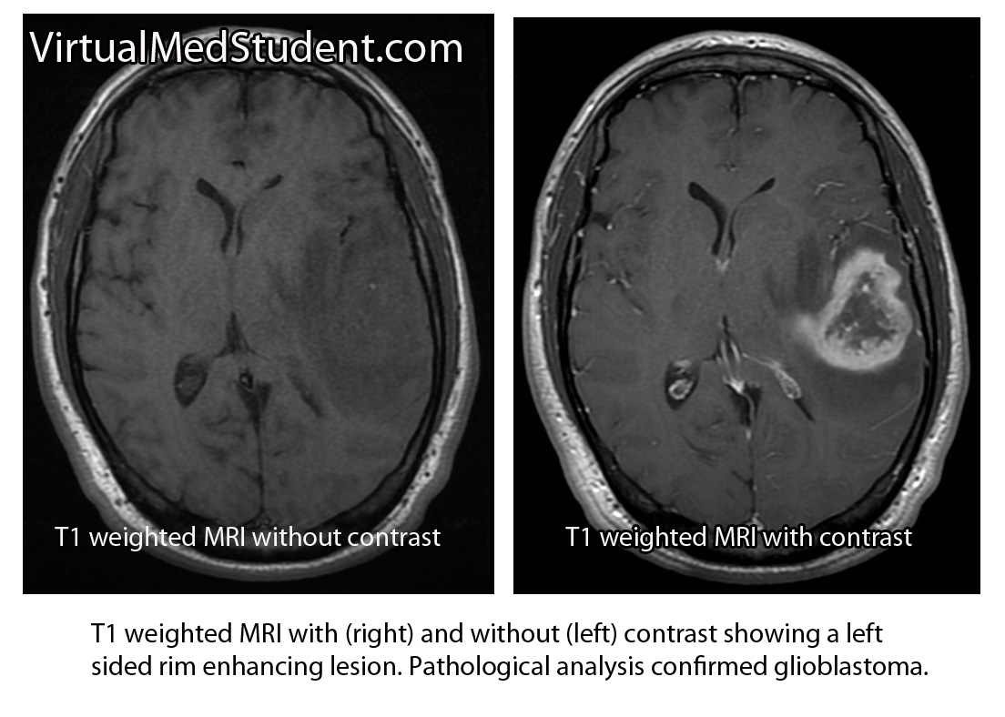
However, glioblastomas also have typical features seen on imaging studies such as MRI. For example, these tumors will “rim-enhance” when a contrast material such as gadolinium is infused into the patient during the scan. Rim enhancement is a result of the contrast material leaking out of all of the blood vessels present within the tumor. It is important to note that other diseases such as abscesses, lymphomas, and other infections can also cause rim-enhancement.

MRI of glioblastoma

Another useful study known as MR spectroscopy measures the relative amounts of different molecules present within the tumomr. In a glioblastoma the amount of lactate, choline, and lipid are all increased. Lactate is a marker of brain tissue that is not receiving enough oxygen, which is common in necrotic tumor areas. Choline is a molecule that is present in cell membranes. When neurons are rapidly dividing, which is what occurs in glioblastoma, the amount of choline present also increases. A different molecule known as N-acetyl aspartate (NAA) is present in mature cells. Therefore, unlike lactate and choline levels, NAA is decreased in glioblastoma because these cells are "immature" (ie: poorly differentiated).

Treatment

Treatment combines a mixture of surgery, radiation therapy, and different chemotherapeutic drugs, the most common being temozolomide (Temodar®). Surgery is only useful when a significant amount of the tumor can be removed. Despite optimal treatment the prognosis for patients with glioblastoma remains extremely poor.

It is also highly important to treat the edema that frequently surrounds the tumor. Steroids, most commonly dexamethasone (Decadron®), are used to decrease the amount of edema, which usually improves symptoms.

Patients are often started on an anti-seizure medication such as levetiracetam (Keppra®) or phenytoin (Dilantin®).

Overview

Glioblastoma is a malignant astrocytic tumor. It is the most common primary brain tumor. It has unique characteristics that distinguish it from more benign brain tumors that also arise from astrocytes. It is treated with a combination of surgery, radiation, and chemotherapy.

References and Resources

The Hangman’s Fracture

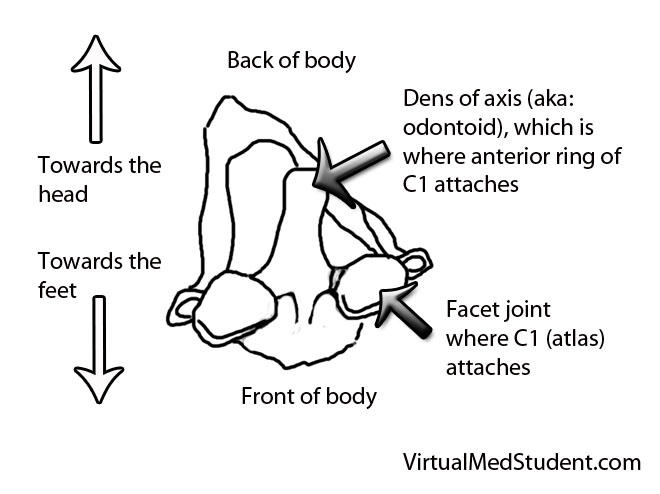
The axis, or second cervical vertebrae (C2), is unique amongst the vertebrae of the spine. It contains a body, which has an elongated structure that extends towards the head known as the dens (aka: odontoid). It also contains a ring-like structure that extends around the spinal cord, which is composed of the pedicles, pars interarticularis, and lamina. It forms joints with the atlas (ie: first cervical vertebrae) above it, and the third cervical vertebrae below it. It also has two foramen transversarium on either side, which are conduits for the vertebral arteries.

A hangman’s fracture is a break in both pedicles and/or pars interarticulari. The terms pedicle and pars interarticularis are not as well defined at C2 as they are for the other vertebrae, and thus have to be more clearly delineated before we discuss the details of a hangman’s fracture.

The pars interarticulari of C2 are the narrow pieces of bone that sit between the superior articulating facets (ie: the portion of bone that allows C2 to form a joint with C1) and the inferior articulating facets (ie: the portion of bone that allows C2 to form a joint with C3) of C2. The pedicles lie more anterior and are defined as the bony "bridges" that lie underneath the superior articulating facet and just medial (ie: closer to the spinal cord) to the transverse foramen, which house the vertebral arteries.

So now let’s get to the good stuff… Why do these fractures happen? Extension of the neck! This is why they are colloquially termed "Hangman’s" fractures; in the old days when a person was hanged the noose would pull the mandible upwards and cause the neck to violently extend. The end result was a tremendous amount of force on the pars interarticulari and pedicles of C2 leading to a fracture.

Since hangings are infrequent in today’s society, a more common cause of hyperextension of the neck is a head hitting the steering wheel or windshield of a car.

The hyperextension also often causes significant anterior ligamentous injury. The anterior longitudinal ligament (ie: the ligament that runs down the front of the spine) and the annulus fibrosis of the C2 disc are often ruptured. These findings are consistent with a hyperextension injury as the ligaments in the front get stretched to the point of rupture.

Signs and Symptoms

Surprisingly, hangman’s fractures rarely cause neurological injury. Most patients are neurologically intact meaning that there is no injury to the spinal cord and/or nerves at the level of the fracture. Typically there is neck pain, which is the most common symptom.

It is important to realize that many patients with hangman’s fractures will also have co-existent head trauma, and roughly a third of patients will have additional spine fractures. So keep a look out for associated injuries!!!

Diagnosis

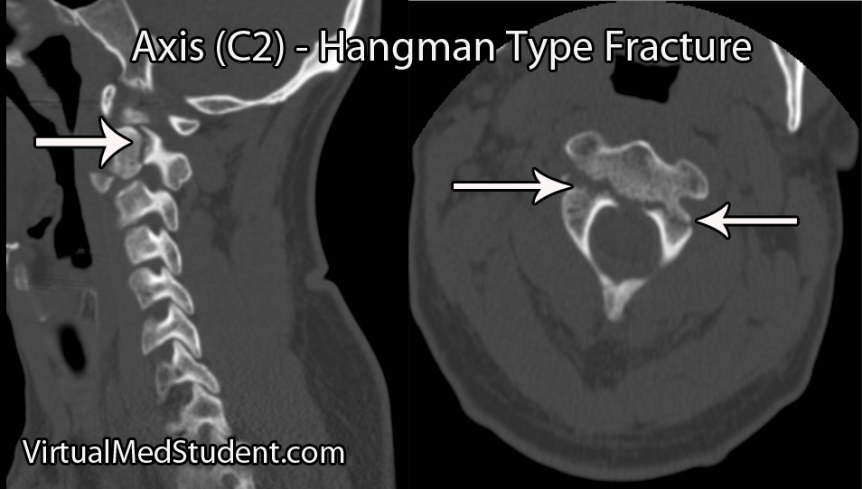
The diagnosis of a hangman’s fracture can be made using x-rays and CT scans. MRI scans are also frequently ordered to determine the extent of co-existent ligamentous and soft tissue injury. A CT angiogram or MR angiogram should also be done to assess for co-existent vertebral artery injury.

Hangman's fracture

There are several different grading systems for hangman’s fractures. They include the Effendi, Francis, and Levine and Edwards classifications.

The Effendi system is based on the orientation of the fracture, as well as the degree of angulation and dislocation between C2 on C3.

The Francis system also takes into account the angulation and displacement between the bodies of C2 and C3, which is measured between the inferior endplate of C2 and the superior endplate of C3.

Perhaps the easiest to implement clinically is the Levine and Edwards classification. A type I Levine fracture is a non-displaced, non-angulated fracture. Type II fractures come in two flavors: type II is a fracture that is significantly angulated (ie: > 11 degrees) and displaced (ie: greater than 3mm) and a type IIa fracture is angulated (ie: greater than 11 degrees), but not significantly displaced. Type III fractures are fracture-dislocations of C2 on C3.

The Levine and Edwards’ Classification System
for Hangman’s Fractures

Type Angulation Displacement Treatment
Type I Minimal Minimal Rigid orthotic
Type II Greater than 11 degrees Greater than 3mm Traction ± rigid orthotic ± surgery
Type IIa Greater than 11 degrees Minimal Traction ± rigid orthotic ± surgery
Type III Minimal to severe Significant (fracture/dislocation) Traction + surgery

Treatment

Most isolated hangman’s fractures can be treated with external immobilization in a rigid cervical collar (ie: Miami J or Philadelphia collar) or in a halo immobilization device.

However, if there is significant ligamentous disruption, severe angulation and/or dislocation, or the inability to obtain adequate alignment of the spine in an immobilization device (ie: rigid collar or halo) then internal surgical fixation and fusion should be performed.

The need for surgery depends on the severity of the fracture and/or the integrity of the associated ligaments and discs.

A surgical approach from the front (aka: an "anterior" approach) may be performed to fuse the C2 and C3 vertebrae by removing the disc material between them. This approach is most often done in the presence of anterior longitudinal ligament rupture and/or intervertebral disc protrusion (ie: a "traumatic" disc).

Surgery from behind may also be used (aka: a "posterior" approach). Usually, the 1st through 3rd cervical vertebrae are incorporated into the fusion process, but some surgeons may opt to fuse to the base of the skull in cases of more severe injuries.

Overview

Hangman’s fractures occur after violent extension of the neck. The pedicles or pars interarticulari are fractured on both sides of the C2 ring. Neurological injury is rare in isolated hangman’s fractures, but frequently there are associated injuries to other bones in the cervical spine, as well as injuries to the brain and face. Diagnosis is made with x-rays, CT scans, and MRI. Treatment is with rigid immobilization of the cervical spine and/or surgical fixation depending on the extent of injury.

Related Articles

References and Resources

Latin for Toothlike: Fractures of the Odontoid Process

The odontoid process (also know as the "dens") is the finger of bone that sticks up from the second cervical vertebrae (ie: the axis).

It articulates via numerous ligaments to the anterior arch/ring of the first cervical vertebrae (ie: the atlas) to form a joint. This joint is what allows you to rotate your head from side to side as if you were nodding “no”.

Axis (C2)

Fractures of the odontoid typically occur after traumatic events. In younger, otherwise healthy individuals tremendous force is necessary to fracture the odontoid. Breaks are typically seen after car, motorcycle, or sporting accidents. In older, osteoporotic people simple ground level falls can result in a fracture. Less commonly, fractures of the odontoid may be caused by tumor chipping away at the underlying bone (a so called “pathologic” fracture).

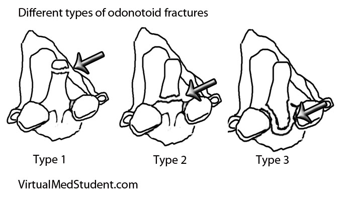
Since the odontoid is a relatively long piece of bone it can fracture at one of several distinct sites. The most commonly used system (Anderson and D’Alonzo) categorizes fractures into one of three types:

  • Type 1 – a fracture at the tip of the odontoid.
  • Type 2 – a fracture at the base of the odontoid.
  • Type 3 – a fracture involving the body of the C2 vertebrae, which includes the odontoid within it.

Odontoid fractures

This grading system is important because it helps predict both stability of the C1-C2 (ie: atlanto-axial) joint and guides potential treatment options.

Signs and Symptoms

Roughly 80% of patients with odontoid fractures do not have any neurological injury to their spinal cord. The remaining patients can exhibit anything from quadriplegia to mild sensory disturbances. Patients with severe cervical spinal cord injury usually are unable to breath (secondary to diaphragm paralysis) and frequently die at the scene of the accident.

Many patients with odontoid fractures will have significant neck pain that radiates up into the scalp. This is usually caused by neck muscles spasming secondary to the injury.

Diagnosis

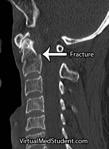
CT of odontoid fracture

CT scans, x-rays, and MRIs are all useful in diagnosing and properly treating odontoid fractures.

CT scans of the cervical spine provide excellent bony detail, and also help illustrate any additional fractures that may be present.

MRI of the cervical spine is useful for assessing any co-existent ligamentous injury. If ligamentous injury is present it drastically alters treatment decisions.

Treatment

Treatment of odontoid fractures is based on both bony and ligamentous injury. The goal of treatment is to stabilize the spine either by allowing the bone to heal on its own, or by fusing the spine artificially using rods, screws, and/or wires.

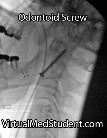
Placement of an odontoid screw is one method of fixing non-displaced type II fractures. However there are numerous contraindications for odontoid screw placement. For example severe angulation of the fractured segment precludes placement of a screw; barrel chested anatomy prevents an adequate angle for screw trajectory in the operating room. In addition, if the transverse ligament is disrupted, bony fixation with an odontoid screw alone will not stabilize the joint.

On the other hand, odontoid screws are beneficial because they typically stabilize the fracture with minimal restriction of neck motion.

Approaching the spine from behind is another option to stabilize odontoid fractures. Fusing the atlas (C1) to C2 or C3 is sometimes used if odontoid screw placement cannot be performed and the injury is deemed unstable. It is important to note that posterior approaches can restrict motion, especially in the high cervical spine.

Non-surgical options must ensure that the patient has minimal to no movement of the neck in order to give the bone an adequate chance to heal on its own. Rigid collars or halos are used to prevent neck motion.

Overview

Odontoid fractures come in three flavors depending on the location of the fracture. Symptoms can be anything from mild neck discomfort to quadriplegia, although neurological injury is surprisingly uncommon. Diagnosis is based on CT and MRI. Treatment is with cervical immobilization for an extended period of time, or surgical fusion. Treatment decisions are based on the degree of both bony and ligamentous injury, as well as the patient’s overall health status.

Related Articles

References and Resources

ICU Notes: The Devil is in the Details

Patients in the intensive care unit (ICU) are usually the sickest in the hospital and require a ton of care. Unlike a normal patient note, which is generally brief and written in the SOAP format, the ICU note is more thorough and exhaustive. Below is a discussion of how an ICU note can be organized so that important information is not missed. Please note that this is not the only way to organize an ICU note, but it is a way that many units use because it is extremely thorough and organized.

Organize by Systems

The beginning of any ICU note should always have the patient’s name, date, time, hospital day, post operative day (if in the surgical ICU), lines (ie: any tubes going into or out of the patient), and drips.

  • Patient’s name
  • Hospital day
  • Post operative day (if applicable)
  • Lines
  • Drips (often abbreviated as “gtts”)

Unlike normal floor notes, which are typically organized by problem, ICU notes are more commonly organized by body system. The systems are usually written in the following order:

So what information should we include in each section?

Neurological

At the bare minimum the neurological part of the note should include the patient’s alertness and orientation (ie: “AAOx3, confused, intubated but following commands, etc.”), and their Glasgow coma scale (GCS) score. If the patient is in the neuro-ICU the exam should be more thorough and include the GCS score by component, as well as a neurological exam. A common question asked by neuro-intensivists is “what can the patient do?” Do they follow all commands? Do they withdraw to pain? Are they decorticate? Decerebrate? Give a description of what the patient can “do” or “not do”.

Less commonly some ICU patients may have an external ventricular drain or lumbar drain used to remove cerebrospinal fluid. If this is the case the output of the EVD or lumbar drain should be recorded. EVDs are set to a specific height above the tragus of the ear; this number is important to record as well.

Finally, specific medicines related to neurological disease should be added. Any medications used to sedate or paralyze the patient should also be included as they may confound the physical exam findings. For example, are they on propofol or fentanyl drips? How much of this medicine is “dripping” into them per hour? Someone on a large amount of fentanyl may not respond much to painful stimuli so it is important to give a description of what may be confounding your examination.

To recap, the neurological section should include the following elements:

  • Physical exam findings
    • Level of alertness and orientation
    • GCS broken down by components
    • Neurological exam (ie: cranial nerves, strength, reflexes, sensation, etc.)
  • Vital signs and tubes
    • EVD and/or lumbar drain output
      • Output over last shift
      • Output over last 24 hours
      • Height of the EVD
  • Neurological medicines
    • Sedatives (ie: propofol, midazolam, etc.)
    • Pain medications
    • Treatment specific medications (anti-seizure meds)

Cardiovascular

In addition to physical exam findings, the cardiovascular section should start off with the heart rate, rhythm, blood pressure, and central venous pressure (if a central line is present). Patients with significant cardiovascular disease may have a pulmonary artery catheter (aka: Swan-Ganz catheter). The data obtained from it should also be included: pulmonary artery pressure, pulmonary artery wedge pressure, cardiac output, cardiac index, central venous pressure, and stroke volume.

The cardiovascular section of the note should also include what drips the patient is on to control blood pressure. Are they on anti-hypertensive medications or pressors?

To recap, the cardiovascular section should include:

  • Physical exam findings
  • Vital signs and tubes
    • Heart rate
    • Heart rhythm (from telemetry)
    • Blood pressure
    • Central venous pressure (from central line or Swan Ganz catheter)
    • Pulmonary artery pressure (from Swan Ganz catheter)
    • Pulmonary artery wedge pressure (from Swan Ganz catheter)
    • Cardiac output and index (from Swan Ganz catheter)
    • Chest tube output (if present)
      • Output over last shift
      • Output over last 24 hours
  • Cardiac specific medications
    • Intravenous drips used to control blood pressure
    • Other cardiac medications (β-blocker, ACEI, etc.)

Respiratory (Pulmonary)

In addition to physical exam findings, the most important thing to include in this section are the ventilator settings. It should be organized in the following way: mode, FiO2, tidal volume, patient’s respiratory rate, ventilator’s rate, peak end expiratory pressure (PEEP), pressure support, peak airway pressure.

After the ventilator settings, the patient’s oxygen saturation should also be stated.

Any laboratory data specific to the pulmonary system is given next. This commonly includes arterial blood gas values written in the following order: pH, PaO2, PaCO2, and HCO3. Chest x-ray results should also be written here.

To recap, the respiratory portion of the note should include:

  • Physical exam findings
  • Ventilator settings
    • Mode
    • FiO2
    • Tidal volume
    • Patient’s respiratory rate
    • Ventilator’s respiratory rate
    • Peak end expiratory pressure (PEEP)
    • Pressure support
    • Peak airway resistance
  • Vital signs
    • Oxygen saturation
  • Laboratory data
    • Arterial blood gas results
    • Chest x-ray results
  • Pulmonary medications
    • Nebulizers, etc.

Renal

The renal section includes an evaluation of the patient’s kidney function. The first piece of data that should be recorded are the "in and outs". The "ins" include the total amount of fluid the patient has received intravenously and orally. The "outs" include the total amount of urine the patient has made. The urine output should be recorded in mL/kg of body weight per hour, shift, and over the last 24 hours.

Laboratory data that should be recorded in the renal section includes the creatinine, blood urea nitrogen (BUN), urinalysis, urine microscopy, and any imaging studies specific to the urinary tract. You may also need to calculate and include the fractional excretion of sodium.

To recap, the renal section of the note should include:

  • Vital signs
    • Ins and outs
      • Output over last shift
      • Output over last 24 hours
  • Laboratory data
    • Creatinine
    • Blood urea nitrogen (BUN)
    • Urinalysis
    • Urine microscopy

Fluids, Electrolytes, and Nutrition (FEN)

The fluids, electrolytes, and nutrition section should have what fluids, both intravenously and orally, the patient is receiving. The patient’s electrolytes should also be recorded in this section. The most common electrolytes that are measured are sodium, potassium, chloride, bicarbonate, phosphorus, calcium, and magnesium. In addition, the patients diet, either orally or intravenously should be discussed along with their nutritional status (usually measured by albumin or prealbumin levels).

To recap the fluids and electrolytes section should include:

  • Intravenous fluids (ie: normal saline, hyper or hypotonic saline, colloids, etc.)
  • Diet
  • Laboratory data
    • Chemistry panel
    • Calcium, magnesium, phosphorus
    • Albumin
    • Prealbumin

Infectious Diseases

The infectious diseases section should always begin with the maximum temperature over the last 24 hours followed by the current temperature.

After that the patient’s white blood cell count should be mentioned. If there are any pending or final culture results they should be included in sequential order. Common data recorded include blood, urine, and sputum cultures. Sometimes cerebrospinal fluid cultures will also be available.

Finally, all antibiotics the patient is receiving should be listed along with how many days they’ve been on each one.

To recap, the infectious disease section should include:

  • Vital signs
    • Maximum temperature
    • Current temperature
  • Laboratory data
    • White blood cell count
    • Culture results
  • Antibiotics

Hematology

Physical exam findings such as bruising, petechiae, oozing from surgical incisions should be mentioned.

The hematology section should have the hemoglobin, hematocrit, platelet count, prothrombin time, INR, and partial thromboplastin time. Other values that are less common, but may be present include iron studies, d-dimer results, fibrinogen levels, and mixing studies.

Since blood clot formation is a huge concern in hospitalized patients, any results related to this should be included. Studies such as lower extremity dopplers, chest CT or ventilation perfusion scans should be included in this section.

To recap, the hematology section should include:

  • Physical exam findings (ie: bruising, oozing, petechiae, etc.)
  • Laboratory data
    • Hemoglobin and hematocrit
    • Platelet count
    • PT, INR, PTT
    • Lower extremity doppler results
    • Chest CT or ventilation/perfusion scan

Gastrointestinal

The gastrointestinal section is devoted to the patient’s bowel function. A discussion of how the patient is being fed and what is being fed to the patient is written in this section. For example, oral versus intravenous feedings. Is a nasogastric tube present?

Laboratory data that should be included here are the patient’s albumin level (measured every few weeks) and the patient’s prealbumin level (usually measured every 48 hours).

Physical examination findings of the gastrointestinal tract should be written as well. They may include abnormal bowel sounds, enlarged liver or spleen, ascites, or blood in the stool.

To recap, the gastrointestinal section should include:

  • Physical exam findings (ie: distended, peritoneal signs, etc.)
  • Laboratory data
    • Liver function tests
    • Amylase and lipase

Endocrine

The most important thing for the endocrine section is the blood sugar results. Other common values that are often ordered for ICU patients include serum osmolality. Any medications used to control glucose levels such as insulin drips or hypoglycemic agents should be recorded.

  • Laboratory data
    • Blood glucose levels (fingerstick, metabolic panel)
  • Medications
    • Insulin drip and rate (if any)
    • Sliding scale insulin
    • Subcutaneous insulin
    • Oral hypoglycemics (ie: metformin, glyburide, etc.)

Psychiatric

The psychiatric portion of the note is often neglected or omitted, but is an important component to at least think about. Many psychiatric issues will be dealt with once the patient is out of the ICU.

Prophylaxis

The prophylaxis section is very important! Most patients in the ICU will be on a medicine to decrease the risk of developing gastric ulcers, as well as several methods to prevent deep venous thrombosis and pulmonary embolism. Any prophylactic measures should be listed and addressed during rounds.

The prophylaxis section includes:

  • Ulcer prophylaxis
    • H2 or proton pump inhibitor
  • Deep venous thrombosis prophylaxis
    • Subcutaneous heparin
    • Compression boots (venodynes)
    • Graduated/compression stalkings

To Sum It All Up…

There are different ways to organize an ICU note. By far the most common way is to organize it by systems. However, various services (ie: neurosurgery, cardiothoracic, etc) will have very specific ways they want data presented depending on what information is most important to them.

Other Content You Will Find Useful…

Systemic Vascular Resistance: Radius, Length, Viscosity

The systemic vascular resistance is the resistance that blood “sees” as it travels throughout the circulatory system of the body.

It is controlled by three different factors: length of the blood vessel (l), radius of the blood vessel (r), and the viscosity of the blood (η). The equation that relates these three factors to resistance is known as Poiseuilles’ equation:

R ≈ (η x l) / r4

Let’s discuss how each of the factors above influences resistance. The longer the vessel, the more likely blood will sludge and stick up against the walls; this tendency intuitively causes an increase in overall resistance. Blood vessel length does not typically change during adulthood, therefore its contribution to vascular resistance in humans is relatively constant.

When blood is more viscous than usual the resistance also increases. For example, molasses flows very slowly because it is highly viscous; blood is no different. There are a few uncommon instances in human physiology where blood viscosity increases; therefore, like vessel length, its contribution to vascular resistance is also relatively constant.

The most important parameter is the radius of the vessel. When the radius of a vessel shrinks, the resistance increases; when the radius of a vessel dilates, the resistance drops. Imagine trying to pump a set amount of fluid through a small opening versus a larger opening. There is much less resistance encountered through the larger opening. The same is true for large and small blood vessels. Even more importantly, as indicated by Poiseuille’s equation above, when the radius of a vessel is halved, the resistance increases by 16 fold! Therefore, even small changes in blood vessel diameter can have dramatic consequences on the bodies’ vascular resistance.

How the Body Controls Resistance

How do you think the body controls resistance most effectively? You guessed it! By controlling the radius of blood vessels. When there is an abnormal drop in vascular resistance, the body compensates by pumping out hormones (ie: epinephrine and norepinephrine); these hormones cause muscle cells surrounding blood vessels to constrict. Constriction leads to a decreased radius of the vessel, which leads to increased resistance.

Clinical Measurements

How do you measure systemic vascular resistance? Well, first off, systemic vascular resistance is not measured, but is calculated from other cardiovascular vital signs such as the mean arterial pressure (MAP), cardiac output (CO), and central venous pressures (CVP). The equation that relates these three values to resistance is:

SVR = [(MAP – CVP) / CO] x 80

Mean arterial pressure is a sort-of average between the systolic and diastolic blood pressure readings. It can be calculated using blood pressures obtained from an arterial catheter or a blood pressure cuff.

Central venous pressures are normally low, and do not contribute much to vascular resistance, but when very accurate measurements are needed they can be obtained from a central venous and/or a pulmonary artery catheter.

Cardiac output can be obtained from a pulmonary artery catheter if accurate values are needed; it can also be roughly estimated from a cardiac echocardiogram (ie: an ultrasound of the heart).

Role in Disease

So what’s the big deal? Why all this talk about vascular resistance? Vascular resistance is important because it is one determinant of blood pressure, and therefore organ perfusion.

The mean arterial pressure is calculated by multiplying the cardiac output by the systemic vascular resistance, and then adding the result to the central venous pressure (MAP = CO x SVR + CVP, a reorganization of the equation above).

Since the mean arterial pressure is the driving force behind the delivery of blood and oxygen to the organs, it is of vital importance that it stays above a certain threshold. Otherwise organs will not receive enough oxygen and will eventually die. One way of ensuring that the mean arterial pressure stays within a set range is for the body to constrict and/or dilate the vessels as needed.

On the flip side, if systemic vascular resistance is too "clamped" down then hypertension occurs. If severe enough, the heart may not be able to generate enough force to eject an adequate amount of blood into the tightened arterial system, which over time could result in systolic heart failure.

Overview

Systemic vascular resistance is the resistance blood sees as it travels throughout the bodies blood vessels. It is influenced by the length and radius of the blood vessel(s), as well as the viscosity of blood. It is calculated from the mean arterial pressure, central venous pressure, and cardiac output. Systemic vascular resistance plays a vital role in maintaining blood pressures within set ranges so that organ perfusion is maximized.

References and Resources

Cardiac Output: Pump, Pump, Squeeze

The cardiac output (CO) measures how much blood the heart pumps per minute. It is directly related to the stroke volume (SV) and heart rate (HR). The stroke volume is the amount of blood in the left ventricle of the heart just before it contracts. The cardiac output is calculated by multiplying the heart rate by the stroke volume (CO = HR x SV).

The cardiac output is related to Ohm’s law, which in electrical terms states that the change in voltage of a circuit is equal to the flow of current, I, multiplied by the resistance, R, of the circuit (ΔV = I x R).

Like electrical current in a circuit, blood flows in a circular pathway from the left ventricle, through the body, where it eventually ends up in the right ventricle. We can change Ohm’s law to govern hemodynamics by stating that the change in voltage is equivalent to the change in pressure between the aorta and right atrium (mean arterial pressure and central venous pressures, respectively), flow of current is equal to the amount of blood pumped per unit time (ie: cardiac output), and resistance is equal to the resistance the blood sees as it travels through the vessels of the body (aka: the systemic vascular resistance).

Therefore, if we re-write Ohm’s law for the hemodynamics of cardiac output we get: central venous pressure (CVP, measured in mmHg) subtracted from the mean arterial pressure (MAP, measured in mmHg) is equal to cardiac output (CO, measured in liters/minute) multiplied by the systemic vascular resistance (SVR, measured in dynes-s / cm5), or:

(MAP – CVP) = (CO x SVR) / 80
(ΔV = I x R)

Doing a simple mathematical rearrangement of the equation above we get:

CO = [(MAP – CVP) / SVR] x 80
*** The 80 in the equation is a conversion factor to convert Wood to metric units ***.

In essence, the cardiac output is directly proportional to the difference in blood pressure between the left (arterial side) and right (venous side) sides of the body, and inversely proportional to the vascular resistance. In other words, if the pressure difference between the arterial and venous side of the body decreases the cardiac output will fall. Along the same lines, if the systemic vascular resistance increases then cardiac output will decrease.

Clinical Measurements

How do you clinically measure the components that constitute the cardiac output?

The most reliable, but invasive way is to use a pulmonary artery catheter, also known as a "Swan Ganz" catheter. The catheter is inserted into the pulmonary artery and advanced until it “wedges” in a small branch of the pulmonary arterial tree. From there a balloon in the tip of the catheter is inflated to keep it in place. At this location the tip of the catheter is effectively measuring the pressure of blood in the left atrium (ie: the "pulmonary artery wedge pressure"). Using the catheter, and various methods such as the thermodilution technique, the cardiac output and/or stroke volume can be measured directly.

Unfortunately, the use of Swan-Ganz catheters are fraught with serious problems such as arrhythmias and infection. Therefore, other less invasive techniques such as echocardiography with doppler (ie: an ultrasound of the heart) can be used to estimate cardiac output and stroke volume.

The other elements of cardiac output can also be measured clinically. The mean arterial pressure is calculated from the systolic and diastolic blood pressures, which are measured from a cuff or arterial catheter. Central venous pressure can be measured with a central venous catheter (ie: a "central line").

Role in Disease

A normal cardiac output is roughly 5.5 L/min for an average sized male and 5.0 L/min for an average sized female. However, in individuals with diseased heart muscle the pumping ability of the heart is reduced. The result is a decrease in cardiac output.

So what’s the big deal? A fall in cardiac output means that the body sees less oxygenated blood per minute. Every organ in the body requires oxygen to function properly. If cardiac output is decreased the organs may not receive enough oxygen to do their job. In super severe cases, when cardiac output approaches zero, cardiogenic shock occurs. If no oxygen reaches the organs multi-organ failure occurs and the patient may die.

Overview

Cardiac output is related to the stroke volume, heart rate, systemic vascular resistance, and difference in pressure between the arterial and venous sides of the body. When cardiac output drops the bodies’ organs receive less oxygen per unit time. In severe cases this may lead to organ death.

Related Articles

References and Resources

Fluid Around the Heart: Pericardial Effusions

The heart is encased in a connective tissue capsule known as the pericardium. The pericardium contains two layers, known as the parietal and visceral pericardium. These layers are like two blankets enveloping the heart. The visceral pericardium sits adjacent to the heart muscle itself and the parietal pericardium sits on top of the visceral pericardium. Because of this arrangement there is a potential space between the two layers.

When fluid (ie: blood, pus, water, etc.) leaks into this space a pericardial effusion is present. Fluid can leak out quickly, in which case the effusion is said to be “acute”; or it can leak out gradually in which case it is said to be “chronic”.

Causes

There are numerous causes of pericardial effusion some of which are listed below:

  • Infections
    • Viral (including HIV, coxsackie, echo, adeno, ebstein-barr, and varicella viruses)
    • Bacterial (including pneumococcus, neisseria meningitides, staphylococcus aureus)
    • Tuberculosis
  • Malignancies (cancers)
  • Autoimmune conditions (including connective tissue disorders, vasculitis, and drug induced)
  • Uremia (from renal failure)
  • Cardiovascular (cardiac surgery, myocardial infarction, aortic dissection, congestive heart failure)
  • Hypothyroidism
  • Cirrhosis (liver problems)
  • Idiopathic (unknown)

Please note that by no means is this list exhaustive, but these are the most common causes of effusions!

Signs and Symptoms

The classic symptom of pericardial effusion is chest pain that is better when the patient sits up and leans forward. However, numerous other symptoms including light headedness, shortness of breath, cough, and palpitations can occur.

Depending on how quickly the effusion develops, patients may spiral into a condition known as “tamponade”. When this occurs the effusion effectively “chokes” the heart muscle causing decreased contractile function. This causes decreased cardiac output and even multi-organ failure if left untreated!

The classic signs of tamponade are hypotension (ie: decreased blood pressure), muffled heart sounds, and increased jugular venous pressures (you can see the jugular veins engorged with blood). These three signs are known as “Beck’s triad”, which is generally a late finding of tamponade (ie: the patient is almost dead!).

Diagnosis

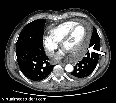
CT scan of pericardial effusion
There are numerous tests that can support the diagnosis. Electrocardiogram may show decreased voltages, and a finding known as “electrical alternans” where the QRS complexes change amplitude and/or direction as a result of the heart “sloshing” around in the effusion. Chest x-ray may show an enlarged heart, but this is neither specific, nor sensitive, for effusion. CT scans can show fluid surrounding the heart. Finally, echocardiography (ie: ultrasound of the heart) can be very useful in delineating not only the presence of, but also the size, and location of the effusion.

Treatment

For chronic effusions with no significant symptoms patients can be treated for the underlying condition causing the effusion. This will sometimes cure the effusion. However, in patients with acute presentations who have signs of cardiovascular instability (ie: low blood pressure, evidence of organ dysfunction from decreased blood flow, etc.) emergent removal of the fluid is performed. The quickest way to do this is to insert a needle under the xyphoid process and aspirate the fluid. In less acute situations, or in recurrent cases, surgical “windows” in the pericardial tissue can be created to allow the effusion to drain.

Overview

Pericardial effusions occur when fluid accumulates between the visceral and parietal pericardial layers surrounding the heart. There are numerous causes. Rapidly expanding effusions can cause cardiac tamponade and lead to cardiovascular collapse. Signs and symptoms include chest pain, shortness of breath, cough, distant heart sounds, and decreased blood pressure. Diagnosis is made by characteristic ECG, echocardiography, and CT scan findings.

References and Resources

  • Imazio M, Brucato A, Mayosi BM, et al. Medical therapy of pericardial diseases: part II: Noninfectious pericarditis, pericardial effusion and constrictive pericarditis. J Cardiovasc Med (Hagerstown). 2010 Nov;11(11):785-94. Review.
  • Khandaker MH, Espinosa RE, Nishimura RA, et al. Pericardial disease: diagnosis and management. Mayo Clin Proc. 2010 Jun;85(6):572-93. Review.
  • Spodick DH. Pericarditis, pericardial effusion, cardiac tamponade, and constriction. Crit Care Clin. 1989 Jul;5(3):455-76.
  • Mookadam F, Jiamsripong P, Oh JK, et al. Spectrum of pericardial disease: part I. Expert Rev Cardiovasc Ther. 2009 Sep;7(9):1149-57.
  • Jiamsripong P, Mookadam F, Oh JK, et al. Spectrum of pericardial disease: part II. Expert Rev Cardiovasc Ther. 2009 Sep;7(9):1159-69.
  • Woo KM, Schneider JI. High-risk chief complaints I: chest pain–the big three. Emerg Med Clin North Am. 2009 Nov;27(4):685-712, x.