Radial Nerve and the Saturday Night Palsy

Radial Nerve Course
In order to appreciate the radial nerve, we have to first understand the brachial plexus. The brachial plexus can be thought of as a massive highway intersection, in which numerous highways come together and then split apart again.

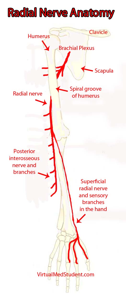
The "highways" merging into the brachial plexus are the 5th, 6th, 7th, and 8th cervical nerve roots, as well as the 1st thoracic nerve root. These nerve "highways" tangle together to form trunks, divisions, cords, and then branches. The radial nerve is one of the branches of the brachial plexus; it gets its input from the 5th, 6th, 7th and 8th cervical nerve roots.

The radial nerve courses along the humerus in the upper arm. It wraps around the humerus in a spot called the spiral groove. Just before wrapping around the humerus, it sends a branch that innervates the triceps muscle (long, medial, and lateral heads) in the upper arm.

After wrapping around the spiral groove, it sends additional branches to the brachioradialis, extensor carpi radialis longus, and extensor carpi radialis brevis muscles.

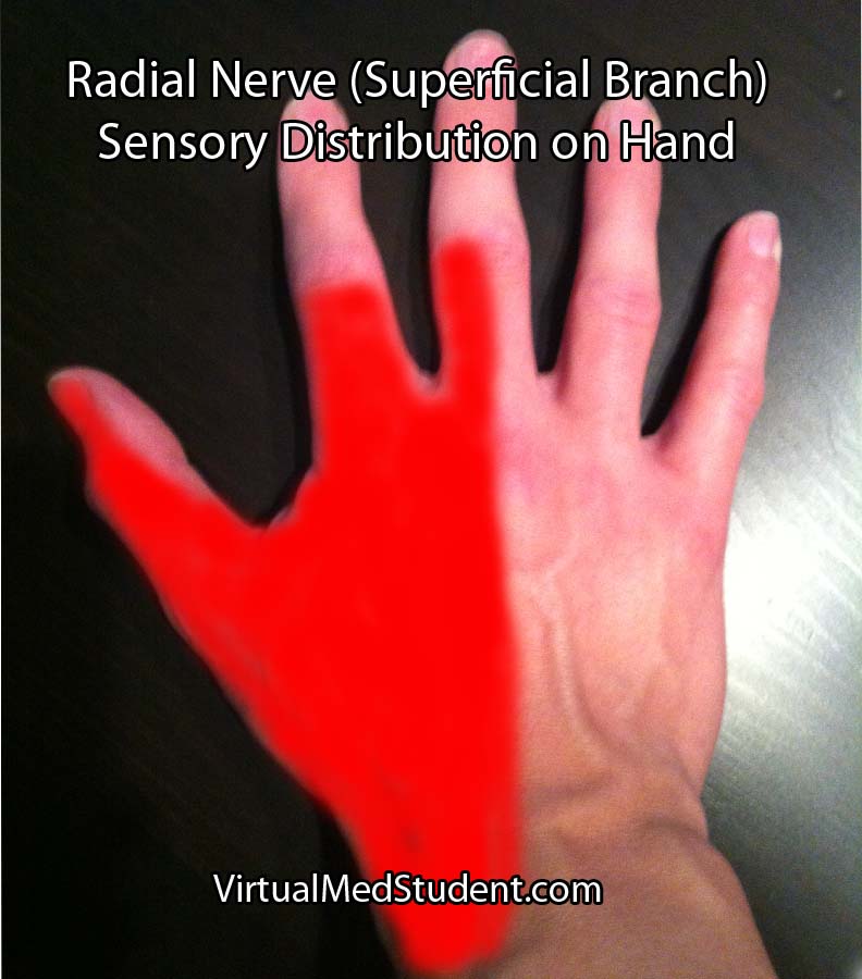
The first major branch in the forearm is known as the superficial radial nerve. This nerve courses along the medial/ulnar aspect of the forearm (ie: the ulnar or medial side of the forearm is closest to your body when your palms are facing forward) and heads straight for the hand. It relays sensory information from the lateral portion of the back of the hand.

The second major branch at the elbow can be thought of as the deep radial nerve, but it is formally known as the posterior interosseous nerve.

The posterior interosseus nerve sends branches to eight muscles in the forearm. They include the supinator (through which the nerve travels via a fibrous tunnel known as the arcade of Frohse), extensor digiti minimi, extensor carpi ulnaris, extensor digitorum, abductor pollicis longus, extensor pollicis longus, extensor pollicis brevis, and extensor indicis.

Muscles Innervated by the Radial Nerve and Its Branches
Muscle Action of Muscle
Triceps brachii – Extension of the forearm away from upper arm
Brachioradialis – Helps flex the forearm closer to the upper arm
– Helps supinate (ie: palm towards the sky)
– Helps pronate (ie: palm towards the floor)
Extensor carpi radialis
(longus and brevis)
– Helps extend the wrist
– Abducts the hand (ie: hand moves away from
the body when the palms are facing forward)
Extensor digiti minimi – Helps extend the little finger (5th digit)
Extensor carpi ulnaris – Helps extend the wrist
– Adducts the hand (ie: hand moves towards
the body when the palms are facing forward)
Supinator – Allows palms to face up towards the sky
Extensor digitorum – Helps with extension of the fingers
(specifically at the metacarpophalngeal joint)
Abductor pollicis longus – Helps abduct the thumb
Extensor pollicis
(longus and brevis)
– Help extend the thumb
Extensor indicis – Helps extend the index finger

Generally speaking, the radial nerve and its branches are involved in muscles that allow joints to extend (ie: widen or separate away from one another).

Importance in Disease

Damage to the radial nerve may take the form of compression or sheering injuries, typically after traumatic events.

The most common site of injury is at the spiral groove of the humerus. The nerve may be damaged if someone breaks their humerus, or if someone leans the back of their arm on something for an extended period of time (ie: a "Saturday night palsy" is the informal term given to a drunk who falls asleep with their arms draped over a chair… they end up waking up with a radial nerve palsy!).

Injury to the nerve at the spiral groove causes a wrist drop, in which the affected person cannot extend their wrist. The triceps are not affected because nerve branches to this muscle are proximal to the spiral groove. In addition, patients also complain of decreased sensation on the back of the hand (see image to right).

Radial Nerve Sensory Distribution in Hand
Additionally, the posterior interosseous branch of the radial nerve may get compressed as it passes through the supinator muscle in the forearm. This is referred to as posterior interosseous nerve syndrome.

The compression occurs at a fibrous portion of the supinator muscle known as the "arcade of Frohse", which causes an inability to extend the fingers at the metacarpophalangeal joints; this is due to dysfunction of the extensor digitorum muscle (extension at the interphalangeal – both distal and proximal – joints is controlled by the lumbricals and interossei muscles which are innervated by the median and ulnar nerves).

In posterior interosseous nerve syndrome it is still possible to extend the wrist because the branches of the nerve to the extensor carpi radialis muscle are unaffected (ie: they branch before the arcade). However, when the wrist is extended it deviates towards the radial side of the forearm because of the unopposed action of the extensor carpi radialis muscles (in other words, the extensor carpi ulnaris is affected and cannot keep the wrist extension neutral).

Sensation is entirely normal when the posterior interosseous nerve gets compressed because it contains no sensory fibers.

Overview

The radial nerve is a terminal branch of the brachial plexus. It sends branches to most of the extensor muscles of the arm and forearm. It also provides sensation to the back side of the hand. If injured it causes either a radial nerve palsy, or posterior interosseous syndrome, in which the affected patient has an inability to extend various joints at the wrist and/or fingers.

Related Anatomy You Should Know About…

References and Resources

  • Ducic I, Felder JM 3rd, Quadri HS. Common nerve decompressions of the upper extremity: reliable exposure using shorter incisions. Ann Plast Surg. 2012 Jun;68(6):606-9
  • Colbert SH, Mackinnon SE. Nerve compressions in the upper extremity. Mo Med. 2008 Nov-Dec;105(6):527-35.
  • Reddy MP. Peripheral nerve entrapment syndromes. Am Fam Physician. 1983 Nov;28(5):133-43.
  • Calfee RP, Wilson JM, Wong AH. Variations in the anatomic relations of the posterior interosseous nerve associated with proximal forearm trauma. J Bone Joint Surg Am. 2011 Jan 5;93(1):81-90.
  • Arle JE, Zager EL. Surgical treatment of common entrapment neuropathies in the upper limbs. Muscle Nerve. 2000 Aug;23(8):1160-74.

Diffuse Axonal Injury: Shearing the Cables

Diffuse axonal injury is a form of traumatic brain injury that occurs under conditions of rapid acceleration and deceleration of the head. This frequently occurs after high impact injuries such as motorcycle crashes or high speed car accidents.

Let’s discuss a few basic brain terms before we dive into why diffuse axonal injury happens. Brain tissue is composed of neurons and glia. The neurons communicate with one another through long extensions known as axons. You can think of an axon as the telephone wire that connects one phone to another. Axons compose the bulk of what is known as “white” matter in the brain; on the other hand, “gray” matter represents clumps of neurons.

Both gray and white matter have different densities associated with them. Because of this, rapid accelerations or decelerations of the head cause the gray matter to move at a greater relative velocity compared to the white matter. If these accelerations are severe enough stretch and shear injury occurs; the end result is that the “wires” get disconnected. This, in a nutshell, is diffuse axonal injury.

Signs and Symptoms

Depending on the severity of diffuse axonal injury patients may present with different signs and symptoms. In fact, diffuse axonal injury is frequently diagnosed when patients “fail” to wake up after a traumatic event despite adequate resuscitative treatment.

In its most severe form, diffuse axonal injury results in coma. Less severe injuries may cause long term cognitive issues and personality changes.

Diagnosis

Diagnosis of diffuse axonal injury is made with an MRI scan. CT scans of the head are frequently ordered early to rule out other treatable causes for the decreased mental status often seen in patients with DAI (ie: epidural, subdural, and intraparenchymal hematomas).

The MRI will reveal restricted diffusion on diffusion weighted (DWI) and apparent diffusion coefficient (ADC) maps of the brain. Gradient echo imaging (GRE) will often show small spots of low intensity consistent with shear/stretch injury.

MRI of diffuse axonal injury

Treatment

Unfortunately, there is no effective treatment for diffuse axonal injury. There is currently no way to “re-wire” the axons. All care for diffuse axonal injury at this point is supportive.

Overview

Diffuse axonal injury occurs after rapid changes in acceleration of the head. The axons, or “wires”, between neurons get stretched, sheared and, effectively disconnected. Coma is the common presenting sign of severe axonal injury. Diagnosis is made with MR imaging. Treatment is supportive since there is currently no way to re-wire the damaged connections.

Other Stuff You Might Want to Learn About

References and Resources

  • Hijaz TA, Cento EA, Walker MT. Imaging of head trauma. J Trauma. 2010 Dec;69(6):1610-8.
  • Andriessen TM, Jacobs B, Vos PE. Clinical characteristics and pathophysiological mechanisms of focal and diffuse traumatic brain injury. J Cell Mol Med. 2010 Oct;14(10):2381-92.
  • Meythaler JM, Peduzzi JD, Eleftheriou E. Current concepts: diffuse axonal injury-associated traumatic brain injury. Arch Phys Med Rehabil. 2001 Oct;82(10):1461-71.
  • Smith DH, Meaney DF, Shull WH. Diffuse axonal injury in head trauma. J Head Trauma Rehabil. 2003 Jul-Aug;18(4):307-16.
  • Osborn AG. Osborn’s Brain: Second Edition. Elselvier, 2017.

Multiple Sclerosis: Multiple Scars in the Central Nervous System

Multiple sclerosis literally means “multiple scars”. And in multiple sclerosis the scaring takes place in the central nervous system. The pathology of why multiple sclerosis occurs is not yet fully understood. We do know that it is an autoimmune condition in which the body attacks itself.

The target of that attack is myelin. Myelin is a fatty substance that insulates neuronal axons. You can think of myelin as the rubber coating around electrical wiring. The insulating role of myelin helps axons send information rapidly from one neuron to the next. Interestingly, multiple sclerosis only affects myelin created by oligodendrocytes, which are glial cells (ie: neuron support cells) found in the central nervous system. Multiple sclerosis does not affect myelin formed by Schwann cells, which are located in the peripheral nervous system.

The damage to myelin is thought to be caused by T-cells. T-cells are a branch of the adaptive immune system. T-cells do not normally invade the central nervous system, but in multiple sclerosis some antecedent event (possibly infection?) allows T-cells to gain access to the CNS. In genetically susceptible people these trapped T-cells "see" myelin as a foreign substance and attack it. The result is inflammation and scaring (in the brain, scaring is called "gliosis").

Signs and Symptoms

Although multiple sclerosis can occur in all races and in both genders, it tends to affect women of European descent more frequently. In addition, patients who live at higher latitudes also seem to be at higher risk.

The classic presentation of someone with multiple sclerosis is numerous neurological complaints spaced out in time. These neurological symptoms can be highly variable. Often times patients will have blurred vision or blindness if demyelination occurs at the optic nerve. This is known as optic neuritis and is a common feature in multiple sclerosis patients.

In addition, weakness of an extremity can occur and can be partial (paresis) or complete (paralysis). Difficulty speaking and swallowing can occur, as can bowel and bladder incontinence. Sensory deficits like numbness and tingling are also common. If the cerebellum or spinocerebellar tracts in the spinal cord are involved loss of coordination can occur (ataxia).

There are many other symptoms that can be seen in multiple sclerosis depending on which part of the central nervous system is involved. Ultimately, if a patient returns with several neurological complaints spaced out in time, then multiple sclerosis should be on the list of possible diagnoses.

Diagnosis

The diagnosis of multiple sclerosis is based on clinical and supporting laboratory evidence. There is no single test that can determine if it is present, but there is a set of guidelines that can help the clinician make the diagnosis, which include:

(1) Signs and symptoms referable to the central nervous system.
    (a) Symptomatic episodes should last at least 24 hours.
    (b) Two or more symptomatic episodes at least one month apart.
(2) Two or more lesions (ie: “scars”) seen on MRI of the brain (see image below).
(3) Physical exam findings supporting central nervous system disease.
(4) Cerebrospinal fluid results consistent with a diagnosis of multiple sclerosis.
(5) No other disease(s) that could explain the symptoms and findings.

MRI of Multiple Sclerosis

Many other tests are often performed to rule out other causes for the patient’s symptoms. Testing for systemic lupus erythematosis, Lyme disease, neuro-syphilis, vitamin B12 deficiency, and thyroid disease are common adjunctive tests.

Treatment

There is currently no cure for multiple sclerosis. However, like many other autoimmune disorders, there are several disease modifying medications that can slow its progression.

The first class of modifying medications are known as the "interferons". Interferon is believed to act through several different mechanisms. It inhibits white blood cell proliferation (T-cells are one type of white blood cell) and decreases antigen presentation by immune cells. It also reduces T-cell migration and switches the body towards an anti-inflammatory state. These effects help decrease the inflammation associated with multiple sclerosis. There are currently three different formulations of interferon on the market for multiple sclerosis. The first two are interferon β-1a formulations known as Avonex® and Rebif®. The third is an interferon β-1b formulation known as Betaseron®.

Another medication known as glatiramer acetate (aka: Copaxone®) acts as a possible myelin "mimick", protecting normal myelin from attack. Glatiramer is a synthetic polymer of four amino acids (ie: the building blocks of protein) found in normal human myelin. The exact mechanism of glatiramer’s actions are unknown.

Natalizumab (aka: Tysabri®) is approved for use in multiple sclerosis. It is a monoclonal antibody that binds to, and inhibits, the function of α4-integrin. α4-integrin is a cell adhesion molecule found on white blood cells that normally allows them to move into body tissues to fight infection. By blocking this movement, natalizumab is thought to decrease the number of T-cells that enter the central nervous system.

Another medication known as mitoxantrone is also used in the treatment of multiple sclerosis. It was originally designed as a cancer medication. Its mechanism of action is to disrupt DNA synthesis and repair by inhibiting a protein known as topoisomerase.

Patients who are acutely symptomatic (ie: having a "flair") are treated with intravenous steroids. Methylprednisolone is the most common steroid used in patients with acute neurological symptoms.

The goal of all of these medications is to dampen the immune response and prevent the abnormal inflammation that occurs in multiple sclerosis. Because of this, many of these medications have side effects such as the risk of infections.

Finally, depending on a patient’s symptoms, therapies designed to mitigate them may be used as well. Medications like baclofen are commonly used for spasticity; dalfampridine (Ampyra®) can help patients walk longer distances. It is important to prescribe medications that can palliate some of the symptoms.

Overview

Multiple sclerosis is an autoimmune condition in which the body attacks the myelin surrounding axons in the central nervous system (ie: the “insulation around the wires”). Symptoms are numerous and depend on what area of the brain and/or spinal cord is being “attacked”. Everything from weakness to blindness can occur. Diagnosis is based on clinical signs and symptoms, as well as supporting evidence such as MRI findings and cerebrospinal fluid abnormalities. Treatment is with various disease modifying agents as well as medications designed to treat symptoms from MS.

Related Articles

References and Resources

Cervical Facet Dislocation: Houston We Have a Problem…

The spine is composed of thirty-three bony elements referred to as vertebrae. Each vertebrae connects with the vertebrae above and below it. These connection points are referred to as “facet” joints.

Each vertebrae has two bony sections known as superior articulating processes, as well as two bony sections known as inferior articulating processes. The inferior articulating processes of one vertebrae connect with the superior articulating processes of the vertebrae directly below it; the connections are held in place by ligaments.

The combination of the superior articulating process, inferior articulating process, and ligaments that connect them together form the facet joint. This arrangement makes the joints look like shingles on the roof of a house (see image below).

Normal Facet Anatomy

Like any other joint in the body, facet joints can dislocate. This is most likely to occur in the cervical spine where the facet joints are more horizontal in orientation.

Facet joint dislocations occur under conditions of severe flexion and/or rotation. The flexion injury causes disruption of the ligaments that stabilize the joint; this allows the inferior articulating process of the top vertebrae to "jump" over the superior articulating process of the bottom vertebrae. The end result is a dislocated (aka: "jumped" or "locked") facet.

Signs and Symptoms

Roughly a quarter of patients will have no signs or symptoms if one facet joint is dislocated between two vertebrae; another quarter will have incomplete spinal cord injuries. Ten percent will be paralyzed or paretic (ie: unable to move their arms or legs) and the remaining forty percent will have injury to the nerve roots exiting the spinal column. Nerve root injuries can cause paresthesias (ie: abnormal sensations) in a dermatomal pattern, as well as decreased reflexes and muscle weakness in the muscle group that the nerve serves.

The stakes for spinal cord injury increase dramatically if both facet joints are dislocated between two vertebrae. Roughly three quarters of these patients will be paralyzed! Very few people escape bilateral jumped facets without neurological injury.

Diagnosis

X-ray jumped facet
Dislocated Facet

Diagnosis can be made with x-ray and CT imaging of the spine. The image will show the inferior articulating surface of the top vertebrae displaced anterior (ie: towards the chest) and inferior (ie: towards the feet) relative to the superior articulating surface of the bottom vertebrae.

The x-ray and CT scan to the right show a jumped facet between the fourth and fifth cervical vertebrae.

Treatment

Treatment of most facet dislocations begins with external traction. This involves placing tongs on the patient’s head and applying weight to gently distract (ie: pull apart) the spine.

Continuous x-rays are taken until the inferior articulating process of the dislocated joint is perched on top of the superior articulating process of the vertebrae below. At this point the weight is gradually reduced until the joint "pops" back into the proper anatomical position.

Since facet joint dislocations involve severe ligamentous injury, patients often require surgical intervention to stabilize the neck, even after reduction with external traction.

Overview

A dislocated facet joint occurs when the inferior articulating process of one vertebrae jumps over the superior articulating process of the vertebrae below it. This usually occurs after severe flexion and rotational injuries. Diagnosis is made on x-ray or CT of the spine. Treatment is usually with a combination of external traction and surgical fusion.

Related Articles

References and Resources

  • Dvorak MF, Fisher CG, Fehlings MG, et al. The surgical approach to subaxial cervical spine injuries: an evidence-based algorithm based on the SLIC classification system. Spine (Phila Pa 1976). 2007 Nov 1;32(23):2620-9.
  • Patel AA, Dailey A, Brodke DS, et al. Subaxial cervical spine trauma classification: the Subaxial Injury Classification system and case examples. Neurosurg Focus. 2008;25(5):E8.
  • Rabb CH, Lopez J, Beauchamp K, et al. Unilateral cervical facet fractures with subluxation: injury patterns and treatment. J Spinal Disord Tech. 2007 Aug;20(6):416-22.
  • Andreshak JL, Dekutoski MB. Management of unilateral facet dislocations: a review of the literature. Orthopedics. 1997 Oct;20(10):917-26.
  • [No authors listed]. Treatment of subaxial cervical spinal injuries. Neurosurgery. 2002 Mar;50(3 Suppl):S156-65.
  • Handbook of Neurosurgery. Sixth Edition. New York: Thieme, 2006. Chapter 25.
  • Baehr M, Frotscher M. Duus’ Topical Diagnosis in Neurology: Anatomy, Physiology, Signs, Symptoms. Fourth Edition. Stuttgart: Thieme, 2005.

The Persistent Carotid-Vertebrobasilar Anastomoses

The post-natal blood vessels of the brain are broadly broken up into the anterior (carotid) and posterior (vertebro-basilar) circulations. After birth, these circulations are, at least in theory, connected together via a circle of blood vessels at the base of the brain known as the Circle of Willis. The vessels that complete this circle are the paired posterior communicating arteries.

Prior to the development of the posterior communicating arteries in-utero, there are a series of blood vessels that appear and disappear as the arterial tree of the brain develops into its post-natal form. These vessels form transient connections between the anterior (carotid) and posterior (vertebro-basilar) circulations.

In some individuals these transient blood vessels do not regress and remain patent. These embryonic connections are collectively known as the "persistent carotid-vertebrobasilar anastomoses".

There are four known anastomoses. They are the persistent trigeminal artery, otic (acoustic) artery, hypoglossal artery, and proatlantal intersegmental artery. Some authors classify fetal posterior communicating arteries as a fifth anastomoses, but given how common these are we have dedicated an entire article to this variant. We will only discuss the remaining four.

The Anastomoses

The most common of the persistent anastomoses is the trigeminal artery. This artery connects the cavernous segment of the internal carotid artery directly to one of the vertebral arteries. Saltzman further categorized these anastomoses into two types. A type I vessel has an absent ipsilateral (ie: same side) posterior communicating artery; whereas a type II has an ipsilateral fetal posterior communicating artery present. Individuals with trigeminal arteries are also at increased risk of developing aneurysms throughout their cerebral vasculature.

The least commonly seen anastomoses is the otic (acoustic) artery. This vessel arises from the petrous internal carotid artery and plugs into the basilar artery. This is the first vessel to regress in-utero and is rare to see in post-natal life.

Trigeminal Artery

The proatlantal intersegmental artery is a link between the cervical carotid artery and the vertebral artery. Like the trigeminal artery, it is further sub-divided into two different types. In a type I intersegmental artery the vessel arises from the internal carotid artery; whereas in a type II intersegemental artery the vessel arises from the external carotid artery.

The hypoglossal artery arises from the cervical internal carotid artery and plugs in to the basilar artery.

Persistent Carotid Vertebrobasilar Anastomoses
Name Vessels Connected
Trigeminal artery Cavernous internal carotid to vertebral artery
Otic (acoustic) artery Petrous internal carotid to basilar artery
Proatlantal intersegmental artery

Type I – cervical internal carotid artery to vertebral artery

Type II – external carotid artery to vertebral artery

Hypoglossal artery Cervical internal carotid artery to basilar artery

Overview

The persistent vertebrobasilar anastomoses are connections between the anterior and posterior cerebral circulations that fail to regress in-utero. Certain types are associated with increased risk of aneurysm formation. They are normally clinically silent and found incidentally during the work-up for other symptoms.

Related Articles

References and Resources

Colloid Cysts of the Third Ventricle

Colloid cysts of the third ventricle are slow-growing, benign cranial tumors. They are believed to be composed of an epithelial wall with either mucous or protein like material trapped inside a spherical structure. However, their exact etiology is still under debate.

They are typically found in the anterior portion of the third ventricle near the foramen of Monroe (the channels that connect the lateral ventricles to the third ventricle). The third ventricle is one of the spinal fluid filled cavities of the brain.

Colloid cysts are “benign” because they are not cancerous (ie: don’t invade other parts of the body); however, they have the potential to block the flow of cerebrospinal fluid, which can lead to acute hydrocephalus and brain herniation. Therefore, in this regards they are certainly not “benign” tumors!

Signs and Symptoms

The most common presenting symptom of a colloid cyst is headache and difficulty walking. Acute hydrocephalus (dilation of the ventricular system secondary to blocked cerebrospinal fluid) can occur if the cyst blocks the flow of cerebrospinal fluid; this can cause nausea, vomiting, headache, and lethargy. Changes in mental status may also be seen in patients with these lesions.

There are numerous reports of patients dying suddenly from colloid cysts of the third ventricle. This is believed to be due to rapid obstruction of cerebrospinal fluid at the foramen of Monroe. The fluid builds up behind the blockage which puts pressure on the brain. Too much pressure can cause the brain to herniate through the base of the skull (see the Monro-Kellie doctrine).

Diagnosis

Colloid Cyst of the 3rd Ventricle CT
MRI of colloid cyst of the third ventricle
Diagnosis can be made with MRI or CT scan. Head CT scans will reveal a hyperdense (ie: bright or white colored) lesion. MRI is beneficial because it provides a superior picture of the regional anatomy around the cyst. Lumbar puncture should never be performed in a patient with a colloid cyst due to the risk of brain herniation.

Treatment

Treatment of colloid cysts is surgical. There are numerous approaches including the use of an endoscope, or the use of stereotactic guidance systems. In patients with contraindications to surgery bilateral cerebrospinal fluid shunts can be placed to prevent acute hydrocephalus from developing.

Overview

Colloid cysts of the third ventricle are "benign" tumors. They have the potential to block the flow of cerebrospinal fluid leading to acute hydrocephalus. The most common symptom is headache followed by gait instability. Diagnosis is made with CT and MRI imaging. Treatment is surgical resection.

Related Articles

References and Resources

Medulloblastoma: Sonic Hedgehog, Wingless, and Prognosis

Medulloblastomas are highly malignant brain tumors. They are the most common primary malignant brain tumor, and the second most common overall brain tumor in children. They are uncommonly seen in adults. Medulloblastomas are believed to arise from the granular cell layer of the cerebellum and are part of a broader category of tumors known as primitive neuroectodermal tumors (PNETs, coolloquially called "peanuts").

The term medulloblastoma is somewhat of a misnomer because it actually comprises several distinct pathologic types. These types include classic medulloblastoma, desmoplastic/nodular medulloblastoma, large cell medulloblastoma, anaplastic medulloblastoma, and medulloblastoma with extensive nodularity.

In addition to their pathologic appearance, medulloblastomas vary in their molecular make-up. There are currently four molecular categories. They include those that belong to the sonic hedgehog gene group (SHH), the wingless gene group (WNT), and two less well understood groups known as "group three" and "group four".

The SHH group contains roughly a third of all medulloblastomas. Aberrant activation of the SHH gene is responsible for the development of all pathologic types of medulloblastomas, but is most commonly seen in anaplastic, desmoplastic, and large cell types.

The least common molecular group is the WNT group. The wingless gene signaling pathway is extremely complicated and outside the scope of this article. Suffice it to say that aberrant activation of the WNT gene can cause medulloblastoma formation, most commonly of the classic variety.

The molecular nature of group three and four is still poorly understood.

As you can see, the classification of medulloblastomas is quite complex! Medulloblastomas can be categorized both molecularly and pathologically. The table below attempts to organize the complex nature of this heterogeneous group of tumors:

Pathologic Type Molecular Type Clinical Features Outcome
Classic SHH, WNT, group 3 and group 4 Midline location, mostly in children < 10 years old, second peak in 20 to 40 year olds Better prognosis
Large cell Group 3, group 4, SHH Uncommon, similar to anaplastic Worse prognosis
Anaplastic Group 3, group 4, SHH Midline with cysts, necrosis, and bleeding within tumor Worse prognosis
Desmoplastic SHH Located in the midline in children and off midline in adults Better prognosis
Extensive nodularity SHH Off midline and nodular architecture Better prognosis

Given the malignant nature of these tumors it is not uncommon for medulloblastomas to seed other areas of the central nervous system. Tumor frequently "coats" the spinal cord. These lesions are known as "drop" metastasis and are seen in 10% to 40% of patients at the time of diagnosis.

Signs and Symptoms

Patients with medulloblastoma can present with a variety of signs and symptoms. Headaches with nausea and vomiting secondary to obstructive hydrocephalus is frequently observed. In addition, signs of brainstem dysfunction including dizziness and trouble with eye movements may occur. Cerebellar signs like ataxia and dysdiadochokinesia are also commonly seen.

Diagnosis

Medulloblastoma MRI
Characteristic imaging findings on MRI and CT scans, especially in the right age groups, can support the diagnosis. However, definitive diagnosis can only be made at the time of surgical resection by an experienced pathologist.

Treatment

Treatment is composed of surgical removal of the tumor, chemotherapy, and radiation. Surgery is always the first treatment because it decreases the disease "burden" so that radiation and chemotherapy can effectively treat any remaining tumor cells.

After surgery patients are classified as either “standard risk patients” or “poor risk patients”. Standard risk patients have complete surgical removal of their tumors and no dissemination of the disease to other areas of the central nervous system (ie: no “drop mets”). Poor risk patients have more than 1.5 cm2 of tumor left after surgery and evidence of dissemination in the cerebrospinal fluid.

Numerous chemotherapeutic medications including carboplatin, etoposide, cisplatin, cyclophosphamide, and vincristine have helped improve survival in poor risk patients. In addition, radiation therapy to the entire cranio-spinal axis has been shown to reduce recurrence rates.

Overview

Medulloblastoma is considered a malignant primitive neuroectodermal tumor. They are the second most common brain tumor in children, and the most common malignant brain tumor in children. They are rare in adults. There are several pathologic and molecular "sub-categories" of medulloblastoma; each category has different clinical features and outcome. Diagnosis is made with characteristic imaging findings in conjunction with pathologic analysis made at time of surgical resection. Treatment consists of surgery, radiation, and chemotherapy.

Related Articles

References and Resources

  • Northcott PA, Hielscher T, Dubuc A, et al. Pediatric and adult sonic hedgehog medulloblastomas are clinically and molecularly distinct. Acta Neuropathol. 2011 Aug;122(2):231-40.
  • Jones DT, Jäger N, Kool M, et al. Dissecting the genomic complexity underlying medulloblastoma. Nature. 2012 Aug 2;488(7409):100-5.
  • Northcott PA, Jones DT, Kool M, et al. Medulloblastomics: the end of the beginning. Nat Rev Cancer. 2012 Dec;12(12):818-34.
  • Byrd T, Grossman RG, Ahmed N. Medulloblastoma-biology and microenvironment: a review. Pediatr Hematol Oncol. 2012 Sep;29(6):495-506.
  • Robertson PL, Muraszko KM, Holmes EJ, et al. Incidence and severity of postoperative cerebellar mutism syndrome in children with medulloblastoma: a prospective study by the Children’s Oncology Group. J Neurosurg. 2006 Dec;105(6 Suppl):444-51.
  • Allen J, Donahue B, Mehta M, et al. A phase II study of preradiotherapy chemotherapy followed by hyperfractionated radiotherapy for newly diagnosed high-risk medulloblastoma/primitive neuroectodermal tumor: a report from the Children’s Oncology Group (CCG 9931). Int J Radiat Oncol Biol Phys. 2009 Jul 15;74(4):1006-11.
  • Packer RJ, Gajjar A, Vezina G, et al. Phase III study of craniospinal radiation therapy followed by adjuvant chemotherapy for newly diagnosed average-risk medulloblastoma. J Clin Oncol. 2006 Sep 1;24(25):4202-8.

Os Odontoideum: Floating Bone of the Axis

In order to understand what an os odontoideum is, we have to first appreciate the anatomy of the first two cervical vertebrae.

The first cervical vertebrae is known as the "atlas". It forms joints with the base of the skull and the second cervical vertebrae, which is also known as the axis. It has a an elongated structure on its ventral aspect called the “odontoid”. The odontoid of the axis connects to the atlas via numerous ligaments. This joint provides most of the flexibility that allows you to move your head in various directions.

An os odontoideum is a failure of the tip of the odontoid (ie: the part closest to the atlas) to fuse with its base on the axis.

Exactly why this occurs is still debated. The first theory is that it represents a congenital failure of the odontoid to fuse properly with the axis. The second, and more supported theory is that it may be caused by a previous fracture in early childhood that failed to heal properly. Regardless of the cause, the end result is a floating mass of bone that represents the superior (ie: top) most portion of the odontoid process.

This mass of bone may be fused to the base of the skull. If this is the case, the term "dystopic" os odontoideum is used. Or it may articulate and move with the atlas; if this is the case, the term "orthotopic" os odontoideum is used.

Signs and Symptoms

Many patients with os odontoideum are asymptomatic. However, because the tip of the odontoid is not technically connected to the base of the axis the patient may have an unstable neck. If the instability is severe, damage to the spinal cord can result causing myelopathy.

Myelopathy can manifest with several symptoms. Patients may have numbness and tingling in the upper and lower extremities. If damage to the nervous tissue responsible for motor movements occurs, patients may complain of weakness (and possibly even paralysis in extreme cases!).

On examination, patients may have both upper and lower motor neuron signs. Upper motor neuron signs refer to exaggerated reflexes – Babinski and Hoffmann signs, and clonus are all examples of this. These findings tend to be seen below the level of the actual spinal cord injury. Lower motor neuron findings typically occur at the level of the spinal cord damage, and consist of flaccid weakness with decreased reflexes.

Diagnosis

Diagnosis of os odontoideum is made by x-rays or CT of the cervical spine. To assess the degree of instability in the joint, some doctors will get flexion and extension x-rays as well.

The image to the right is a CT of the cervical spine that illustrates the missing portion of the odontoid process (marked by arrows in the image). A normal CT of the cervical spine is shown to the left for comparison.

Os Ondontoideum

Some patients may also get an MRI to assess for spinal cord and ligamentous injury, especially when symptoms or physical examination findings are present.

Treatment

Treatment depends on whether or not symptoms are present, and whether or not the cervical spine is unstable. Many patients without symptoms may be followed with serial X-rays or CT scans to assess for progression of instability.

If significant instability exists, or the patient has signs and symptoms consistent with spinal cord injury, then surgical stabilization is performed. There are numerous ways to achieve stabilization in this region surgically, which are outside the scope of this article. Regardless of which method is used, the end result is stabilization of the joint between the first and second cervical vertebrae.

Overview

Os odontoideum is an absence of part of the odontoid process. It may be due to a congenital malformation, or an early childhood fracture that fails to heal properly. Symptoms, when present, are due to spinal cord injury (ie: myelopathy) and consist of weakness, numbness, tingling, and other signs of spinal cord dysfunction. Imaging with x-rays or CT scan can show the bony defect. MRI is occasionally used to assess the spinal cord itself. Treatment depends on whether or not symptoms or significant instability is present. The best treatment options are surgical stabilization of the joint between C1 and C2 using one of several potential methods.

Related Articles

References and Resources

Cervical Myelopathy: A Squished Spinal Cord

Cervical myelopathy refers to a constellation of signs and symptoms that occur when the spinal cord is squeezed and unable to function properly. Myelopathy is most commonly caused by cervical stenosis, which is a general term used to describe a decrease in the cross sectional area of the spinal canal

Stenosis comes in two flavors: congenital or acquired. In congenital cervical stenosis the spinal canal is developmentally narrower than normal. Congenital stenosis is not considered hereditary, but is present from birth, and is a result of “abnormal” development.

Acquired stenosis is more common, and typically occurs in people over the age of 50. This type of stenosis is caused by a combination of factors and pathologic changes.

Degenerative cervical disease is the most common cause of acquired cervical stenosis and myelopathy. It has several distinct features, which include: loss of disc height, disc herniation, osteophyte formation, and resultant deformity (ie: kyphosis, or a forward leaning cervical spine). Let’s discuss each of these features in more detail…

The first feature in acquired degenerative stenosis is a loss of intervertebral disc elasticity and height. You can think of the intervertebral discs like shock absorbers for the spinal column. In the cervical spine, a healthy disc is slightly taller at its front most portion and slightly shorter at its back most portion. This gives the normal cervical spine a lordotic curvature.

As someone ages, the discs tend to dry out and the height differential between the front and back of the disc is lost. This can cause the cervical spine to bow forward resulting in a loss of lordosis. The new forward lean to the spine is called a kyphotic deformity. This deformity can cause the cord to stretch over the bones of the spine resulting in myelopathy.

The second factor that contributes to acquired degenerative stenosis is when the back most section of the disc weakens and pooches out towards the spinal cord. This is known as a disc herniation. Disc herniations narrow the cross sectional diameter of the spinal canal and contribute to stenosis.

Finally, as the discs age and dry out they lose their elasticity, and therefore lose their ability to absorb shock. As a result, the bones and ligaments take on more of the burden. They attempt to fight back by forming bony growths known as osteophytes. The osteophytes further narrow the spinal canal and contribute to stenosis.

Let’s bring it all together… Acquired degenerative cervical stenosis occurs when a combination of kyphotic deformity, disc herniations, and osteophytic growths narrow the spinal canal and cause compression of the spinal cord.

Other less common causes of cervical stenosis include ossification of the posterior longitudinal ligament. Additionally, traumatic injuries of the cervical spine can cause immediate cervical stenosis and cord injury. And even less commonly, tumors in the cervical spine can push on the cord and cause stenosis.

It is important to note that cervical stenosis does not always cause myelopathy. In fact, some patients can have a very "tight" cervical cord, but have no signs or symptoms of cord compression.

Signs and Symptoms

Patient’s that are myelopathic present with a constellation of signs and symptoms depending on how "squished" their cord is. Myelopathic patients present with a combination of gait dysfunction, clumsiness when using their hands, weakness, abnormally brisk reflexes, spasticity, and Lhermitte sign.

Signs and Symptoms
of Myelopathy:

– Gait dysfunction
– Clumsiness/weakness
– Brisk reflexes

– Spasticity
– Sensation problems
– Lhermitte’s sign

Reflexes are typically brisk, especially in the legs. Examples of brisk reflexes include clonus (a sustained contraction that causes the foot to "beat" several times), upgoing plantar response (ie: the big toes extend upwards, as opposed to curling down when the sole of the foot is stroked), and Hoffmann’s sign (ie: the thumb curls in when the tip of the index or middle finger is flicked).

Patients who have myelopathy tend to walk with a broad based gait as if steadying themselves. They often complain of feeling like they are walking “drunk”. Clumsiness is also a common symptom of myelopathy. Patients will typically complain of problems writing, using a utensil, holding a coffee cup, or performing other fine motor movements.

Finally, a phenomenon known as Lhermitte’s sign can occur. Lhermitte’s sign occurs when someone flexes their head forward and a shock like sensation runs down the neck and back into the extremities. It is a result of compression on the back most portion of the spinal cord, which is an area that relays sensation to the brain.

The Nurick grading system (table below) was designed to assess the severity of myelopathy. It is useful when guiding treatment decisions. The Japanese Orthopedic Association (JOA) score, European Myelopathy Score, Myelopathic Disability Index, and Ranawat Classification are other myelopathy grading scales that are commonly used in clinical practice.

The Nurick Grading System for Myelopathy
Grade 0 Nerve root impingement may be present (ie: radiculopathy), but no cord symptoms are present
Grade 1 Able to walk normally, but may have signs (but not symptoms) of cord compression
Grade 2 Some difficulty with walking, but still able to work
Grade 3 Unable to walk to the point of not being able to work
Grade 4 Can only walk when assisted
Grade 5 Confined to a wheelchair or bed bound

Diagnosis and Classification

Diagnosis of cervical stenosis is based on imaging (ie: CT, MRI, etc). Remember that stenosis does not necessarily translate into myelopathy! Patient’s should be symptomatic as well! A stenotic spine is radiographically present when the diameter of the spinal canal in the antero-posterior plane is less than 10mm.

An alternative way of diagnosing cervical stenosis is using Pavlov’s ratio. This is defined as the ratio of the canal diameter to the diameter of the adjacent vertebral body; any ratio less than 0.8 is worrisome.

Treatment

Treatment of cervical stenosis depends on several factors. First and foremost are symptoms. If a patient is not myelopathic, then frequently the best course of action is to monitor the patient until signs or symptoms of myelopathy appear.

If symptoms are present, the discussion becomes whether or not to reconstruct and decompress the front part of the spine (ie: an anterior approach), the back part of the spine (ie: a posterior approach), or both.

Anterior approaches come in one of two flavors. The first is removal of the disc with placement of a bone graft; this is formally known as a cervical discectomy and fusion. When the bones of the spine are causing the compression a more extensive surgery known as a corpectomy is performed; in this procedure the entire vertebral body is removed and a cage is placed in its place to reconstruct the spine.

Posterior approaches include laminoplasty and laminectomies with or without instrumented fusion. Laminoplasty involves splaying open the lamina so that the spinal cord can drift back. Laminectomies involve un-roofing the bone behind the spinal cord. Laminectomies are frequently done with an instrumented fusion in which rods and screws are placed to help the patient maintain a normal cervical alignment (ie: lordosis).

Surgical Treatment of Myelopathy
Deformity Compression Surgical Approach
Kyphotic Anterior and only 3 spinal levels or less involved Anterior (anterior cervical discectomies and/or corpectomies)
Kyphotic Anterior and posterior Combined anterior (anterior cervical discectomies and/or corpectomies) and posterior (laminectomies with or without instrumented fusion)
Lordotic Anterior and only 3 spinal levels or less involved Anterior (anterior cervical discectomies and/or corpectomies)
Lordotic Posterior Posterior (laminoplasty or laminectomies with or without posterior instrumented fusion)
Lordotic Anterior and posterior Combined anterior (anterior cervical discectomies and/or corpectomies) and posterior (laminectomies with or without instrumented fusion)

The choice of surgical options is highly dependent on the degree of deformity, instability, and where specifically (ie: what levels) in the spine the cord compression is located.

Ultimately, the surgical treatment of cervical stenosis and myelopathy must be tailored to each individual patient based on many factors.

Overview

Cervical stenosis is abnormal narrowing of the spinal canal. It has numerous causes, of which the most common one is acquired degenerative stenosis. Stenosis can cause compression of the spinal cord, which can cause signs and symptoms of myelopathy. Treatment for symptomatic patients involves decompressing the spinal cord via either an anterior, posterior, or combined approach.

Related Articles

References and Resources

Hydrocephalus is Greek for “Water Head”

Hydrocephalus literally means "water head". It is a term used to describe a pathological increase in the amount of cerebrospinal fluid (CSF) within the ventricles (fluid filled cavities) of the brain.

In order to understand hydrocephalus we have to first appreciate the cerebrospinal fluid pathway and ventricular system of the brain. The brain has four ventricles: a pair of lateral ventricles, a third ventricle, and a fourth ventricle. They are connected to one another through narrow channels. The fourth ventricle drains into the subarachnoid space around the upper spinal cord. The spinal fluid travels down into the lumbar cistern and then back up again where it is absorbed by the arachnoid granulations overlying the cerebral hemispheres.

CSF pathway

Cerebrospinal fluid is created at a rate of roughly 500 mL per day. It is primarily secreted by specialized cells within the walls of the ventricles known as choroid plexus. As you can imagine, if it is secreted at 500 mL per day there must be an equal amount of re-absorption. This re-absorption occurs in the subarachnoid space by venous structures known as "arachnoid villi". Re-absorption does not occur in the ventricles themselves; this is an important point to keep in mind as we discuss the difference between communicating and non-communicating forms of the disease.

Hydrocephalus occurs when excess cerebrospinal fluid backs up. It is called "communicating" hydrocephalus if all of the ventricles are enlarged. Otherwise it is known as "non-communicating" hydrocephalus. There is another informal type of hydrocephalus that is known as "ex-vacuo"; it occurs when dilation of the ventricular system is a result of brain tissue loss, rather than a pathological increase in the amount of cerebrospinal fluid.

The term hydrocephalus usually implies an abnormally high pressure within the ventricular system. However, a different type of hydrocephalus known as "normal pressure" hydrocephalus defies this rule.

Communicating hydrocephalus usually develops when the arachnoid villi get "gunked up". Abnormal materials (such as blood in subarachnoid hemorrhage or proteins after meningitis) can block the arachnoid villi and prevent re-absorption. The cerebrospinal fluid then backs up and causes the entire ventricular system to enlarge.

Non-communicating hydrocephalus is slightly different because the re-absorption pathway is functioning properly, but CSF backs up behind a "road block" in one of the channels connecting the individual ventricles. "Road blocks" can be anything from tumors to developmental narrowing of the channel itself. For example, colloid cysts of the third ventricle can occlude the foramen of Monroe (the channel between the lateral and third ventricles); when this occurs CSF produced in the lateral ventricles is not able to flow into the third ventricle. CSF then backs up in the lateral ventricle(s) resulting in pathologic dilation.

Overall, there are crap tons of causes for both communicating and non-communicating hydrocephalus. Some patients have congenital forms secondary to improper development of the CSF pathways. Others may develop hydrocephalus later in life as a result of infection, tumor formation, or aneurysm rupture.

Signs and Symptoms

The signs and symptoms depend on the age of the patient and the rapidity of onset. In newborns hydrocephalus often presents as failure to thrive with an abnormally enlarging head.

If hydrocephalus develops rapidly, the increase in intracranial pressure (as a result of cerebrospinal fluid putting pressure on the brain) leads to nausea, vomiting, headache, and if severe enough, coma and potentially death!

Normal pressure hydrocephalus (NPH) is a unique type of hydrocephalus in which there is no elevation in pressure (hence the term "normal pressure"). The classic symptoms of NPH are difficulty walking, dementia, and urinary incontinence.

Diagnosis

Non-communicating hydrocephalus
Communicating Hydrocephalus

Diagnosis of hydrocephalus is made with CT scan coupled with clinical evidence of increased pressure within the ventricular system.

The scan will show an abnormally enlarged ventricular system. MRI scans with contrast are also routinely done to evaluate for tumors as a cause of hydrocephalus.

Treatment

Treatment consists of "shunting" the cerebrospinal fluid to another part of the body, usually the peritoneal cavity.

Other techniques such as third ventriculostomy, in which a surgically made "hole" is placed between the third ventricle and the subarachnoid space is also sometimes employed. This is very effective in cases of non-communicating hydrocephalus secondary to aqueductal stenosis (ie: the small passage between the third and fourth ventricles).

The ultimate treatment depends on the type of hydrocephalus and whether symptoms are severe enough to warrant surgical intervention.

Overview

Hydrocephalus is a pathological accumulation of cerebrospinal fluid. It can be communicating, non-communicating, or normal pressure. It can cause numerous signs and symptoms, but headache, nausea and vomiting are amongst the most common. Diagnosis is with CT scanning and clinical evidence of increased intracranial pressure. Treatment consists of shunting the CSF to another part of the body where it can be reabsorbed.

Related Articles

References and Resources