Sir William Ogilvie and His Syndrome: Colonic Pseudo-Obstruction

Ogilvie syndrome, which is also known as acute colonic pseudo-obstruction, is a pathological dilation of the colon (ie: “large” intestine) in the absence of a mechanical lesion responsible for the dilation. This differs from colon obstruction caused by tumors, volvulus, strictures, or infection (ie: "mechanical" causes).

The underlying reason why Ogilvie syndrome occurs is not fully understood. The current hypothesis is that the autonomic nervous system, which is partially responsible for gut motility, stops working properly.

Therefore, in order to understand Ogilvie syndrome, we have to understand a little about the autonomic nervous system. The autonomic nervous system is broken up into two parts: sympathetic and parasympathetic. The sympathetic nervous system is responsible for the “fight or flight” response and the parasympathetic system is responsible for the “rest and digest” response. When it comes to the colon, the parasympathetic system increases gut motility and the sympathetic system decreases motility.

Ok, so what is happening in colonic pseudo-obstruction? It is believed that there is an over-abundance of sympathetic input to the colon. This causes a failure of the gut to contract (aka: peristalsis) and therefore feces is not pushed through the GI tract.

If the colon stops contracting then it starts to expand as mucous and stool accumulate. For unknown reasons, the most common part of the colon that is involved in Ogilvie syndrome is the cecum (first portion of the large colon) and the ascending colon.

Signs and Symptoms

Acute colonic pseudo-obstruction causes belly pain, nausea, and vomiting. In addition, since the colon is not contracting, many patients have obstipation (ie: a fancy term for intractable constipation).

If medical attention is not sought soon, the colon may perforate, which can cause peritonitis (infection of the abdominal cavity). Fever, chills, and septic shock may occur in the setting of colonic perforation and peritonitis.

Diagnosis
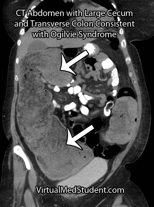
Ogilvie Syndrome CT Abdomen
Diagnosis is made when there is evidence of an enlarged colon with no mechanical obstruction. In essence, diagnosing Ogilvie syndrome is more about ruling out other causes of colon obstruction.

CT scans, abdominal x-rays, barium swallow studies, and colonoscopies are commonly used as adjunctive means for diagnosing pseudo-obstruction.

Treatment

Treatment of Ogilvie syndrome is primarily conservative. This means making the patient NPO (ie: nothing by mouth) and placing a nasogastric tube (NGT) to suction. The NGT helps "decompress" the colon from above until the pseudo-obstruction resolves.

If conservative measures fail then a medication known as neostigmine can be given. Neostigmine is a parasympathetic "mimic". It blocks the breakdown of acetylcholine by inhibiting an enzyme known as acetylcholine esterase. Acetylcholine is the main neurotransmitter of the parasympathetic nervous system. Therefore, if there is more acetylcholine, then more gut motility occurs.

Another medication known as methylnaltrexone may also be useful in cases of acute colonic pseudo-obstruction caused by opioids.

In cases that are refractory to conservative and medical therapies the use of colonoscopic decompression is extremely effective. Patient’s who have evidence of colonic perforation require prompt surgical exploration.

Overview

Ogilvie syndrome, or acute colonic pseudo-obstruction, is a pathologic dilation of the colon that occurs in the absence of a mechanical cause. It is believed to be the result of autonomic nervous system dysfunction. Diagnosis is made with x-rays and CT scans of the abdomen. Treatment is with nasogastric decompression, neostigmine, methylnaltrexone, colonoscopic decompression, or surgery in advanced cases.

References and Resources

  • Hsu HL, Wu YM, Liu KL. Ogilvie syndrome: acute pseudo-obstruction of the colon. CMAJ. 2011 February 22; 183(3): E162.
  • Eisen GM, Baron TH, Dominitz JA, et al. Acute colonic pseudo-obstruction. Gastrointest Endosc. 2002 Dec;56(6):789-92.
  • Saunders MD. Acute colonic pseudo-obstruction. Gastrointest Endosc Clin N Am. 2007 Apr;17(2):341-60, vi-vii.
  • Saunders MD, Kimmey MB. Colonic pseudo-obstruction: the dilated colon in the ICU. Semin Gastrointest Dis. 2003 Jan;14(1):20-7.
  • Ponec RJ, Saunders MD, Kimmey MB. Neostigmine for the treatment of acute colonic pseudo-obstruction. N Engl J Med. 1999 Jul 15;341(3):137-41.
  • Laine L. Management of Acute Colonic Pseudo-Obstruction. N Engl J Med 1999; 341:192-193.
  • Thomas J, Karver S, Cooney GA, et al. Methylnaltrexone for opioid-induced constipation in advanced illness. N Engl J Med. 2008 May 29;358(22):2332-43.
Hypertrophic Cardiomyopathy: Athletes and Genetic Mutations

Hypertrophic cardiomyopathy occurs when the size of heart muscle cells increase (aka: hypertrophy). Genetic mutations in DNA that codes for heart muscle cell proteins are responsible for the development of hypertrophic cardiomyopathy. Most of these mutations are in DNA that code for sarcomere proteins (ie: myosin, actin, troponin, etc.). The mutated proteins cause decreased contractile function. As a result, the muscle cell hypertrophies (enlarges) in an attempt to overcome the decreased contractility. The result is a disorganized pattern of muscle cell fibers with intervening fibrosis (ie: scar tissue).

Signs and Symptoms

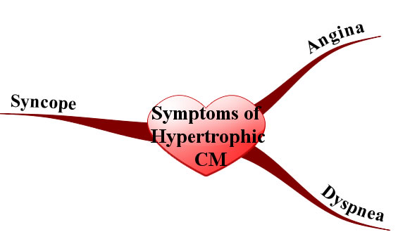
Since the myocardium is hypertrophied there is less ventricular compliance (ie: the heart becomes stiff). This stiffness decreases the filling capacity of the ventricle. The result is diastolic dysfunction, or a decreased ability of the heart to fill during its relaxation phase. High diastolic pressures occur leading to the back-up of blood into the left atrium, pulmonary veins, and pulmonary capillaries. Excess fluid in the pulmonary capillaries causes pulmonary edema with resultant shortness of breath and exercise intolerance.

In addition, angina (chest pain) can occur even without co-existing coronary artery disease because the increased muscle mass of the ventricle results in a higher oxygen demand. Under strenuous conditions the hypertrophied muscle cannot get enough oxygen, which causes chest pain.

Symptoms and Signs of Hypertrophic Cardiomyopathy
Syncope (ie: fainting) is another common symptom that is usually due to arrhythmias caused by the abnormal myocyte architecture.

Physical exam can reveal an S4 gallop (aka: atrial gallop), which is caused by the atrium forcing blood into a stiff left ventricle during the "atrial kick" at the end of diastole.

Murmurs can also be heard, usually mitral regurgitation and a systolic outflow obstruction murmur. Mitral regurgitation occurs because the hypertrophied ventricular septum acts as a barrier to blood flow into the aorta. As a result, during systole blood will flow backwards through the mitral valve into the left atrium. Blood flowing across the septal barrier into the aorta will create an obstruction murmur. The obstruction murmur worsens with valsalva, which distinguishes it from the murmur of aortic stenosis.

Diagnosis

The work-up is very similar to dilated cardiomyopathy, except ancillary studies are usually not helpful. Echocardiography (ie: ultrasound of the heart) is the gold standard and will show the hypertrophic myocardium. ECG will often reveal left ventricular hypertrophy and left atrial hypertrophy. Arrhythmias may sometimes be observed on ECG as well. Prominent Q-waves can be seen in the lateral leads (ie: V4-V6) and inferior leads (II, III, aVF); this is the result of greater depolarization of the hypertrophied septum (remember depolarization of the septum starts on the left side and moves rightward creating a downward deflection in leads on the opposite side of the body, before the left ventricle "overpowers" the ECG findings).

Treatment

β-blockers are the mainstay of treatment. They decrease the heart rate and allow increased diastolic filling times, which leads to decreased outflow obstruction; they also decrease myocardial oxygen demand leading to decreased anginal symptoms.

Prevention of fatal arrhythmias is important in hypertrophic cardiomyopathy. Medical management of arrhythmias is accomplished with amiodarone and/or disopyramide. In some patients, strong consideration should be given to an implantable cardiac defibrillator, especially those at high risk of sudden death. Surgery with partial myomectomy to remove some of the hypertrophied muscle can also be done if the patient is unresponsive to medical management.

Since hypertrophic cardiomyopathy is caused by genetic mutations, genetic counseling should be offered to children of affected parents. First degree relatives should undergo screening with echocardiography as well.

Unlike dilated cardiomyopathy, diuretics should be used sparingly because they can worsen outflow obstruction by causing decreased venous return to the left ventricle. Digoxin is also contraindicated because it can worsen outflow obstruction. When considering treatment options it is important to remember that the problem in hypertrophic cardiomyopathy is diastolic, not systolic dysfunction.

Prognosis depends on the type and severity of the genetic mutation involved. Some mutations result in minimal morbidity and a normal life span, whereas others can cause significant heart failure symptoms. Overall mortality is roughly 5% per year secondary to ventricular fibrillation; therefore, even minimally symptomatic patients must be monitored closely.

Overview

The cause of hypertrophic cardiomyopathy is genetic. Diagnosis is made with echocardiography (ie: ultrasound of the heart). Treatment is generally with beta blockers, amiodarone, implantable cardiac defibrillators (ICD), and myomectomy in select patients. All 1st degree relatives should be offered genetic counseling and undergo screening echocardiography. Prognosis is variable and depends on the mutation type.

Related Articles

References and Resources

  • Bos JM, Ommen SR, Ackerman MJ. Genetics of hypertrophic cardiomyopathy: one, two, or more diseases? Curr Opin Cardiol. 2007 May;22(3):193-9.
  • Bo CY, López B, Coelho-Filho OR, et al. Myocardial fibrosis as an early manifestation of hypertrophic cardiomyopathy. N Engl J Med. 2010 Aug 5;363(6):552-63.
  • Kumar V, Abbas AK, Fausto N. Robbins and Cotran Pathologic Basis of Disease. Seventh Edition. Philadelphia: Elsevier Saunders, 2004.
  • Lilly LS, et al. Pathophysiology of Heart Disease: An Introduction to Cardiovascular Disease. Seventh Edition. Lippincott Williams and Wilkins, 2006.
  • Flynn JA. Oxford American Handbook of Clinical Medicine (Oxford American Handbooks of Medicine). First Edition. Oxford University Press, 2007.

Restrictive Cardiomyopathy: Amyloid, Diastolic Dysfunction, and Kussmaul’s Sign

Restrictive cardiomyopathy is the least common type of cardiomyopathy. The restriction refers primarily to diastolic dysfunction (ie: problems with relaxation of the heart); systolic function is generally well preserved. From a clinical perspective the restrictive cardiomyopathies present similarly to the hypertrophic cardiomyopathies.

There are two main causes of restriction: infiltration of the heart muscle with abnormal substances or scarring of the heart muscle. Each of these causes has many underlying etiologies, a few of which are listed below.

   (1) Infiltration
        (a) Amyloidosis
        (b) Sarcoidosis
        (c) Storage diseases
            (i) Hemochromatosis
            (ii) Glycogen storage diseases
   (2) Fibrosis and scarring
        (a) Post-radiation
        (b) Endomyocardial fibrosis

Regardless of the cause, the heart muscle gets "gunked up" with stuff that shouldn’t be there. The result is decreased contractile ability.

Signs and Symptoms

Patient’s with restrictive cardiomyopathy often present with signs and symptoms of diastolic dysfunction. Pulmonary edema (ie: fluid in the lungs) results in dyspnea (difficulty breathing), paroxysmal nocturnal dyspnea (waking up gasping for air), and orthopnea (inability to lay flat secondary to shortness of breath). Reduced cardiac output results in fatigue, dizziness, and weakness.

On physical exam, Kussmaul’s sign can sometimes be appreciated. Kussmaul’s sign occurs when you can see the jugular vein distend when a patient inspires (breathes in). Normally jugular venous distension decreases with inspiration as venous return to the heart increases due to decreased intrathoracic pressure. However, in restrictive cardiomyopathy the ventricle cannot accommodate the increased blood flow and it backs up into the jugular veins during inspiration, hence the paradoxical worsening.

In addition, signs of systemic volume overload can occur including hepatomegaly (ie: an enlarged liver), ascites (ie: fluid in the abdomen), and bilateral lower extremity edema (ie: leg swelling). Arrhythmias are also common because amyloid deposits can wreak havoc on the conduction system of the heart.

Diagnosis and Work-Up

It is important to distinguish restrictive cardiomypathy from restrictive pericarditis, which is treatable. The gold standard test is a myocardial biopsy, which will reveal infiltrating substances in restrictive cardiomyopathy. MRI and CT can show a thickened pericardium, which is more consistent with restrictive pericarditis.

Treatment and Prognosis

Treatment is aimed at the underlying cause. For example, chemotherapy for multiple myeloma or phlebotomy for hemochromatosis can slow disease progression. Since diastolic function is most affected, treatment is aimed at ensuring adequate filling time for the left ventricle. Diuretics should be used cautiously because patient’s are dependent on higher blood volumes to fill as much of the restricted ventricle as possible. Digoxin and vasodilator therapies are usually not helpful since systolic function is usually well-preserved.

Prognosis is generally poor unless treatment of the underlying condition is curative.

Overview

Restrictive cardiomyopathy is caused by infiltrative processes (most commonly amyloidosis). Physical exam reveals signs of diastolic dysfunction resulting in blood backing up in the lungs and body. The gold standard for diagnosis is myocardial biopsy, although this is not routinely performed in clinical practice. Treatment is aimed at the underlying cause, if identifiable. In addition, since diastolic dysfunction is present, treatments should be used that allow the heart adequate time to fill.

Related Articles

References and Resources

Dilated Cardiomyopathy: Poor Pump, An S3, and Crackles

Cardiomyopathy = cardio (heart) + myo (muscle) + pathy (pathology). In other words, cardiomyopathies are pathologic processes that affect the heart muscle. Dilated cardiomyopathy, which this article is about, is the most common form of cardiomyopathy. It has some known causes, but interestingly the majority of cases have no known cause and may in fact be inherited. In order to be diagnosed with dilated cardiomyopathy you must have left ventricular dilation and a low ejection fraction on echocardiography.

   (1) Idiopathic (ie: no known cause) and/or genetic
   (2) Alcoholism (chronic)
   (3) Inflammatory
       (a) Infectious
           (i) Viral
                 1. Adenovirus
                 2. Coxsackie virus
                 3. Parvovirus
                 4. HIV
           (ii) Protozoan
                 1. Trypanosomiasis (Chagas’ disease)
       (b) Non-infectious
           (i) Collagen vascular disorders
           (ii) Sarcoidosis
   (4) Drug/medicine related
       (a) Chemotherapeutics (daunorubicin/doxorubicin)
       (b) Cocaine
       (c) Methamphetamines
       (d) Heavy metals
   (5) Metabolic
       (a) Hypothyroidism
       (b) Hypocalcemia (chronic)
       (c) Hypophosphatemia (chronic)
   (6) Neuromuscular diseases

Regardless of the cause, the left ventricle of the heart dilates, which decreases its ability to pump effectively.

Signs and Symptoms

The symptoms of dilated cardiomyopathy are directly related to the decreased pumping ability of the heart (ie: systolic dysfunction). Blood backs up into the rest of the body starting with the lungs. This causes pulmonary edema, which can manifest as orthopnea (ie: inability to sleep flat due to shortness of breath), dyspnea (ie: shortness of breath with exertion), and paroxysmal nocturnal dyspnea (ie: waking up in the middle of the night short of breath). Patients also complain of exercise intolerance, dizziness, and fatigue.

The physical exam for someone with dilated cardiomyopathy will often reveal an S3 gallop (aka: ventricular gallop). An S3 gallop is caused by excess blood in the left ventricle after systole; during diastole the blood from the left atrium rushes into a relatively full left ventricle creating the S3 gallop, which can be heard with a stethoscope.

In addition, “crackles” may be heard in the lung fields secondary to pulmonary edema. If right sided heart failure has also occurred (usually after many years of left sided cardiomyopathy) there may be signs of systemic volume overload. These signs include hepatomegaly (a larger than normal liver), bilateral lower extremity edema (ie: pitting edema), and jugular vein distension.

Diagnosis

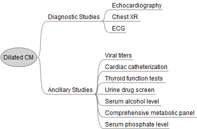
Work Up for Dilated Cardiomyopathy
Echocardiography (ie: an ultrasound of the heart) is the gold standard test and will traditionally show a dilated ventricle(s) with a depressed ejection fraction (EF < 55%).

Additional studies can be ordered depending on the clinical scenario. It is especially important to not miss alcoholism or hypothyroidism as these can be easily treated. Cardiac catheterization is often performed to determine if there is co-existing (or causative) coronary artery disease.

Treatment

Treatment for patients with dilated cardiomyopathy consists of the similar treatments used for other heart failure patients. Many patients will be on an angiotensin converting enzyme inhibitor (ACEI, lisinopril is a commonly used one), or angiotensin receptor blocker (ARB) and a beta blocker (carvedilol is commonly used due to its beneficial lipid profile compared to other beta blockers). Other considerations include spironolactone (an aldosterone receptor antagonist).

ACEI/ARBs, beta blockers, and spironolactone improve survival rates in patients with dilated cardiomyopathy. In addition, an implantable cardiac defibrillator (ICD) should be considered in all patients with an ejection fraction of less than 35% because it has been shown to reduce death from abnormal heart rhythms.

Blood thinning medications like warfarin are indicated if the patient has a thrombus (ie: a “blood clot”) seen on echocardiogram, atrial fibrillation, or previous embolic event, although some physicians may recommend thinning the blood prophylactically if ventricular function is severely impaired (EF < 30%).

Symptomatic management consists of diuretics for volume overload (ie: pitting edema, shortness of breath secondary to pulmonary edema, etc.) and digoxin to increase cardiac contractility and improve forward blood flow.

Curative treatment is a heart transplant. Overall prognosis without a transplantation is poor. Over 50% of non-transplant patients are deceased at 5 years compared to 25% of transplanted patients.

Overview

There are many causes of dilated cardiomyopathy some of which are reversible. An S3 gallop and symptoms of volume overload are often seen on physical exam. Echocardiography is the gold standard for diagnosis. It is important to treat with at least a beta blocker and ACEI; spironolactone is another option. Symptomatic management includes diuretics and digoxin. Prognosis is poor without transplant.

Related Articles

References and Resources

  • Wexler RK, Elton T, Pleister A, et al. Cardiomyopathy: an overview. Am Fam Physician. 2009 May 1;79(9):778-84.
  • Abdo AS, Kemp R, Barham J, et al. Dilated cardiomyopathy and role of antithrombotic therapy. Am J Med Sci. 2010 Jun;339(6):557-60.
  • Fatkin D, Otway R, Richmond Z. Genetics of dilated cardiomyopathy. Heart Fail Clin. 2010 Apr;6(2):129-40.
  • Kumar V, Abbas AK, Fausto N. Robbins and Cotran Pathologic Basis of Disease. Tenth Edition. Philadelphia: Elsevier Saunders, 2004.
  • Lilly LS, et al. Pathophysiology of Heart Disease: A Collaborative Project of Medical Students and Faculty. Seventh Edition. Lippincott Williams and Wilkins, 2006.
  • Flynn JA. Oxford American Handbook of Clinical Medicine (Oxford American Handbooks of Medicine). First Edition. Oxford University Press, 2007.

Cervical Myelopathy: A Squished Spinal Cord

Cervical myelopathy refers to a constellation of signs and symptoms that occur when the spinal cord is squeezed and unable to function properly. Myelopathy is most commonly caused by cervical stenosis, which is a general term used to describe a decrease in the cross sectional area of the spinal canal

Stenosis comes in two flavors: congenital or acquired. In congenital cervical stenosis the spinal canal is developmentally narrower than normal. Congenital stenosis is not considered hereditary, but is present from birth, and is a result of “abnormal” development.

Acquired stenosis is more common, and typically occurs in people over the age of 50. This type of stenosis is caused by a combination of factors and pathologic changes.

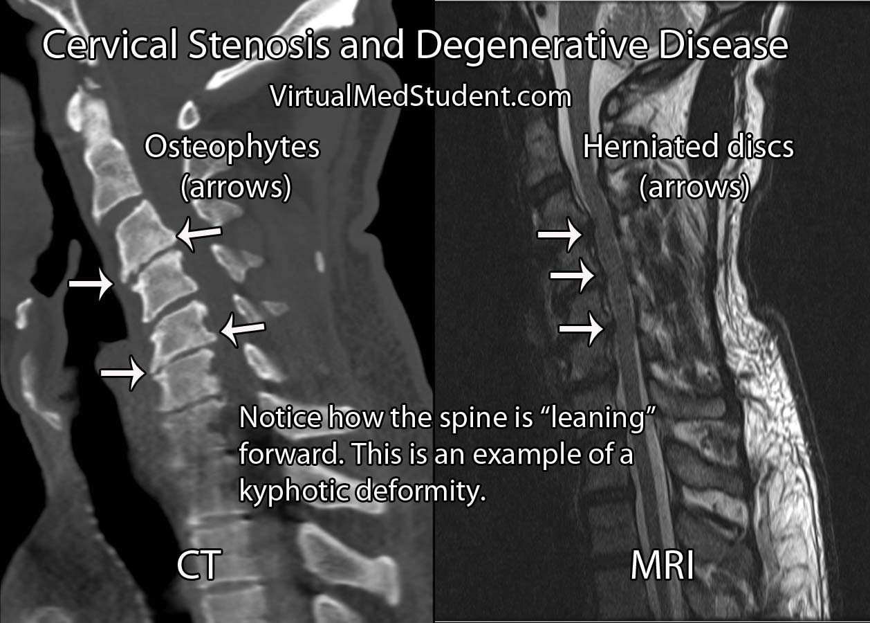
Degenerative cervical disease is the most common cause of acquired cervical stenosis and myelopathy. It has several distinct features, which include: loss of disc height, disc herniation, osteophyte formation, and resultant deformity (ie: kyphosis, or a forward leaning cervical spine). Let’s discuss each of these features in more detail…

The first feature in acquired degenerative stenosis is a loss of intervertebral disc elasticity and height. You can think of the intervertebral discs like shock absorbers for the spinal column. In the cervical spine, a healthy disc is slightly taller at its front most portion and slightly shorter at its back most portion. This gives the normal cervical spine a lordotic curvature.

As someone ages, the discs tend to dry out and the height differential between the front and back of the disc is lost. This can cause the cervical spine to bow forward resulting in a loss of lordosis. The new forward lean to the spine is called a kyphotic deformity. This deformity can cause the cord to stretch over the bones of the spine resulting in myelopathy.

The second factor that contributes to acquired degenerative stenosis is when the back most section of the disc weakens and pooches out towards the spinal cord. This is known as a disc herniation. Disc herniations narrow the cross sectional diameter of the spinal canal and contribute to stenosis.

Finally, as the discs age and dry out they lose their elasticity, and therefore lose their ability to absorb shock. As a result, the bones and ligaments take on more of the burden. They attempt to fight back by forming bony growths known as osteophytes. The osteophytes further narrow the spinal canal and contribute to stenosis.

Let’s bring it all together… Acquired degenerative cervical stenosis occurs when a combination of kyphotic deformity, disc herniations, and osteophytic growths narrow the spinal canal and cause compression of the spinal cord.

Other less common causes of cervical stenosis include ossification of the posterior longitudinal ligament. Additionally, traumatic injuries of the cervical spine can cause immediate cervical stenosis and cord injury. And even less commonly, tumors in the cervical spine can push on the cord and cause stenosis.

It is important to note that cervical stenosis does not always cause myelopathy. In fact, some patients can have a very "tight" cervical cord, but have no signs or symptoms of cord compression.

Signs and Symptoms

Patient’s that are myelopathic present with a constellation of signs and symptoms depending on how "squished" their cord is. Myelopathic patients present with a combination of gait dysfunction, clumsiness when using their hands, weakness, abnormally brisk reflexes, spasticity, and Lhermitte sign.

Signs and Symptoms
of Myelopathy:

– Gait dysfunction
– Clumsiness/weakness
– Brisk reflexes

– Spasticity
– Sensation problems
– Lhermitte’s sign

Reflexes are typically brisk, especially in the legs. Examples of brisk reflexes include clonus (a sustained contraction that causes the foot to "beat" several times), upgoing plantar response (ie: the big toes extend upwards, as opposed to curling down when the sole of the foot is stroked), and Hoffmann’s sign (ie: the thumb curls in when the tip of the index or middle finger is flicked).

Patients who have myelopathy tend to walk with a broad based gait as if steadying themselves. They often complain of feeling like they are walking “drunk”. Clumsiness is also a common symptom of myelopathy. Patients will typically complain of problems writing, using a utensil, holding a coffee cup, or performing other fine motor movements.

Finally, a phenomenon known as Lhermitte’s sign can occur. Lhermitte’s sign occurs when someone flexes their head forward and a shock like sensation runs down the neck and back into the extremities. It is a result of compression on the back most portion of the spinal cord, which is an area that relays sensation to the brain.

The Nurick grading system (table below) was designed to assess the severity of myelopathy. It is useful when guiding treatment decisions. The Japanese Orthopedic Association (JOA) score, European Myelopathy Score, Myelopathic Disability Index, and Ranawat Classification are other myelopathy grading scales that are commonly used in clinical practice.

The Nurick Grading System for Myelopathy
Grade 0 Nerve root impingement may be present (ie: radiculopathy), but no cord symptoms are present
Grade 1 Able to walk normally, but may have signs (but not symptoms) of cord compression
Grade 2 Some difficulty with walking, but still able to work
Grade 3 Unable to walk to the point of not being able to work
Grade 4 Can only walk when assisted
Grade 5 Confined to a wheelchair or bed bound

Diagnosis and Classification

Diagnosis of cervical stenosis is based on imaging (ie: CT, MRI, etc). Remember that stenosis does not necessarily translate into myelopathy! Patient’s should be symptomatic as well! A stenotic spine is radiographically present when the diameter of the spinal canal in the antero-posterior plane is less than 10mm.

An alternative way of diagnosing cervical stenosis is using Pavlov’s ratio. This is defined as the ratio of the canal diameter to the diameter of the adjacent vertebral body; any ratio less than 0.8 is worrisome.

Treatment

Treatment of cervical stenosis depends on several factors. First and foremost are symptoms. If a patient is not myelopathic, then frequently the best course of action is to monitor the patient until signs or symptoms of myelopathy appear.

If symptoms are present, the discussion becomes whether or not to reconstruct and decompress the front part of the spine (ie: an anterior approach), the back part of the spine (ie: a posterior approach), or both.

Anterior approaches come in one of two flavors. The first is removal of the disc with placement of a bone graft; this is formally known as a cervical discectomy and fusion. When the bones of the spine are causing the compression a more extensive surgery known as a corpectomy is performed; in this procedure the entire vertebral body is removed and a cage is placed in its place to reconstruct the spine.

Posterior approaches include laminoplasty and laminectomies with or without instrumented fusion. Laminoplasty involves splaying open the lamina so that the spinal cord can drift back. Laminectomies involve un-roofing the bone behind the spinal cord. Laminectomies are frequently done with an instrumented fusion in which rods and screws are placed to help the patient maintain a normal cervical alignment (ie: lordosis).

Surgical Treatment of Myelopathy
Deformity Compression Surgical Approach
Kyphotic Anterior and only 3 spinal levels or less involved Anterior (anterior cervical discectomies and/or corpectomies)
Kyphotic Anterior and posterior Combined anterior (anterior cervical discectomies and/or corpectomies) and posterior (laminectomies with or without instrumented fusion)
Lordotic Anterior and only 3 spinal levels or less involved Anterior (anterior cervical discectomies and/or corpectomies)
Lordotic Posterior Posterior (laminoplasty or laminectomies with or without posterior instrumented fusion)
Lordotic Anterior and posterior Combined anterior (anterior cervical discectomies and/or corpectomies) and posterior (laminectomies with or without instrumented fusion)

The choice of surgical options is highly dependent on the degree of deformity, instability, and where specifically (ie: what levels) in the spine the cord compression is located.

Ultimately, the surgical treatment of cervical stenosis and myelopathy must be tailored to each individual patient based on many factors.

Overview

Cervical stenosis is abnormal narrowing of the spinal canal. It has numerous causes, of which the most common one is acquired degenerative stenosis. Stenosis can cause compression of the spinal cord, which can cause signs and symptoms of myelopathy. Treatment for symptomatic patients involves decompressing the spinal cord via either an anterior, posterior, or combined approach.

Related Articles

References and Resources

Pelvic Inflammatory Disease: Gonococcus, Chlamydia, and the Chandelier Sign

Pelvic inflammatory disease (PID) refers to infection of the uterus, fallopian tubes, and/or ovaries. The most common infections that cause pelvic inflammatory disease are neisseria gonorrhoeae and chlamydia trachomatis. In fact, both bacteria may be present in the same patient! However, it is not uncommon for other pathogenic bacteria to be present as well. The exact role of most of these other pathogenic bacteria is not well understood.

In order for these organisms to gain access to the uterus, fallopian tubes, and ovaries there must be some breakdown of the normal barriers that separate the vagina from the upper genital tract. Interestingly, a significant number of cases of pelvic inflammatory disease occur just after menstruation when the possibility of pathogenic bacteria gaining access to the upper genital structures is at its greatest. In addition, normal vaginal bacteria can act as a barrier to invading pathogenic strains. Therefore, in some patients with PID the normal vaginal bacterial landscape has been altered in some way, which allows the overgrowth of pathogenic bacteria.

Patients who are at risk for pelvic inflammatory disease are generally those women who are at increased risk of sexually transmitted diseases. Therefore, women with multiple sexual partners, younger age (most cases occur between 15 and 25 years of age), previous history of sexually transmitted disease, and those that do not use barrier contraception (ie: condoms) are all at increased risk.

Signs and Symptoms

Bilateral lower quadrant abdominal pain is a common presenting symptom. Infection and the resulting inflammation that occurs can make having sex painful (ie: "dyspareunia") and can make urinating painful (ie: "dysuria"). In addition, abnormal vaginal odors and discharges are also possible. Like most other serious infections, systemic signs such as fever, nausea, vomiting, and lethargy may also occur.

On pelvic examination patients are extremely tender to palpation of the uterus and ovaries. This is often referred to as the “Chandelier sign” because women will “reach for the chandeliers” because of the discomfort during the exam.

Complications

Complications can be severe if left untreated. Pelvic inflammatory disease can lead to scarring of the fallopian tubes and possible infertility. The risk of ectopic pregnancies is increased in women with a history of PID. Abscesses can form in the ovaries and tubes, as well as in the peritoneal cavity (ie: abdominal cavity). In addition, inflammation of the liver’s capsule can occur in a complication known as Fitz-Hugh-Curtis syndrome. Pelvic inflammatory disease can also lead to infection of the peritoneal cavity (aka: peritonitis).

Diagnosis

Diagnosis of pelvic inflammatory disease is based on clinical signs and symptoms. If lower abdominal discomfort with pain during pelvic examination (ie: cervical motion, uterine, and/or ovarian tenderness) is present, patients should be treated as though they have pelvic inflammatory disease.

There are numerous other criteria that can “support” the diagnosis, but are not necessary to have. They include other signs of infection like abnormal vaginal discharge, fever, elevated white blood cell count, and evidence of gonorrhea or chlamydial infection from laboratory tests.

Definitive diagnosis is made either by biopsy of the upper genital tract, imaging (ie: CT or MRI) that shows edematous fallopian tubes, or disease seen laparoscopically.

Treatment

Treatment of pelvic inflammatory disease is based on the severity of the clinical signs and symptoms, as well as the likely underlying organisms. Patients who are stable can be given oral antibiotics.

Generally a single dose of a third generation cephalosporin like ceftriaxone is given to cover gonorrhea; another antibiotic called doxycycline is given to cover the possibility of co-existent chlamydial infection. Metronidazole may be added to these medications if anaerobic bacteria are suspected.

If the patient appears very sick, is pregnant, unlikely to continue treatment as an outpatient (ie: homeless, destitute, etc), or unable to take oral antibiotics they should be hospitalized and treated with intravenous (IV) antibiotics. Usually a second generation cephalosporin like cefoxitin plus doxycycline (either IV or oral) is given. Clindamycin and gentamicin, as well as ampicillin-sulbactam (Unasyn®) can also be used in certain circumstances. In general, there are numerous antibiotic regimens that can be considered.

Overview

Pelvic inflammatory disease is an infection of the upper genital tract in females. This includes the uterus, fallopian tubes, and/or ovaries. Gonorrhea and chlamydia are the two most commonly isolated bacteria, but many infections are caused by other pathogenic bacteria. Diagnosis is made on clinical signs and symptoms. Treatment is with at least two antibiotics: one to cover gonorrhea and one to cover chlamydia.

Related Articles

References and Resources

Hydrocephalus is Greek for “Water Head”

Hydrocephalus literally means "water head". It is a term used to describe a pathological increase in the amount of cerebrospinal fluid (CSF) within the ventricles (fluid filled cavities) of the brain.

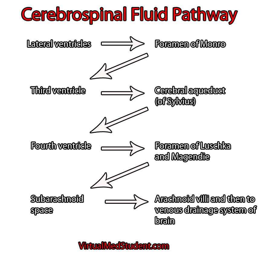
In order to understand hydrocephalus we have to first appreciate the cerebrospinal fluid pathway and ventricular system of the brain. The brain has four ventricles: a pair of lateral ventricles, a third ventricle, and a fourth ventricle. They are connected to one another through narrow channels. The fourth ventricle drains into the subarachnoid space around the upper spinal cord. The spinal fluid travels down into the lumbar cistern and then back up again where it is absorbed by the arachnoid granulations overlying the cerebral hemispheres.

CSF pathway

Cerebrospinal fluid is created at a rate of roughly 500 mL per day. It is primarily secreted by specialized cells within the walls of the ventricles known as choroid plexus. As you can imagine, if it is secreted at 500 mL per day there must be an equal amount of re-absorption. This re-absorption occurs in the subarachnoid space by venous structures known as "arachnoid villi". Re-absorption does not occur in the ventricles themselves; this is an important point to keep in mind as we discuss the difference between communicating and non-communicating forms of the disease.

Hydrocephalus occurs when excess cerebrospinal fluid backs up. It is called "communicating" hydrocephalus if all of the ventricles are enlarged. Otherwise it is known as "non-communicating" hydrocephalus. There is another informal type of hydrocephalus that is known as "ex-vacuo"; it occurs when dilation of the ventricular system is a result of brain tissue loss, rather than a pathological increase in the amount of cerebrospinal fluid.

The term hydrocephalus usually implies an abnormally high pressure within the ventricular system. However, a different type of hydrocephalus known as "normal pressure" hydrocephalus defies this rule.

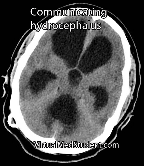
Communicating hydrocephalus usually develops when the arachnoid villi get "gunked up". Abnormal materials (such as blood in subarachnoid hemorrhage or proteins after meningitis) can block the arachnoid villi and prevent re-absorption. The cerebrospinal fluid then backs up and causes the entire ventricular system to enlarge.

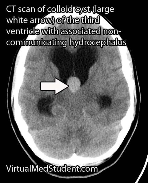
Non-communicating hydrocephalus is slightly different because the re-absorption pathway is functioning properly, but CSF backs up behind a "road block" in one of the channels connecting the individual ventricles. "Road blocks" can be anything from tumors to developmental narrowing of the channel itself. For example, colloid cysts of the third ventricle can occlude the foramen of Monroe (the channel between the lateral and third ventricles); when this occurs CSF produced in the lateral ventricles is not able to flow into the third ventricle. CSF then backs up in the lateral ventricle(s) resulting in pathologic dilation.

Overall, there are crap tons of causes for both communicating and non-communicating hydrocephalus. Some patients have congenital forms secondary to improper development of the CSF pathways. Others may develop hydrocephalus later in life as a result of infection, tumor formation, or aneurysm rupture.

Signs and Symptoms

The signs and symptoms depend on the age of the patient and the rapidity of onset. In newborns hydrocephalus often presents as failure to thrive with an abnormally enlarging head.

If hydrocephalus develops rapidly, the increase in intracranial pressure (as a result of cerebrospinal fluid putting pressure on the brain) leads to nausea, vomiting, headache, and if severe enough, coma and potentially death!

Normal pressure hydrocephalus (NPH) is a unique type of hydrocephalus in which there is no elevation in pressure (hence the term "normal pressure"). The classic symptoms of NPH are difficulty walking, dementia, and urinary incontinence.

Diagnosis

Non-communicating hydrocephalus
Communicating Hydrocephalus

Diagnosis of hydrocephalus is made with CT scan coupled with clinical evidence of increased pressure within the ventricular system.

The scan will show an abnormally enlarged ventricular system. MRI scans with contrast are also routinely done to evaluate for tumors as a cause of hydrocephalus.

Treatment

Treatment consists of "shunting" the cerebrospinal fluid to another part of the body, usually the peritoneal cavity.

Other techniques such as third ventriculostomy, in which a surgically made "hole" is placed between the third ventricle and the subarachnoid space is also sometimes employed. This is very effective in cases of non-communicating hydrocephalus secondary to aqueductal stenosis (ie: the small passage between the third and fourth ventricles).

The ultimate treatment depends on the type of hydrocephalus and whether symptoms are severe enough to warrant surgical intervention.

Overview

Hydrocephalus is a pathological accumulation of cerebrospinal fluid. It can be communicating, non-communicating, or normal pressure. It can cause numerous signs and symptoms, but headache, nausea and vomiting are amongst the most common. Diagnosis is with CT scanning and clinical evidence of increased intracranial pressure. Treatment consists of shunting the CSF to another part of the body where it can be reabsorbed.

Related Articles

References and Resources

Subarachnoid Hemorrhage: Aneurysms, Vasospasm, and Hyponatremia

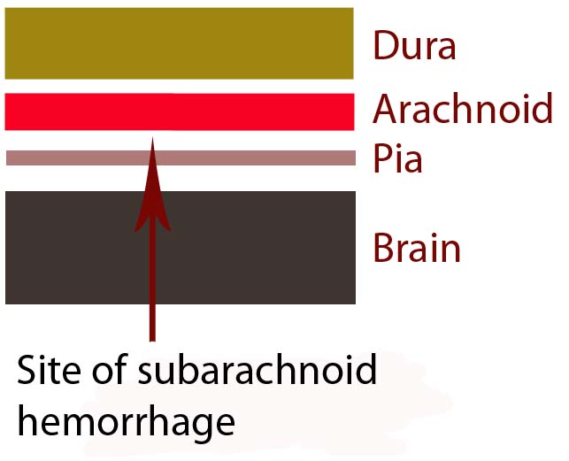
In order to understand subarachnoid hemorrhage we have to first appreciate the layers that make up the brain and its surrounding tissues. The brain itself has three protective layers: dura mater, the arachnoid, and the pia.

The dura is a thick layer of fibrous tissue immediately below the skull. Below the dura is the arachnoid, which is a layer of delicate web-like tissue (hence the name "arachnoid"). Finally, below the arachnoid is the pia mater. The pia is a very thin layer that is directly adjacent to brain tissue.

The subarachnoid space, or the region between the arachnoid tissue and the pia contains cerebrospinal fluid, which acts like a liquid shock absorber for the brain. Also contained within the subarachnoid space are blood vessels that penetrate down into the brain tissue. Sometimes these blood vessels "leak", which can cause a "sub-arachnoid" hemorrhage.

Brain Layers

The most common cause of subarachnoid hemorrhage is traumatic injury; the most common non-traumatic cause is a ruptured aneurysm.

An aneurysm is an abnormal ballooning out of a blood vessel’s wall. The balloon’s dome is much weaker than the rest of the vessel wall. These weak areas can rupture allowing blood to leak out of into the subarachnoid space.

Other causes of subarachnoid hemorrhage include idiopathic (ie: unknown) causes, arteriovenous malformations, vessel dissections, and very rarely tumors. Regardless of the cause, blood will pool in the subarachnoid space.

The remainder of this article will focus on the most common non-traumatic cause of subarachnoid hemorrhage – aneurysm rupture.

Signs and Symptoms

The classic symptom of a subarachnoid hemorrhage is a horrific headache described as the “worst headache of their life". Photophobia, nausea, vomiting, and nuchal rigidity are also common. Seizures may also occur. In addition, depending on how severe the subarachnoid hemorrhage is, patients may have decreased levels of consciousness; some patients become comatose, and many die before reaching medical attention.

The patient’s clinical status is graded according to the Hunt and Hess system. It only applies to patients in whom subarachnoid hemorrhage is caused by rupture of an aneurysm. This grading system was initially established to help determine mortality and clinical outcomes. In modern practice, these numbers are likely high given modern improvements in critical care and neurosurgical intervention since Hunt and Hess first developed their grading system.

Hunt and Hess Clinical Grading Scale
Grade Patient’s Clinical Status Associated Mortality
1 Mild headache and/or nuchal rigidity 1%
2 Cranial nerve dysfunction, moderate to severe headache and/or nuchal rigidity 5%
3 Mild focal neurological deficit, lethargic, confused 19%
4 Stuporous, moderate to severe hemiparesis, early decerebrate posturing 40%
5 Coma, decerebrate posturing 77%

The world federation of neurological surgeons also has a clinical score based on the Glasgow Coma Scale (GCS). It associates the patient’s GCS with the likelihood of death.

World Federation of Neurological Surgeons Grading System
  GCS Major focal deficit Mortality
1 15 No 5%
2 13-14 No 9%
3 13-14 Yes or No 20%
4 7-12 Yes or No 33%
5 3-6 Yes or No 77%

It is very important to think about the possibility of subarachnoid hemorrhage in patients presenting with these signs and symptoms. Prompt diagnosis and treatment is necessary in order to prevent devastating consequences!

Complications

Blood in the subarachnoid space is very irritating to the brain and cerebral blood vessels. Because of this, several complications can occur.

One of the most common complications is known as "vasospasm." Vasospasm occurs when the blood vessels of the brain spasm several days after the initial hemorrhage. When this occurs blood is no longer able to flow past the blockage; if this occurs for a long enough period of time a stroke can occur. The peak period for vasospasm occurs between 3 and 14 days after the initial bleed.

Another complication of subarachnoid hemorrhage is known as cerebral salt wasting. This occurs when a patient urinates excessive amounts of sodium causing the blood level of sodium to drop precipitously. Because of the excessive urination the patient also becomes dehydrated. Aggressive fluid and salt resuscitation must be given to prevent profound hyponatremia (ie: decreased sodium levels in the blood), which can cause seizures, coma, and death.

In addition, for unknown reasons, many patients with subarachnoid hemorrhage also shower their cerebral hemispheres with micro-thrombi (ie: clots), which can lead to many small strokes. The reason why patients with subarachnoid hemorrhage become coagulopathic is still an area of intense research.

Diagnosis

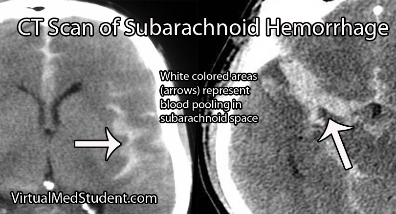
Subarachnoid hemorrhage is most commonly diagnosed by head CT. CT scans are fast and readily pick up the extravasated blood, which layers in the subarachnoid space (see image below). If there is a high clinical index of suspicion but CT of the head is negative than a lumbar puncture should be performed. If the spinal fluid has xanthochromima (a product of red blood cell breakdown) this is highly concerning for subarachnoid hemorrhage.

CT Scan of Subarachnoid Hemorrhage

Subarachnoid hemorrhage is not a diagnosis per say, but rather the result of some underlying pathology (ie: aneurysm, trauma, etc.). Many of these pathologies are treatable; therefore, it is important to figure out what caused the subarachnoid hemorrhage.

Since many are the result of ruptured aneurysms there are several other tests that are often done. The first test is a CT angiogram (CTA). In this test a radio-opaque material is injected into the blood vessels and a CT is performed.

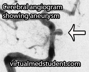
Cererbral Angiogram with Aneurysm
Dye in the dome of the aneurysm will appear as an abnormality helping to confirm the presence, and more importantly the location of the aneurysm.

A more invasive procedure known as a "cerebral angiogram" (image to the right) is also often performed.

In this test, a catheter is inserted into blood vessels in the groin and then threaded up into the blood vessels of the brain. Radio-opaque material is injected and x-rays can pick up abnormalities in the vessel.

The benefit of doing a cerebral angiogram is that it is diagnostic, and treatment can frequently be offered through the catheter itself.

Treatment

There are three main components of treating a subarachnoid hemorrhage: treating the underlying cause, preventing a "re-bleed", and preventing secondary complications.

Since many subarachnoid hemorrhages are caused by aneurysm rupture we’ll discuss the treatment for this common cause. Aneurysms are treated either “open” or “closed”.

“Open” refers to a surgical procedure in which part of the skull is removed. The surgeon then dissects down to the aneurysm. Once identified, a clip is placed around the neck of the aneurysm (ie: you can think of the clip as putting a knot in the neck of a balloon). This stops blood from flowing into the aneurysm, and therefore prevents re-rupture.

“Closed” treatment refers to endovascular technology in which a small micro-catheter is threaded from the blood vessels in the groin into the cerebral vasculature. The aneurysm is located via angiogram. Through a hole in the microcatheter tip the physician then fills the aneurysm dome with small metallic coils.

Once blood is in the subarachnoid space secondary complications often result. One of these complications is referred to as "vasospasm". Medications such as oral nimodipine and intra-arterial nifedipine are used to reduce the amount of vasospasm by inhibiting smooth muscle contraction in the wall of the blood vessel.

Overview

Subarachnoid hemorrhage occurs most commonly after an intracerebral aneurysm ruptures, although other causes exist. Regardless of the cause, blood spills out into the subarachnoid space. Symptoms include a horrible headache, focal neurological deficits (ie: weakness, difficulty speaking, etc.), and coma. If an aneurysm is the cause, it is secured with clipping or coiling. Preventing secondary complications such as vasospasm is also an important component of treatment.

References and Resources

Cholelithiasis (Gallstones): Female, Fat, Fertile, and Forty

Gallstones (aka: cholelithiasis) are stones that form – you guessed it – in the gallbladder. In order to understand gallstones we have to understand how bile is formed. The gallbladder functions as a bile storage bin that releases its contents in response to signals from the intestine and stomach (ie: usually after a meal rich in fats). Bile is formed in the liver and is a unique mix of bile acids, cholesterol, bilirubin, water, phospholipids, and other electrolytes. The most important component is the bile acids. They help keep bile in its liquid state.

With a basic understanding of bile we can now address the problem of gallstone formation. There are different types of gallstones, and the most common type are cholesterol stones. When there is too much cholesterol in bile, the bile acids can no longer keep it soluble. The result? The excess cholesterol precipitates out and forms a stone.

The second type of gallstones are pigmented stones, which come in two flavors: brown and black. Brown stones form as a result of biliary tract infection. Black stones occur when the body breaks down red blood cells excessively, such as in hemolytic anemias. Black stones form from the excess bilirubin (a breakdown product of red blood cells), which gets sent to the liver and ultimately ends up in bile. An excessive amount will do exactly like cholesterol does, and precipitate to form a stone.

Signs and Symptoms

The common risk factors associated with gallstone formation are the “4 Fs”:

– Female
– Fat (overweight/obese, to use the politically "correct" term)
– Fertile
– Forty

Gallstones tend to develop in this demographic more often than in other people. Any one of these factors puts you at increased risk of developing gallstones. Other common risk factors include the following: oral contraceptive use, rapid weight loss, hemolytic diseases/disorders, small bowel resection, total parenteral nutrition (ie: nutrition given through an IV), Crohn’s disease, cystic fibrosis, and Native American ethnicity.

Gallstones by themselves are usually not symptomatic. In fact, many people are walking around with gallstones and don’t even know they have them! However, in a subset of the population symptoms develop. The main symptom associated with gallstones is biliary colic, which is pain that occurs after a meal (especially meals rich in fats). In addition, nausea, vomiting, and fatty food intolerances may also occur.

Complications

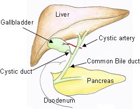
Biliary Tract
There are many potential complications of gallstones. The most common complication is acute cholecystitis. In acute cholecystitis a gallstone lodges in the cystic duct (see image) during contraction of the gallbladder. This leads to inflammation of the gallbladder wall. If left untreated, acute cholecystitis can lead to infection of the gallbladder.

The second complication of gallstones is choledocholithiasis. This is a fancy term for a gallstone lodged in the common bile duct. This can lead to jaundice, pain, pancreatitis, and potentially a very dangerous condition known as acute cholangitis. Acute cholangitis is infection of the biliary tree and can occur secondary to an obstructing stone in the common bile duct.

To review, the common complications of gallstones are:

(1) Biliary colic
(2) Acute cholecystitis
(3) Choledocholithiasis
(4) Acute cholangitis
(5) Pancreatitis

Diagnosis

Since most gallstones are cholesterol, they are radiolucent, meaning that they are not detectable by x-rays. The test that is often done first with any suspected biliary pathology is a right upper quadrant ultrasound. If stones are present they will cast a “shadow” that can be picked up on the ultrasound. Often times additional studies are performed if another diagnosis is being considered. Some clinicians will get hepatitis studies to rule out liver damage. Pancreatic function is also commonly tested.

Treatment

Most gallstones do not require treatment because they are asymptomatic. However, if symptoms develop a trial of lifestyle modification is often tried. For example, avoiding fatty foods can limit biliary colic.

The definitive treatment for symptomatic gallstones, or if any of the complications above develop, is a cholecystectomy. Cholecystectomy is the medical term for removal of the gallbladder. It is done either "open" (ie: with a large incision) or laparoscopically depending on the patient and surgeon’s preferences.

Some patients may not be able to undergo surgery. In these patients a medication known as ursodiol may be given. It is a bile acid mimic that helps liquify bile.

Overview

Cholelithiasis (gallstones) is caused by precipitation of cholesterol or bilirubin. Gallstones are usually asymptomatic, although they can sometimes lead to biliary colic, acute cholecystitis, acute cholangitis, choledocholithiasis, and pancreatitis. Treatment, when symptomatic, is usually lifestyle modifications; if dangerous symptoms or complications occur treatment is with cholecystectomy (ie: removal of the gallbladder).

References and Resources

  • Venneman NG, van Erpecum KJ. Pathogenesis of gallstones. Gastroenterol Clin North Am. 2010 Jun;39(2):171-83, vii.
  • van Erpecum KJ. Biliary lipids, water and cholesterol gallstones. Biol Cell. 2005 Nov;97(11):815-22.
  • Stinton LM, Myers RP, Shaffer EA. Epidemiology of gallstones. Gastroenterol Clin North Am. 2010 Jun;39(2):157-69, vii.
  • Gurusamy KS, Davidson BR. Surgical treatment of gallstones. Gastroenterol Clin North Am. 2010 Jun;39(2):229-44, viii.
  • O’Neill DE, Saunders MD. Endoscopic ultrasonography in diseases of the gallbladder. Gastroenterol Clin North Am. 2010 Jun;39(2):289-305, ix.
  • Flynn JA. Oxford American Handbook of Clinical Medicine (Oxford American Handbooks of Medicine). First Edition. Oxford University Press, 2007.
  • Kumar V, Abbas AK, Fausto N. Robbins and Cotran Pathologic Basis of Disease. Seventh Edition. Philadelphia: Elsevier Saunders, 2004.
  • Blackbourne LH. Surgical Recall, North American Edition (Recall Series). Ninth Edition. Philadelphia: Lippincott Williams and Wilkins, 2009.

Carotid Stenosis: TIAs, Strokes, and Amaurosis Fugax

Carotid stenosis is narrowing of the carotid arteries. This is usually due to atherosclerotic disease present in the internal carotid arteries.

The internal carotid arteries supply a significant portion of the brain with blood, nutrients, and oxygen. If carotid stenosis is present it can result in transient ischemic attacks and possibly stroke.

Signs and Symptoms

Carotid stenosis may be either symptomatic or asymptomatic. Symptomatic carotid stenosis presents in one of three ways: transient ischemic attacks, stroke, or amaurosis fugax.

Transient ischemic attacks (TIA) occur when blood to the brain is blocked temporarily. TIAs cause the same symptoms as a stroke, but the symptoms resolve over 24 hours and there is no permanent brain injury.

A sub-category of TIA is a symptom known as amaurosis fugax, which is a temporary loss in vision, usually in one eye. It is described by patients as a curtain being drawn over the affected eye. It is caused by a small blood clot that breaks off from an atherosclerotic plaque present in the internal carotid artery. This small clot enters the retinal artery and occludes blood flow causing temporary blindness.

Stroke is by far the scariest problem associated with carotid stenosis. When a stroke occurs it can cause severe and irreversible brain injury. Strokes can cause life changing symptoms such as paralysis, aphasia (inability to speak or understand language), and even death!

Carotid Stenosis

Diagnosis

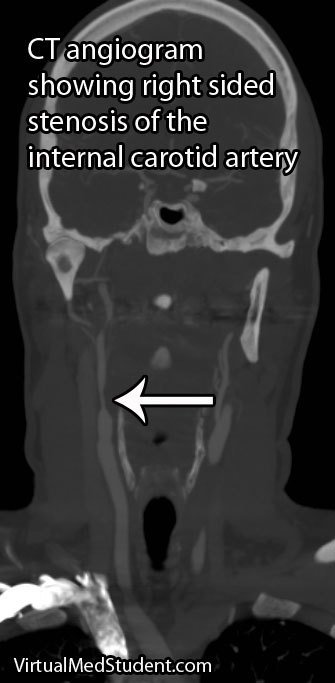
Diagnosis of carotid stenosis is based on imaging studies. The most commonly employed studies are carotid ultrasound, CT angiogram, MR angiogram, and formal angiography.

Treatment

Treatment is dependent on the location and degree of stenosis. Lesions that are near the origin of the internal carotid artery are frequently fixed with surgery in a procedure known as an endarterectomy. In a carotid endarterectomy the artery is surgically opened and the atherosclerotic material is "scooped" out.

The benefit of carotid endarterectomy on stroke risk for symptomatic patients is dependent on the degree of narrowing in the vessel. A landmark study in 1991 showed that carotid endarterectomy was very beneficial in preventing stroke in already symptomatic patients when the degree of narrowing was high (defined as 70 to 99%). A different study looked at lesser degrees of stenosis (50 to 69%) and found a benefit, albeit less robust. There is no clear benefit to having surgery in patients who have narrowing that is 50% or less. Surgery in asymptomatic patients with higher degrees of stenosis follows a more complicated algorithm.

If the diseased segment is higher up on the internal carotid, and surgical access is anatomically difficult, then procedures like carotid stenting or angioplasty can be performed. Stenting is a procedure in which a tiny metal tube-like device is threaded up through the femoral artery in the groin and then opened to “re-expand” the diseased arterial segment.

Patients who may not tolerate a procedure are often managed medically with antiplatelet medications such as aspirin, aspirin/dipyridamole combo (Aggrenox®), ticlopidine (Ticlid®) or clopidogrel (Plavix®). Currently there is no "best" medication to prescribe; the choice is highly dependent on the individual patient and physician.

Overview

Carotid stenosis refers to narrowing of the internal carotid artery. It is usually due to atherosclerotic disease. It can cause transient ischemic attacks, stroke, and amaurosis fugax. Diagnosis is with CT angiogram, carotid ultrasound, and/or formal invasive angiography. Treatment is highly variable, but usually involves a combination of surgery (endarterectomy), stenting, angioplasty, and antiplatelet medications like aspirin.

References and Resources

  • Barnett HJ, Taylor DW, Eliasziw M, et al. Benefit of carotid endarterectomy in patients with symptomatic moderate or severe stenosis. North American Symptomatic Carotid Endarterectomy Trial Collaborators. N Engl J Med. 1998 Nov 12;339(20):1415-25.
  • Chambers BR, Donnan GA. Carotid endarterectomy for asymptomatic carotid stenosis. Cochrane Database Syst Rev. 2005 Oct 19;(4):CD001923.
  • [No authors listed]. Beneficial effect of carotid endarterectomy in symptomatic patients wth high-grade carotid stenosis. North American symptomatic carotid endarterectomy trial collaborators. N Eng J Med. 1991 Aug 15;325(7):445-53.
  • Biller J, Feinberg WM, Castaldo JE, et al. Guidelines for carotid endarterectomy: a statement for healthcare professionals from a Special Writing Group of the Stroke Council, American Heart Association. Circulation. 1998 Feb 10;97(5):501-9.