Atlanto-Occipital Dislocation (Internal Decapitation)

Atlanto-occipital dislocations (AODs) occur when there is severe damage to the ligaments that connect the skull to the cervical spine. The most common cause of an AOD is trauma.

There are several classifications of atlanto-occipital dislocations. In type 1 dislocations the base of the skull moves anteriorly (ie: forward) relative to the dens of the second cervical vertebrae. In type 2 dislocations the skull moves superiorly (ie: upwards) relative to the cervical spine. In type 3 dislocations the skull moves posteriorly (ie: backwards) relative to the cervical spine.

Often times more than one classification may exist in a single patient. For example, a patient may have both a type 1 and type 2 AOD if the patient’s skull has moved forward and upwards relative to the cervical spine.

Signs and Symptoms

Most cases of complete atlanto-occipital dislocation are fatal. Severe damage to the spinal cord around the cranio-cervical junction leads to respiratory paralysis and death if mechanical ventilation is not started rapidly. Patients are often quadriplegic (ie: unable to move their upper and lower extremities) because of damage to the descending motor axons of the corticospinal tract. Incomplete injuries can also occur with varying degrees of cervical spine injury.

The lower cranial nerves are sometimes involved in the injured segment. The most commonly injured cranial nerves are the abducens (VI), vagus (X), and hypoglossal (XII) nerves. Abducens nerve injury causes an inability to deviate the eye outwards. Damage to the hypoglossal nerve causes deviation of the tongue towards the side of the injured nerve (“lick your wound”).

Surprisingly, some patients with atlanto-occipital dislocation may have no neurologic symptoms! It is therefore important to rule out ligamentous injury, especially in patients who have had severe head and neck trauma.

Diagnosis and Classification
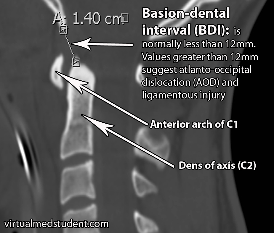
BDI for AOD
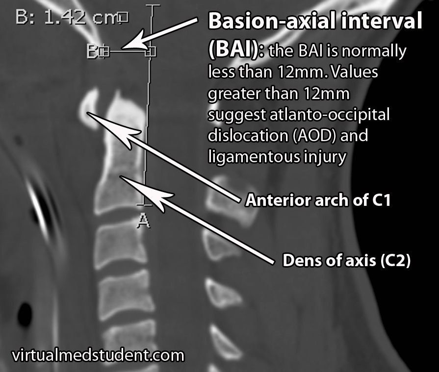
BAI AOD
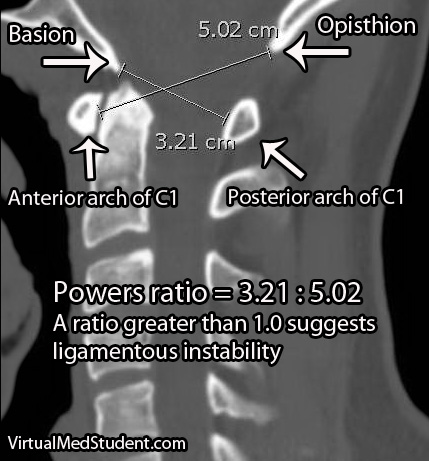
Power's Ratio
Diagnosis is made after appropriate imaging studies are obtained. Plain films and CT scans of the cervical spine are the most commonly obtained initial imaging studies.

There are several methods for determining if atlanto-occipital dislocation has occurred. The first is the “BAI-BDI method”. Two distances are measured. The BDI, or basion-dental interval, measures the distance between the most anterior and inferior portion of the skull (ie: “basion”) and the tip of the dens, which is a superior extension of the second cervical vertebrae. The BDI should normally be less than 12mm. In the image below the BDI is 14mm and is indicative of atlanto-occipital dislocation.

The second distance that is measured is the BAI, or basion-axial interval. The BAI is the distance between the basion and a line drawn straight up from the posterior (ie: back) portion of the dens. The normal interval is less than 12mm. In the image below the BAI is 14.2mm indicating likely atlanto-occipital dislocation.

A second method of assessing atlanto-occipital dislocation is known as Power’s ratio. Power’s ratio is most useful if the dislocation is anterior (ie: the head moves forward relative to the spine). Two distances are measured. The first is the distance between the opisthion and the anterior ring of C1; the second is the distance between the basion and the posterior ring of C1. The second distance is then divided by the first to obtain the ratio. A “normal” ratio is less than 1.0; if the ratio is greater than 1.0 then suspicion for dislocation should be high.

X-rays and CT show bony anatomy very well, but do not demonstrate ligamentous injury. Many patients with suspected AOD may also get an MRI to assess ligamentous damage. MRI will usually show severe injury to the ligaments that connect the first and second cervical vertebrae to the skull base.

There are two commonly used classification systems for describing atlanto-occipital dislocations. The Traynelis system is purely descriptive and is based on which way the head moves relative to the spine.

Traynelis Classification
Type 1 Head moves forward relative to the spine
Type 2 Head moves upwards relative to the spine
Type 3 Head moves backwards relative to the spine

The Bellabarba system is based on CT findings and is used to determine stability; it is also useful because it helps guide treatment decisions. A stable injury is one in which the dislocation is within 2mm of a normal BDI/BAI value and does not get worse with a traction test. Unstable injuries are present when the dislocation gets worse with a traction test; these injuries may be reduced at baseline (ie: within 2mm of a normal BDI/BAI) or grossly abnormal. That being said, it is important to note that very few doctors perform traction tests in patients with suspected AOD as this can worsen the condition!

Bellabarba Classification
Type Description Stability
Type 1 Alignment within 2mm of normal BAI/BDI interval; distracts less than 2mm with traction Stable
Type 2 Reduced at baseline (ie: within 2mm of normal BDI/BAI), but significant distraction with traction test Unstable
Type 3 Severely misaligned relative to normal BDI/BAI values Unstable

Treatment

Treatment of atlanto-occipital dislocations involves spinal immobilization. This is usually done via a surgical procedure known as an occipital-cervical fusion in which the back of the skull is fused to the cervical spine. Some patients are also put in a halo immobilization device.

Overview

Atlanto-occipital dislocation occurs after severe injury to the ligaments that connect the skull to the cervical spine. It most commonly occurs after severe head or neck trauma. Death secondary to ventilatory failure, quadriplegia, and cranial nerve deficits are common symptoms. Diagnosis is based on characteristic findings found on CT, x-rays, and MRI scans. Treatment is with spinal immobilization, either with a halo apparatus, or a surgical procedure known as occipital-cervical fusion.

Some More Useful Learning Material…

References and Resources

  • Garrett M, Consiglieri G, Kakarla UK, et al. Occipitoatlantal dislocation. Neurosurgery. 2010 Mar;66(3 Suppl):48-55.
  • Greenberg MS. Handbook of Neurosurgery. Sixth Edition. New York: Thieme, 2006. Chapter 25.
  • Deliganis AV, Mann FA, Grady MS. Rapid diagnosis and treatment of a traumatic atlantooccipital dissociation. AJR Am J Roentgenol. 1998 Oct;171(4):986.
  • Traynelis VC, Marano GD, Dunker RO, et al. Traumatic atlanto-occipital dislocation. Case report. J Neurosurg. 1986 Dec;65(6):863-70.
  • Bellabarba C, Mirza SK, West GA, et al. Diagnosis and treatment of craniocervical dislocation in a series of 17 consecutive survivors during an 8-year period. J Neurosurg Spine. 2006 Jun;4(6):429-40.
  • Horn EM, Feiz-Erfan I, Lekovic GP, et al. Survivors of occipitoatlantal dislocation injuries: imaging and clinical correlates. J Neurosurg Spine. 2007 Feb;6(2):113-20.
Letters of Intent to Trap Unwary Physicians

The following article is courtesy of Dennis Hursh, author of The Final Hurdle – A Physician’s Guide to Negotiating a Fair Employment Agreement. Dennis is an attorney who focuses on review and negotiation of physician employment agreements.

The contracting process for a physician employment agreement sometimes (but not always) begins with a letter of intent, so it makes sense for you to be aware of a common trap that many physicians find themselves in after they sign a letter of intent.

A letter of intent (“LOI”) is simply a very brief summary of the main terms of what the parties assume will be in a binding physician employment agreement. The purpose of an LOI is to make sure that the both parties are “on the same page” as far as the major terms of the agreement they hope to form.

For example, in a physician employment agreement, if you expect to be paid $300,000 a year but the employer is expecting to pay $200,000 a year, there probably isn’t any value in continuing negotiations. A letter of intent can save a lot of pain and aggravation (not to mention attorney’s fees) by avoiding negotiations that are not likely to lead to a signed deal.

Since the purpose of a LOI in physician employment agreements is just to determine if further negotiations are in order, the provisions in a LOI are generally not legally binding. Accordingly, it is very common for physicians to treat a LOI as an unimportant formality. Having been told that the document is not legally binding, they sign the LOI even though it contains some terms that they hope to negotiate.

You must remember that the purpose of a LOI is to make sure that you and your potential employer are on the same page with respect to the major terms of the physician employment agreement you hope to conclude. Although there will be many terms and conditions of the final agreement that the parties will negotiate, you should assume that anything you agree to in the LOI is “off the table”. Accordingly, do not sign a letter of intent unless you are completely comfortable with the compensation and other material terms set forth in the LOI. Legally binding or not, make sure you aren’t “agreeing” to something that you don’t want in the final agreement.

Footnotes, References, and Resources

(1) Some provisions of the LOI are typically legally binding. Specifically, the LOI will most likely provide that each party is responsible for its own attorney’s fees, that the negotiations will remain confidential, and that the physician will negotiate exclusively with this employer for some period of time. These provisions generally are legally binding. In other words, even if you do not sign a physician employment agreement, you are still bound to pay your own attorney’s fees and keep the terms of the negotiations confidential.

(2) For additional information about physician employment agreements, see www.TheFinalHurdle.com and The Physician Contract Blog.

The Anterior Choroidal Artery: Small but Mighty

The anterior choroidal arteries are small, but vital blood vessels in the brain. They are branches of the internal carotid arteries. They arise proximal to the splitting of the internal carotid into the anterior and middle cerebral arteries.

The anterior choroidal arteries deliver blood to vital brain structures. These structures include the posterior limbs of the internal capsules, portions of the thalami, optic tracts, middle third of the cerebral peduncles, portions of the temporal lobes (ie: parts of the pyriform cortex, uncus, and amygdala), substantia nigra, portions of the globus pallidus, as well as the choroid plexus in the lateral ventricles.

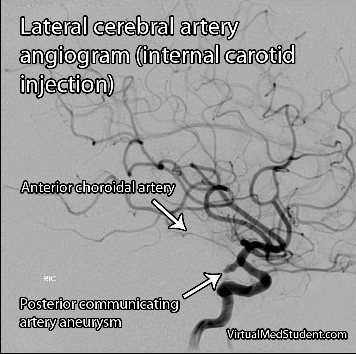
The anterior choroidal artery forms connections (anastamoses) with the posterior lateral choroidal arteries. Cerebral angiograms are the best way to visualize the anterior choroidal arteries.

Importance in Disease

Blockage of the anterior choroidal artery can cause a stroke. The most common symptoms of an anterior choroidal stroke are hemiparesis (weakness on the opposite side of the body), hemianesthesia (decreased sensation on the opposite side of the body), and a homonymous hemianopsia (loss of a portion of the visual field of both eyes). High blood pressure is the most common underlying factor in people with anterior choroidal artery strokes.

The hemiparesis is a result of damage to the posterior limb and genu of the internal capsule. The posterior limb contains the corticospinal tracts, which send information about movement from the brain to the spinal cord.

The hemianesthesia is a result of damage to the ventral posterolateral nucleus of the thalamus. This nucleus contains neurons that receive information from the spinal cord about sensation from the body. This symptom is less common than weakness, and occurs in roughly half of patients with an anterior choroidal stroke.

Cerebral angiogram showing anterior choroidal artery.

The final symptom, homonymous hemianopsia, is caused by damage to the optic tracts and lateral geniculate nucleus of the thalamus. Patients lose the ability to see objects on the left or right side (depending on which anterior choroidal artery is involved) in both eyes. This is an even more uncommon symptom, which occurs in less than 10% of patients with an anterior choroidal artery stroke.

Strokes of the anterior choroidal artery rarely cause all three symptoms. This is because the brain tissue served by the anterior choroidals also receives blood flow from other arteries.

Aneurysms of the anterior choroidal arteries are rare and will not be discussed in this article.

Overview

The anterior choroidal arteries are paired structures that arise from the internal carotid arteries. They supply blood to many important structures within the brain. Stroke is the most common pathological disease related to this blood vessel and frequently causes weakness of the opposite side of the body. High blood pressure is the most common underlying disease seen in people who have a stroke in this vascular distribution.

Other Stuff Worth Looking At…

References and Resources

  • Pezzella FR, Vadalà R. Anterior choroidal artery territory infarction. Front Neurol Neurosci. 2012;30:123-7. Epub 2012 Feb 14.
  • Bruno A, Graff-Radford NR, Biller J, et al. Anterior choroidal artery territory infarction: a small vessel disease. Stroke. 1989 May;20(5):616-9.
  • Baehr M, Frotscher M. Duus’ Topical Diagnosis in Neurology: Anatomy, Physiology, Signs, Symptoms. Fourth Edition. Stuttgart: Thieme, 2005.
  • Nolte J. The Human Brain: An Introduction to its Functional Anatomy. Sixth Edition. Philadelphia: Mosby, 2008.
  • Baskaya MK, Coscarella E, Gomez F, et al. Surgical and angiographic anatomy of the posterior communicating and anterior choroidal arteries. Neuroanatomy (2004) v3:38-42.

Respiratory Acidosis: Breathe Darn You!

A respiratory acidosis occurs when a person hypoventilates (ie: breathes too slow or too shallow). The result is an increase in PaCO2 (ie: the amount of CO2 dissolved in blood). The increase in plasma CO2 causes the blood to become acidic, which is manifest by a drop in the bodies’ pH. The reason blood becomes more acidic under these conditions is based on Le Chatelier’s principle. To understand this principle better let’s look at the equation that governs CO2 and HCO3 formation:

HCO3 + H+ <—> H2CO3 <—> CO2(g) + H2O

You’ll notice that CO2 (on the right most part of the equation) is what is exhaled via the lungs. When a patient is hypoventilating there is more CO2 than normal in the blood stream. The body compensates by turning this CO2 into HCO3 and H+. The resulting increase in H+ causes the acidosis (decrease in pH).

Causes

What causes someone to hypoventilate? There are many causes! All of them relate to a decreased ability of the patient to breath at a rate sufficient to remove carbon dioxide from the blood stream.

Medications that slow respiratory rate (ie: morphine and other pain medications) are notorious culprits. Poor pulmonary mechanics from obesity or neuromuscular disease (ie: amyotrophic lateral sclerosis) can also cause decreased respiratory rates. Lung and chest wall diseases are also common causes of respiratory acidosis and include pneumonia, pneumothorax, and decreased respiratory rate secondary to pain from rib fractures.

When assessing someone who has a respiratory acidosis ask this question first: what is causing the patient to have a decreased respiratory rate? Look for signs of external chest wall trauma, pneumonia, etc. Look through the medication record (how much pain medication have they gotten?) to get an idea of what medications could be causing their decreased ventilatory drive.

In general, the most common causes of hypoventilation are:

  • Medicines (especially pain medications)
  • Airway obstruction
  • Central nervous system disease (ie: diaphragmatic paralysis from cervical spinal cord trauma)
  • Chest wall problems (pneumo/hemothorax, flail chest, broken ribs, etc.)
  • Nerve and muscle diseases
  • Lung diseases (ie: pneumonia, restrictive lung diseases, etc.)

Acute Versus Chronic and Kidney Compensation

A respiratory acidosis can be either acute or chronic. The difference depends on how much the kidney compensates for the change in pH. How exactly does the kidney compensate? It decreases its secretion of HCO3 (aka: bicarbonate ion) into the urine. This helps offset the acidosis, and brings the bodies pH back towards normal limits.

How do we determine if the kidney is acutely or chronically compensating? We measure the bicarbonate level (one of the results in a "chemistry panel"). The kidney is acutely compensating if the HCO3 level is increased 1 to 2 mmol/L per every 10 mmHg increase in the PaCO2 level (normal PaCO2 level is 40 mmHg). The kidney is chronically compensating if the HCO3 level is increased 3 to 4 mmol/L per every 10 mmHg increase in PaCO2.

For example, if a patient’s PaCO2 on blood gas analysis is found to be 60 mmHg (a normal level is 40) we would say there is a 20 mmHg increase present (ie: the patient is unable to eliminate 20 mmHg of excess CO2 from the blood stream via the lungs). If the HCO3 (determined by a chemistry panel) is at 27 (for argument sake we’ll say a normal bicarbonate level is 23) then that represents a 4 mmol increase in the bicarbonate level for the 20 mmHg increase in CO2, or approximately 2 mmol increase in bicarb per 10 mmHg increase in CO2. This would mean the patient’s kidney is acutely compensating for the respiratory acidosis.

  Bicarbonate Level (HCO3)
Acute Kidney Compensation Increased by 1-2 mmol/L for every 10 mmHg increase in the PaCO2
Chronic Kidney Compensation Increased by 3-4 mmol/L for every 10 mmHg increase in the PaCO2

Why is it important to determine if acute or chronic kidney compensation is occurring? For starters, it gives the clinician a better idea of what may be causing the respiratory acidosis.

If the kidney is acutely compensating we know that the problem is new. The patient is likely having an acute issue (ie: trauma to the chest that caused multiple rib fractures). If the compensation is chronic then we know that the patient has been breathing at a slower than normal rate for a prolonged period of time. This may be seen in long standing neuromuscular diseases that cause poor pulmonary mechanics, obesity, etc.

Treatment

Treatment is straightforward: eliminate the underlying cause! If the patient received too much morphine give some naloxone to wake them up. Sometimes patients cannot maintain an adequate respiratory rate on their own, and mechanical ventilation is required. Once the patient is adequately ventilated the respiratory acidosis should resolve.

Overview

A respiratory acidosis occurs when a patient is unable to remove CO2 from the bloodstream secondary to a decreased respiratory rate (ie: hypoventilation). There are numerous causes including neuromuscular diseases, pain medication, and chest trauma. The kidney can acutely or chronically compensate for a respiratory acidosis depending on how long it has been present. Treatment is to fix the underlying cause.

You Are Just Getting Started… Learn Some More!

References and Resources

Respiratory Alkalosis: PaCO2 and Some Rapid Breathing

A respiratory alkalosis occurs when a person breathes too rapidly. The result is a decrease in PaCO2 (ie: the amount of CO2 dissolved in the blood). This causes the blood to become alkalotic (less acidic), which is manifest by an increase in the blood’s pH. The reason the blood becomes less acidic is based on Le Chatelier’s principle. If we take a look at the following equation:

HCO3 + H+ —> H2CO3 –> CO2(g) + H2O

You’ll notice that CO2 (on the right most part of the equation) is what is exhaled via the lungs. When a patient is hyperventilating there is much less CO2 than normal in the blood stream. The body compensates by combining HCO3 and H+ to form more CO2. The resulting decrease in H+ causes the alkalosis (ie: rise in pH).

Causes of Respiratory Alkalosis

So what could cause someone to hyperventilate? The most common things are pain, anxiety, and fever. If the patient is in the ICU and being mechanically ventilated then a respiratory alkalosis may develop if the ventilator is set to give too many breaths per minute.

The most common causes of hyperventilation are:

  • Fever
  • Pain
  • Anxiety
  • Overventilating a mechanically ventilated patient

Acute Versus Chronic and Kidney Compensation

A respiratory alkalosis can be either acute or chronic. The difference depends on how much the kidney compensates for the change in pH. How exactly does the kidney compensate? It dumps HCO3 (aka: bicarb) into the urine. This helps offset the alkalosis and brings the bodies pH back to normal limits.

How do we determine if the kidney is acutely or chronically compensating? We measure the bicarb level. The kidney is acutely compensating if the HCO3 level is decreased 1 to 2 mmol/L per every 10 mmHg drop in the PaCO2 level. The kidney is chronically compensating if the HCO3 level is decreased 4 to 5 mmol/L per every 10 mmHg drop in PaCO2.

For example, if a patient’s PaCO2 on blood gas analysis is found to be 20 mmHg (a normal level is around 40) we would say there is a 20 mmHg drop present. If the HCO3 (determined by a chemistry panel) is at 19 (for argument sake we’ll say a normal bicarbonate level is 23) then that is a 4 mmol drop in the bicarb level for the 20 mmHg drop in CO2, or approximately 2 mmol drop in bicarb per 10 mmHg drop in CO2. This would mean the patient’s kidney is acutely compensating for the respiratory alkalosis.

  Bicarbonate Level (HCO3)
Acute Compensation Decreased by 1-2 mmol/L for every 10 mmHg decrease in the PaCO2
Chronic Compensation Decreased by 4-5 mmol/L for every 10 mmHg decrease in the PaCO2

Why is it important to determine if acute or chronic compensation is occurring? For starters, it gives us a better idea of what may be causing the respiratory alkalosis. If the kidney is acutely compensating we know that the respiratory alkalosis is new. The patient is likely having an acute reaction to something (ie: pain, anxiety, panic attack, etc.). If the compensation is chronic then we know that the patient has been breathing at a faster than normal rate for a prolonged period of time. This may be seen in pregnancy, COPD, and emphysema.

Treatment

Treatment is very straightforward: eliminate the underlying cause! If the patient appears in pain then give pain medication. Fever? Hit them with some acetaminophen. If the patient is mechanically ventilated then decrease the respiratory rate. Once the stimulus for hyperventilating is removed the respiratory alkalosis should improve.

Overview

A respiratory alkalosis occurs when a patient is breathing too rapidly, which cause too much CO2 to be removed from the bloodstream. There are numerous causes including anxiety, pain, and fever. The kidney can acutely or chronically compensate for a respiratory alkalosis depending on how long it has been present. Treatment is to fix the underlying cause.

A Little More Learning…

References and Resources

Amyotrophic Lateral Sclerosis (Lou Gehrig’s Disease)

The pathology of amyotrophic lateral sclerosis (ALS) is not well known. There are familial forms, but they account for only 10% of cases. The familial form of amyotrophic lateral sclerosis is genetically heterogenous. This means that there are genes on different chromosomes that have been implicated in the disease process.

One of the most well studied is a gene known as superoxide dismutase (SOD) on chromosome 21. There are over 100 mutations of this gene. The defective protein that occurs from these mutations is believed to cause oxidative injury and/or toxic protein aggregates that damage motor neurons.

The remaining 90% of cases are sporadic. There is still debate about what causes most cases. Studies looking at cytoskeletal abnormalities, inflammation, and excitotoxicity from overactive glutamate stimulation have all been proposed as contributing factors.

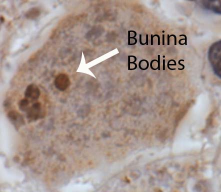
Bunina Bodies
Regardless of the cause, amyotrophic lateral sclerosis causes the death of motor neurons in both the brain and spinal cord. These motor neurons are commonly referred to as upper motor neurons (if in the brain) and lower motor neurons (if in the brainstem or spinal cord).

The dying neurons are replaced with proliferating astrocytes in a process known as gliosis. This proliferation of glial cells leads to "glial scarring".

In addition, amyotrophic lateral sclerosis neurons have intracellular inclusions when viewed under the microscope. These inclusions are composed of different abnormal protein molecules that can be phosphorylated or ubiquinated. A specific type of inclusion known as a Bunina body is commonly seen.

Signs and Symptoms

Damage to the upper and lower motor neurons gives ALS its characteristic clinical presentation. Patients present with upper motor neuron findings: spastic weakness and hyper-reflexia (increased reflexes). Lower motor neuron death results in a flaccid weakness, atrophy of the muscles, muscular fasciculations (ie: abnormal twitching), and hypo-reflexia (decreased reflexes). Thus patients with ALS have a unique mix of both spastic and flaccid weakness, muscular atrophy, and hyper-reflexia (hyperactive reflexes usually predominate).

There are also many sub-categories of ALS. For example, "ALS Plus Syndrome" is characterized by the classic motor findings of amyotrophic lateral sclerosis plus dementia, Parkinsonism, autonomic nervous system instability, and sensory disturbances. Another sub-category known as bulbar palsy occurs when the symptoms of ALS predominately affect the face/cranial musculature. Cranial muscular weakness can cause difficulty swallowing (ie: dysphagia) and speaking (ie: dysarthria).

Diagnosis

Amyotrophic lateral sclerosis, for the most part, remains a clinical diagnosis. It is based upon finding the classic motor neuron findings on physical exam with symptoms that worsen over time. Unfortunately, there is still no laboratory test that definitively diagnoses the disease.

The El Escorial World Federation of Neurology criteria help guide the clinical diagnosis. In order to qualify for a diagnosis of amyotrophic lateral sclerosis a patient must meet the following criteria: evidence of lower motor neuron pathology on physical exam, electrophysiological, or neuropathological testing, and progressive worsening / spread of symptoms, as well as evidence of upper motor neuron disease on physical exam. In addition to the above, there must also be no evidence to support another possible explanatory diagnosis for the symptoms.

There are adjunctive tests that can support the diagnosis of amyotrophic lateral sclerosis. These include electromyography (EMG). EMG looks at the electrical activity produced in muscles. In amyotrophic lateral sclerosis, the EMG will generally show findings of denervation (ie: muscles that do not have nerves connecting to them). Fasciculation potentials are often seen on the EMG as well. Interestingly, nerve conduction studies are often normal in amyotrophic lateral sclerosis.

Finally, blood testing can help support the diagnosis. For example, creatine kinase (a marker of muscle damage) may be elevated, which is a result of denervation.

Other laboratory tests are often sent. Lyme disease antibodies, anti-nuclear antibodies, HIV, vitamin B12, thyroid function tests, calcium, and phosphorus are often sent to rule out other causes for the symptoms.

Treatment

Treatment for amyotrophic lateral sclerosis currently consists of a medication known as riluzole. This medication has been shown to improve survival rates.

In addition, supportive care is extremely important. Most patients eventually require tracheostomy and gastric tube placement in order to breath and eat. Motorized wheel chairs can help patients maintain some independence.

Regardless of treatment, the prognosis is extremely guarded. Most patients are deceased within 5 years of their diagnosis. Death is usually related to pulmonary complications like pneumonia.

Overview

Amyotrophic lateral sclerosis is a neurodegenerative disease that results in death of upper and lower motor neurons. Signs and symptoms are a bizarre mix of flaccid weakness with increased reflexes. Diagnosis is based on physical exam findings and symptoms that worsen over time. In addition, there should be no evidence that other causes are responsible. Treatment is with a medication known as riluzole, which has been shown to slow the progression of disease. Supportive treatment is extremely important for quality of life.

Learn More…

References and Resources

  • Brooks BR. Managing amyotrophic lateral sclerosis: slowing disease progression and improving patient quality of life. Ann Neurol. 2009 Jan;65 Suppl 1:S17-23.
  • Baehr M, Frotscher M. Duus’ Topical Diagnosis in Neurology: Anatomy, Physiology, Signs, Symptoms. Fourth Edition. Stuttgart: Thieme, 2005.
  • Kumar V, Abbas AK, Fausto N. Robbins and Cotran Pathologic Basis of Disease. Seventh Edition. Philadelphia: Elsevier Saunders, 2004.
  • Simon RP, Aminoff MJ, Greenberg DA. Clinical Neurology, Seventh Edition (LANGE Clinical Medicine). Seventh Edition. New York: McGraw Hill, 2009.
  • Boillée S, Vande Velde C, Cleveland DW. ALS: a disease of motor neurons and their nonneuronal neighbors. Neuron. 2006 Oct 5;52(1):39-59.
  • Brooks BR. El Escorial World Federation of Neurology criteria for the diagnosis of amyotrophic lateral sclerosis. J Neurol Sci. 1994 Jul;124 Suppl:96-107.

Denis’ Three Column Spine: A Simple Way to Think About Spinal Stability

The three column model of the spine was first introduced by Dr. Francis Denis in his aptly named paper, "The Three Column Spine and its Significance in the Classification of Acute Thoracolumbar Spinal Injuries". This paper, published in 1983, proposed a new biomechanical model for spinal stability that challenged Dr. Frank Holdsworth’s two column model from the 1960s. Although replaced by more modern grading scales (ie: the TLICS model) the three column spine is a simple way to think about spinal biomechanics.

Denis’ three column model proposes that the thoracolumbar spine can be divided into three columns. The first column includes the anterior longitudinal ligament (ALL) up to the first half of the bony vertebral body. The second column includes the second half (ie: more posterior half) of the vertebral body, up to, and including the posterior longitudinal ligament (PLL). The third column includes the pedicles, spinal cord/thecal sac, lamina, transverse processes, facet joints, spinous process, and the posterior ligaments (ie: supraspinous, interspinous, and ligamentum flavum).

The purpose of Denis’ model was to delineate which injuries to the thoracolumbar spine were considered "unstable". This delineation was important because it determined which patient’s required operative treatment of their spine.

Simply stated, an unstable spine was present if two or more of the columns were involved in the injury. However, it is important to note that every rule can be broken, so not all injuries follow this rule. Part of the "art" of practicing spine surgery is determining which two-column injuries can be left alone and managed non-operatively, and which truly require operative intervention.

Types of Injuries

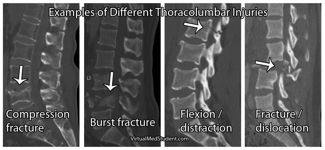
Based on Denis’ model he classified specific types of injuries. Injuries to the anterior column only were called "compression fractures". Damage to the anterior and middle columns were known as "burst fractures". Injury to the middle and posterior column were known as "flexion-distraction" injuries, or more colloquially as "seat-belt type" injuries. Finally, damage to all three columns were classified as "fracture-dislocation" injuries.

Classification of Thoracolumbar Injuries Using the Three Column Model

Columns Name Stability
Anterior Compression fracture Stable
Anterior and Middle Burst fracture Unstable
Middle and Posterior Flexion-distraction injury (aka: seat-belt type) Unstable
Anterior, middle, and posterior Fracture-dislocation Unstable

Examples of Thoracolumbar Spine Fractures

Clinical Significance

In very simple terms, unstable fractures require definitive treatment. Treatment for unstable spine injuries is usually operative, although rigid immobilization with bracing is sometimes used in certain circumstances.

The treatment of each fracture type is beyond the scope of this article (and discussed in more detail elsewhere on this site), but suffice it to stay that for the most part (and again, every rule was made to be broken) two or three column injures = unstable = surgery.

Overview

Denis’ three column model of the spine revolutionized the way we think about spinal biomechanics and stability. He divided the spine into three columns. Although it has been largely replaced by more modern grading scales such as the thoracolumbar injury classification and severity score (TLICS) it is still a nice way to “think” about spinal integrity.

Other Common Spine Injuries…

References and Resources

The Internal Capsule: Some Pricey Brain Real Estate

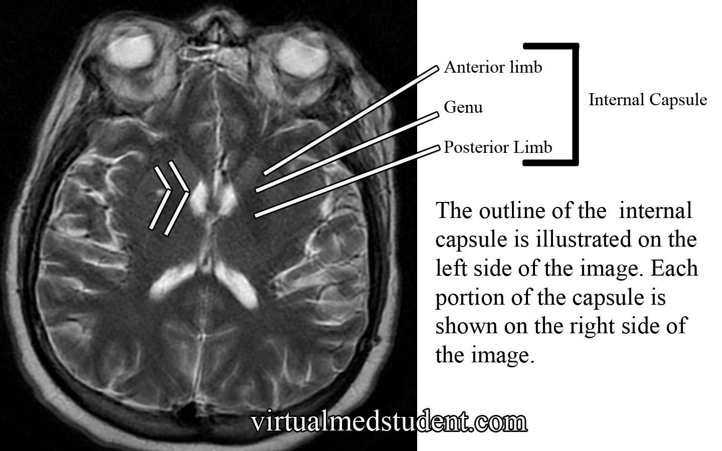
The internal capsule is one pricey piece of brain real estate! It contains all of the pathways that allow information to be transferred between the cerebral cortex and the spinal cord, brainstem, and subcortical structures (ie: thalamus, basal ganglia). It is divided into an anterior limb, posterior limb, and genu (ie: the area where the anterior and posterior limbs meet).

The anterior limb contains axons that send information between the thalamus and the cingulate gyrus and pre-frontal cortex. It also contains axons in the frontopontine pathway (ie: axons going from the frontal cortex to a portion of the brainstem known as the pons).

The genu contains the corticobulbar tract, which originate in the motor areas of the frontal lobes and extend to the cranial nerve nuclei in the brainstem. It also contains axons that connect the motor section of the thalamus (ie: VA and VL nuclei) with the motor areas of the frontal cortex.

The posterior limb contains the corticospinal tract, which are axons that come from the motor area of the frontal cortex and travel all the way to the anterior horns of the spinal cord where α-motor neurons are located. The posterior limb also contains sensory information coming from the body via the medial lemniscus and the anterolateral (aka: spinothalamic tract) systems.

Internal Capsule MRI

The blood supply to most of the internal capsule comes from the lenticulostriate arteries. These small arteries originate from the first portion of the middle cerebral artery. Two other important arteries also supply portions of the internal capsule: the anterior choroidal artery and the recurrent artery of Heubner. The anterior choroidal artery is a branch of the internal carotid. It supplies the inferior portion of the posterior limb. The recurrent artery of Heubner is a branch of the anterior cerebral artery. It supplies the inferior portions of the anterior limb and the genu.

Anatomy of the Internal Capsule
Division Major Communication Tracts Blood Supply
Anterior limb

– Tracts between the frontal lobe and pons (brainstem)

– Tracts between the thalamus and prefrontal cortex

– Tracts between the thalamus and cingulate gyrus

– Lenticulostriate arteries (branches of the middle cerebral artery)

– Recurrent artery of Heubner (branch of the anterior cerebral artery)

Genu – Tracts between the motor cortex in the frontal lobe and the cranial nerve nuclei in the brainstem (aka: corticobulbar tract)

– Lenticulostriate arteries (branches of the middle cerebral artery)

– Recurrent artery of Heubner (branch of the anterior cerebral artery)
Posterior limb

– Tracts between the motor cortex of frontal lobe and anterior horn of spinal cord (aka: corticospinal tract)

– Medial lemniscus tract (a continuation of the dorsal columns), which carries information about light touch, vibration, and pressure sensation from the body and spinal cord.

– Anterolateral (aka: spinothalamic) tract, which carries pain and temperature information

– Lenticulostriate arteries (branches of the middle cerebral artery)

– Anterior choroidal artery (branch of the internal carotid)

Importance in Disease

Thalamic Hemorrhage
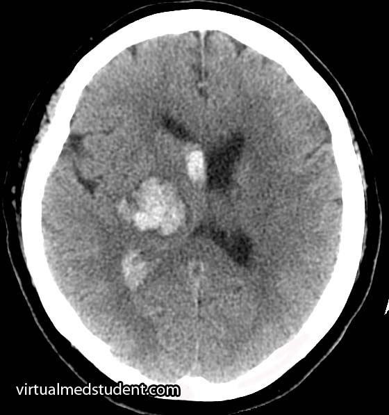
Thalamic intracerebral hematoma
compressing the posterior limb
of the internal capsule

Damage to the internal capsule can be devastating neurologically because it contains so many vital tracts.

For example, a stroke of the anterior choroidal artery can lead to posterior limb damage. This can cause paralysis of the contralateral (ie: opposite) arm and leg secondary to interruption of the corticospinal tract.

Posterior limb disruption can also cause co-existent sensory deficits including an inability to feel light touch, pain, and temperature due to damage of the spinothalamic and medial lemniscal pathways.

Hypertensive hemorrhages in the thalamus or basal ganglia can compress the adjacent fibers of the internal capsule leading to similar clinical findings. The head CT to the right shows a thalamic hemorrhage secondary to severely elevated blood pressure. The patient had compression of the posterior limb of the internal capsule. As a result she was unable to move her left arm and leg, and could not feel pain or light touch on the left side of her body.

Overview

The internal capsule is a vital structure. It contains many communication pathways between the brain’s cortex, brainstem, spinal cord, and subcortical nuclei (ie: thalamus, basal ganglia). Its blood supply comes from branches of the middle cerebral artery (ie: lenticulostriates), anterior cerebral artery (ie: recurrent artery of Heubner), and the internal carotid (ie: anterior choroidal artery). Lesions in this area caused by strokes or hypertensive hemorrhages can have devastating clinical consequences.

Other Pertinent Articles…

References and Resources

  • Greenberg MS. Handbook of Neurosurgery. 9th Edition. New York: Thieme, 2006. Chapter 25.
  • Chowdhury F, Haque M, Sarkar M, et al. White fiber dissection of brain; the internal capsule: a cadaveric study. Turk Neurosurg. 2010 Jul;20(3):314-22. doi: 10.5137/1019-5149.JTN.3052-10.2.
  • Simon RP, Aminoff MJ, Greenberg DA. Clinical Neurology, Seventh Edition (LANGE Clinical Medicine). Seventh Edition. New York: McGraw Hill, 2009.
  • Nolte J. The Human Brain: An Introduction to its Functional Anatomy. Sixth Edition. Philadelphia: Mosby, 2008.
  • Bickley LS, Szilagyi PG. Bates’ Guide to Physical Examination and History Taking. Ninth Edition. New York: Lippincott Williams and Wilkins, 2007.

Peroneal (Fibular) Nerve and the Dropped Foot

The peroneal nerve is one of the branches of the sciatic nerve. It receives most of its innervation from the L4, L5, and S1 nerves. The common peroneal nerve (aka: common fibular nerve) wraps around the outside of the knee over the head of the fibula. It then branches into two separate nerves: the superficial peroneal nerve and the deep peroneal nerve.

The superficial peroneal nerve innervates two muscles: peroneus longus and brevis (aka: fibularis longus and brevis). These muscles allow you to evert (ie: move your foot outwards) and plantarflex (ie: help you step on the gas pedal) the foot. This nerve also provides the sensation to the outside half (ie: lateral half) of the lower leg, as well as the top of most of the foot (ie: the dorsum of the foot).

The deep peroneal nerve innervates three muscles in the leg: tibialis anterior, extensor digitorum longus, and extensor hallucis longus. Tibialis anterior allows you to dorsiflex (ie: lift your foot off the ground) and invert (ie: bringing your big to closer to the middle of the body) your foot. Extensor digitorum longus helps you extend your toes (ie: the opposite of curling them), as well as evert, and dorsiflex your foot. The third muscle, extensor hallucis longus, allows you to extend your big toe.

The deep peroneal nerve also innervates two muscles in the foot: extensor digitorum brevis (also helps to extend the toes) and extensor hallucis brevis (also helps to extend the big toe).

Importance in Disease

The peroneal nerve is most frequently compressed over the fibular head. Compression typically affects the deep peroneal nerve rather than the common or superficial nerve; however, all of the nerves may be involved.

When the deep peroneal nerve is compressed, the foot is unable to dorsiflex secondary to dysfunction of the tibialis anterior muscle. Additionally, the patient is unable to extend the toes. Sensation may be decreased on a small patch of skin between the big toe and second toe; pain in the area of the lateral lower leg may also be present.

When the superficial peroneal nerve is compressed, the peroneus longus and brevis muscles are affected. Dysfunction of these muscles prevents the patient from everting their foot. Sensation over the lateral half of the lower leg and top of the foot may also be decreased.

Placing the nerve under passive, or active, stretch by placing the patient’s foot in inversion will often reproduce the symptoms. Percussing the nerve over the fibular head (Tinel’s test) can reproduce the symptoms.

It is important to distinguish a peroneal nerve palsy from a herniated L4-L5 disc. A patient with a herniated L4-L5 disc – causing an L5 radiculopathy – will not only have difficulty dorsiflexing the foot and toes (secondary to dysfunction of the L5 component of the peroneal nerve), but will also have difficulty inverting the foot (secondary to dysfunction of the L5 component of the tibial nerve).

Wait a second! This still shouldn’t make sense if you are actually thinking about it, and this is where things get tricky… Both the anterior tibialis (deep peroneal nerve innervated) and the posterior tibialis (tibial nerve innervated) help invert the foot. So how do we know that the weakness in inversion is related to an L5 disc herniation, a peroneal nerve palsy, or a tibial nerve palsy? You need to have the patient attempt to invert the foot while plantarflexing! The posterior tibialis is an inverter and plantarflexor of the foot so if dorsiflexion is weak (L5 –> deep peroneal nerve –> anterior tibialis) and the patient cannot invert (L5 –> tibial nerve –> posterior tibialis OR L5 –> deep peroneal nerve –> anterior tibialis) well while the foot is plantarflexed you probably have an L5 radiculopathy and not a peroneal nerve palsy.

To make it a bit easier clinically… A foot drop WITH inversion weakness is most likely an L5 radiculopathy (most commonly from a herniated disc) because you are getting the invertors for both the deep peroneal nerve (tibialis anterior muscle) AND the tibial nerve (tibialis posterior muscle), which both have get fibers from the L5 nerve root. However, injury to the sciatic nerve proximally in the leg could also give you this, but it would be associated with significant plantarflexion weakness too (due to gastroc weakness from S1)! So therefore a foot drop with eversion weakness and toe extension weakness is also suspicious for a peroneal nerve injury.

Overview

The peroneal nerve splits at the level of the knee from the sciatic nerve. It then further divides into the superficial and deep peroneal nerves. The superficial branch controls the evertors of the foot (peroneus longus and brevis) and provides sensation over the lateral aspect of the lower leg and top of the foot. The deep branch controls the dorsiflexors of the foot, and the extensor muscles of the toes. The common peroneal, and either of its branches, is most commonly compressed near the fibular head.

More Important Anatomy to Master…

References and Resources

  • Greenberg MS. Handbook of Neurosurgery. Sixth Edition. New York: Thieme, 2006. Chapter 25.
  • Prakash, Bhardwaj AK, Devi MN. Sciatic nerve division: a cadaver study in the Indian population and review of the literature. Singapore Med J. 2010 Sep;51(9):721-3.
  • Yuen EC, So YT. Sciatic neuropathy. Neurol Clin. 1999 Aug;17(3):617-31, viii.
  • Simon RP, Aminoff MJ, Greenberg DA. Clinical Neurology, Seventh Edition (LANGE Clinical Medicine). Seventh Edition. New York: McGraw Hill, 2009.
  • Netter FH. Atlas of Human Anatomy: with Student Consult Access (Netter Basic Science). Fifth Edition. Philadelphia: Saunders Elsevier, 2010.
  • Bickley LS, Szilagyi PG. Bates’ Guide to Physical Examination and History Taking. Ninth Edition. New York: Lippincott Williams and Wilkins, 2007.

The Frustrations of Dealing with a Demanding Patient

Dealing with a demanding patient can be extremely frustrating! But before we delve further into how to handle these pesky buggers, let’s take a moment to reflect on what constitutes a “demanding” patient.

Demanding patients exhibit a variety of behaviors and attitudes. Patients may be rude, down right nasty, non-compliant, litigious, passive-aggressive, or inconsiderate of your time. In essence, a "demanding" patient is one that requires a tremendous amount of investment on the doctor’s part.

It is important that being sick can make people nasty! I know I turn into a real asshole when I am sick (just ask my wife). Not to mention many patients are on medications (ie: steroids) or illicit drugs, which can alter their behavior. Therefore, it is extremely important to not take the threats of demanding patients personally. In fact, placating a demanding patient can be difficult, if not impossible!

But fret not! There are many ways to deal with a demanding patient! I recommend learning as many different ways as possible because each method is a "weapon" in your arsenal. One method may work on patient A, but not on patient B. Remember that no two patients are the same! Enough intro, let’s get into some real world approaches and solutions…

The first type of demanding patient is the "confused" patient. This patient is "demanding" because they just don’t seem to get it. They are lost in the massive sea that is healthcare and no one has bothered to explain why they are adrift! Ask the patient to tell you, in their own words, why they are in the hospital or clinic. If they cannot, a few minutes (literally minutes!) explaining what is happening to them can turn this difficult patient into a bucket of smiles!

Educating your patients about their disease is paramount to being a great physician! I cannot tell you how many patients have literally ZERO idea why they are taking such and such medication. Very few have actually seen their x-rays or CT scans. Show them and educate them; I guarantee you will develop instant rapport if you do this.

Sometimes, no matter how much time you spend, or how much you educate a patient there are some people who will continue to be a pain in the tookus! I call this patient the "asshole". These patients can be particularly tricky to deal with, and many of them have underlying personality disorders (remember you psych rotation!). However, many will respond to the "tough love" approach. These patients typically need firm, stern, and direct confrontation otherwise they will continue to walk all over doctors, nurses, and other members of the healthcare team. Remember that you didn’t go to medical school and spend countless hours studying to be told by the patient what tests should be done or what treatment should be administered (and just so you know this isn’t advice they will likely give you in medical school!). A little bit (and I do mean a little bit) of paternalism should still exist in medical care, in my honest opinion.

When all else fails and the patient continues to be overly demanding and unsatisfied with their care a simple, "you are free to get a second opinion somewhere else" will often free both the patient and the doctor from turning a nasty situation into a total crap storm. If they take you up on the offer you’re free of their painful tactics, if not, they may re-think their demanding ways and become a more amenable patient.

And perhaps my best piece of advice… Ask a nurse! Nurses are the front line and deal with ghastly behavior on the regular! When you learn some nursing skills as a medical student it makes you look like a rock star, and will endear you to the health care team you work with on a daily basis.

Overall, the doctor patient relationship is exactly what it sounds like, a relationship! The patient does not have a right to abuse their doctor. In my personal experience I have found that some patients respond extremely well to firm and direct discussion, others to education, and others need to be coddled a bit.

It is important to recognize that there are a thousand different ways to approach a demanding patient. In fact, there are books written about it! The best piece of advice I can give is to learn many different methods and practice them! Interacting with patients is part of the “art of medicine”… and with a little bit of practice (and perhaps luck) you wont get any poo thrown at you!

Related Content

References and Resources