Pager PTSD… A Beep that Won’t Go Away

It’s Friday night and I’m not on call. It’s a welcome break from a hectic and stressful 80+ hour work week. We are about to sit down to watch a movie – popcorn included of course – and enjoy a nice relaxing evening. The microwave starts beeping to let us know that the popcorn is ready. As the beeping continues my heart rate and blood pressure increase ever so slightly, and I reach down towards my left hip. But something is amiss…

The beep of our microwave is eerily similar to the sound my pager makes when I’m on call at the hospital. Of course, the weird thing is that tonight I’m not on call! Yet I still reflexively reach towards my hip, my mind assured that there is a threat to someone’s life, a threat that I might be able to fix. It’s “pager PTSD”, a condition that seems to afflict many young medical trainees.

I’ve tried battling this baffling condition, but to no avail. Perhaps a different beep tone on my pager’s settings would do the trick? It worked for a little while, but invariably my mind would pick out the tone in a favorite TV show and I’m right back to square one… Reaching for that damn left hip.

I’ve even tried putting my pager on vibrate mode. This also worked for a little while until my psyche accommodated… The gentle pull of a blanket, or even my shirt moving in the wind would mimic the vibration, and of course I would reach down. Clearly, someone needed my help, and if I wasn’t there to answer the chirp (or this time vibration) of the pager something catastrophic might happen.

A close friend of mine has a similar story… Although his is a bit more intense! As a soldier in Afghanistan, he would carry a 9mm handgun on his left hip. In hairy situations he’d reach towards his "nine" in case he needed to quickly defend himself. After his second tour he confided that whenever he heard a loud sound, like a car backfiring, or an ambulance siren, he would instinctively reach towards his left hip, hoping that his gun was there to keep him safe.

Unlike my buddy, we are never personally in danger when our pager’s start chirping. And please don’t get me wrong, most pages a doctor receives are not "life and death" situations. In fact, most are easy to handle, a potassium replacement order here, or a Tylenol order there. However, the real problem lies in the fact that every time the pager starts beeping it’s dealer’s choice. It might be something simple, or it might be someone bleeding to death. You don’t know until you reach down and check the message sitting on your left hip.

It’s funny, but humans are not that much different than animals. We can be conditioned to experience a surge of catecholamines. Like a mouse that gets an electric shock a couple of seconds after a tone is played. As the tone goes off the mouse gets visibly nervous, its heart rate and blood pressure start to rise until the shock is administered. It’s similar in pager PTSD, although the mouse is some poor intern, nervously navigating the hospital, waiting for the next medical emergency to present itself.

As I’ve become more senior in my training I fortunately no longer have the visceral reaction when my pager beeps. This is probably because I am more confident in my ability to handle most of the things that might be waiting for me. But interestingly, I still find myself reaching for my left hip even when I am not on call… It’s ingrained in the deepest part of my being. It’s part of being a doctor…

Related Articles

Middle Cerebral Artery: A Common Site for Stroke

Middle cerebral artery (MCA) strokes occur when the MCA or its branches are occluded. With occlusion, blood, and along with it, oxygen and nutrients fail to reach the brain. If blood flow is not restored quickly the affected brain tissue dies leading to permanent neurological injury.

Risk factors for MCA strokes are the same for other strokes. They include hypertension, diabetes, smoking, atrial fibrillation, and a whole slew of hypercoagulable states (ie: pathologic increases in the bodies’ propensity to form blood clots).

In order to understand MCA strokes we have to first appreciate the anatomy of the MCAs, as well as the brain that they serve. The MCAs are subdivided into four parts. The M1 segment is one of the terminal branches of the internal carotid artery. The MCAs then become progressively more narrow and form more and more branches as they reach out towards the cortical surfaces of the brain. The most distal portions of the MCA are deemed the M4 branches. The lateral lenticulostriate arteries are small branches that branch from the M1 segments; these small arteries feed some of the deeper structures of the brain.

Importantly, the MCAs and their branches provide blood flow to an extremely large portion of the brain. These areas include the lateral and inferior frontal lobes, superior portion of the temporal lobes, insula, and lateral parietal lobes. Branches of the first portion of the MCAs (aka: the lateral lenticulostriate arteries) also provide blood flow to the deep sections of the brain including the putamen, head and body of the caudate, external globus pallidus, and parts of the posterior limb of the internal capsule.

The segmental anatomy of the MCAs is important because strokes that occur in the outer segments (ie: M3 and M4) cause less neurological injury than the inner MCA segments (ie: M1 and M2). This is because less total brain volume is affected by outer segment strokes.

The most common cause of MCA strokes are clots that break off and travel from the heart or the carotid arteries to the MCA (aka: emboli). Less commonly, a blood clot will form directly in the MCA itself (aka: thrombus). Atherosclerotic disease is the most common cause of thrombus formation in the MCA and atrial fibrillation (an abnormal heart rhythm) is the most common cause of emboli from the heart.

Signs and Symptoms

Middle cerebral artery strokes present with one of two types of syndromes depending on which MCA – right or left – is involved, as well as which segments of the MCA are involved.

In the worst case scenario, a stroke of the right or left M1 segment of the MCA causes weakness of the opposite side of the body. This is a result of cortical damage to the primary motor cortex as well as possible infarction of the posterior limb of the internal capsule (ie: the location of descending motor tracts). MCA strokes usually present with face and arm weakness that is worse than leg weakness. Remember that the motor cortex that controls leg function is served by the anterior cerebral arteries.

M1 strokes also cause decreased ability to feel sensation on the opposite side of the body as a result of damage to the parietal lobes.

Damage to the optic radiations, which course in the parietal (Baum’s loop) and temporal lobes (Meyer’s loop) can cause problems with vision. Finally, injuries to the frontal eye fields cause the eyes to deviate towards the side of the stroke (ie: the frontal eye fields normally allow you to make fast eye movements in the opposite direction, therefore damage prevents patients from looking to the non-affected side).

Right and left sided M1 strokes will give you all of the above symptoms; however, laterality can be determined based on specific symptoms caused by only a left or a right sided strokes.

The poor patients with left M1 strokes have a decreased ability to speak and/or understand language because of damage to Broca’s area in the left frontal lobe (speech production) and Wernicke’s area in the left temporal lobe (speech comprehension). Remember this is in addition to all the other stuff above.

Right M1 strokes can cause "anosognosia", in which the patient is unaware of certain deficits they may have; these patients also often fail to recognize the left side of their body (ie: they "neglect" or fail to appreciate the entire left side of the world) and may have difficulty appreciating people or objects presented in their left visual field.

Right MCA stroke
Less "severe" cases, in which M2, M3 or M4 branches are affected can produce a variety of signs and symptoms depending on the specific branches involved.

Diagnosis

Diagnosis of MCA strokes are based on symptoms, CT scans, and MRI images. CT and MR angiograms frequently show the blocked blood vessel causing the stroke. CT perfusion scans are a newer technology that give information regarding the amount of blood flow to affected brain tissue.

Treatment

Treatment depends on the timing of the stroke. If the patient presents within 3 hours of symptom onset, and the head CT reveals no bleeding, than intravenous tissue plasminogen activator (tPA) may be given to help "break" up the blood clot causing the stroke.

Other treatments using catheter based approaches are frequently used in patients who are unable to receive tPA. Such treatments include mechanical clot removal with special catheter and wire devices. In addition, in patients more than 3 hours, but less than 6 hours out from symptom onset intra-arterial (not to be confused with intravenous) tPA may be used.

Less commonly, large "malignant" MCA strokes may cause significant swelling, which can put pressure on the brainstem. These patients sometimes undergo an open surgical procedure known as a "craniectomy", in which the bone overlying the affected brain tissue is removed. This surgery allows the edematous brain tissue to swell outwards preventing it from herniating downwards towards vital brainstem structures.

Overview

Middle cerebral artery strokes are most commonly caused by blood clots that break off from the heart or carotid artery. Symptoms of MCA strokes depend on the segment involved, as well as which MCA (right versus left) is involved. Diagnosis is made with a combination of MRI, CT, and symptomatology. Treatment consists of intravenous or intra-arterial tPA and/or mechanical clot removal depending on the time frame of the symptoms.

Related Articles

References and Resources

Hemangiopericytoma: A Tumor of Pericytes

In order to understand hemangiopericytomas we have to define a few terms. The first term is mesenchyma. Mesenchyma is a word used to describe the different tissues that provide structure to the bodies’ organ systems. A type of mesenchymal cell known as a “pericyte” provides structural support to blood vessels. When pericytes go haywire they form hemangiopericytomas.

Hemangiopericytomas can occur anywhere blood vessels are located, but are most commonly located in the lower extremities, pelvis, head, and neck.

Intracranial hemangiopericytomas are uncommon. They represent less than 1% of tumors within the confines of the skull. They typically arise from blood vessels adjacent to the dura (ie: lining of the brain) and often form dural attachments. They are therefore commonly lumped into the category of “dural-based tumors”, but should be distinguished from their more benign meningeal cousins (ie: meningiomas).

Since hemangiopericytomas are mesenchymal in origin, they typically have lots of reticulin (a collagen fiber) that envelopes individual cells (see pathology slide). They are highly cellular tumors, and vascular channels in the shape of "staghorns", may be seen under the microscope. Actively dividing cells (aka: "mitotic" figures) are commonly seen and are a testament to their more malignant nature. Unlike meningiomas, calcifications are absent.

Hemangiopericytomas test positive for vimentin (a marker of connective tissue), Ki-67 (a marker of proliferation), vascular endothelial growth factor (VEGF, a marker of blood vessel proliferation), CD34 (a marker of blood and vascular cell lineage), and reticulin (a collagen fiber). These tumors do not stain positive for epithelial membrane antigen. Genetic mutations have been found on different chromosomes , but the importance of these abnormalities is not well understood.

Intracranial hemangiopericytomas are considered malignant tumors. This means that they can spread to other areas of the body. In addition, hemangiopericytomas that have been removed surgically have a high recurrence rate.

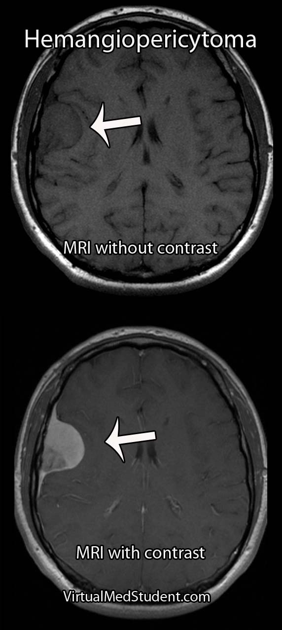
Hemangiopericytoma

Signs and Symptoms

Hemangiopericytomas are relatively slow growing and often do not cause symptoms until they become quite large. However, once they start to compress adjacent brain tissue they may cause headaches, seizures, confusion, weakness, or visual problems.

Diagnosis

MRIs and CT scans of the brain typically reveal a contrast enhancing dural-based lesion. Cerebral angiograms show a highly vascular tumor with blood supply coming from the dura, as well as the underlying brain tissue.

Based on imaging alone, hemangiopericytomas are often mistaken for meningiomas. Subtle characteristics such as a lack of calcification seen on CT scans may help distinguish one from the other, but this is not reliable.

The only reliable way to diagnose hemangiopericytoma is to look at a specimen of the tumor under a microscope. Special stains and features of the tumor can help delineate it from a meningioma (see pathology section above).

Did I Hear Someone Say “Treatment”?

Intracranial hemangiopericytomas should be surgically resected when feasible. Unfortunately, even after complete resection, they frequently recur and/or spread to other areas of the body.

Because of their aggressive nature, patients with hemangiopericytomas should also have adjuvant radiation therapy. Radiation treatment after surgical removal of the tumor has been shown to lengthen survival and slows (but doesn’t appear to prevent) the time to recurrence.

The role of chemotherapy is less clear and is still being investigated. At this point, chemotherapy is typically used in patients where radiation and surgery have failed to control the disease.

Let’s Recap It…

Intracranial hemangiopericytomas are malignant dural-based tumors that arise from pericytes. They are highly vascular tumors that enhance on MRI and CT scans. Symptoms are variable and depend on the size and location of the tumor. Treatment is with surgical removal followed by radiation therapy. Recurrence rates are high despite optimal treatment.

You Should Learn About This Stuff Too…

Want Some More?

Spinal Shock and Neurogenic Shock: The Battle Begins

Endless hours of studying, relentless exams, and the never-ending confusion between two perplexing phenomena: spinal shock and neurogenic shock. So, buckle up (so you don’t end up in spinal shock) and prepare for the journey through the world of spinal and neurogenic shock as we break down their differences.

The Tale of Two Shocks

Picture this: you’re in the ER, and a patient comes in with a recent spinal cord injury. You rack your brain to differentiate between spinal shock and neurogenic shock, but all you can remember is the cranial nerves mnemonic and your chief resident is about to pimp the you know what out of you… Let’s make it easy!

  • Spinal Shock: Think of it as your body’s initial reaction to “breaking up” with your spinal cord. It’s a temporary and unexpected “break-up shock” that leaves your reflexes, motor function, and sensation feeling lost and numb (physically, not emotionally) below the level of injury.
  • Neurogenic Shock: Now, imagine your body losing its balance between the sympathetic and parasympathetic nervous systems after a spinal cord injury. It’s like a tug-of-war, but the parasympathetic system wins, causing blood vessels to dilate and blood pressure to drop. Your heart, not knowing how to cope, slows down in response (bradycardia). Neurogenic shock occurs because the descending sympathetic fibers of the cord are injured, whereas the parasympathetic supply to the body provided by the vagus nerve (ie: “wandering nerve”) off of the brainstem is still intact and is now un-inhibited.

Diagnostic Dilemmas: The Hints are There You Just Have to Look

When you’re trying to diagnose spinal or neurogenic shock, look for these clues:

  • Spinal Shock: Your patient’s reflexes have gone on vacation, and they’re not telling you when they’ll be back. The muscles are flaccid, and sensations are playing hide-and-seek. But fear not, because those reflexes will eventually return. The delayed plantar response and bulbocavernosus reflex (don’t ask us how someone figured this one out!) are two of the earlier reflexes to come back from vacation. There is a lot of debate about how to define spinal shock and how long spinal shock lasts. However, you cannot prognosticate about the severity of cord injury until at least one reflex has returned.
  • Neurogenic Shock: Here, your patient’s blood pressure is lower than your motivation on a Monday morning, and their heart rate is slower than a sloth doing yoga. The skin may be warm and dry, resembling a cozy blanket you wish you were under instead of being in the ER.

Treatments: The Cures

Now that you’ve (hopefully) identified which shock you’re dealing with, it’s time to strategize and take action:

  • Spinal Shock: When faced with this shock, channel your inner superhero and protect the injured spinal cord at all costs! Immobilize the spine, maintain blood pressure, and ensure proper oxygenation to minimize further damage. Neurosurgical consultation is often indicated if the spinal column is unstable and requires surgical fixation.
  • Neurogenic Shock: Roll up your sleeves and get ready for some serious hemodynamic management. Rehydrate your patient with IV fluids, bring out the vasopressors to constrict those dilated blood vessels, and, if necessary, consider a temporary pacemaker to speed up the slow-motion heart rate.

Remember that both neurogenic and spinal shock are often occuring at the same time! Additionally, remember that neurogenic shock can also co-exist with other types of shock like hypovolemic shock in polytrauma patients.

As future health practitioners, you’ll face many confusing and challenging scenarios, like differentiating between spinal shock and neurogenic shock. But remember, amidst the stress, it’s essential to find some humor and light-heartedness. After all, laughter is the best medicine, and knowing the difference between these two conditions will not only help your patients, but also save you from those embarrassing moments during rounds. So, hold your head high, and step into the world of medicine with a smile on your face and the ability to distinguish between spinal and neurogenic shock in your ever-expanding medical knowledge toolbox.

More Fun Spinal Cord Pathology

References and Resources

Acoustic Neuroma, and Really We Mean Vestibular Schwannoma

Both the term “acoustic” and “neuroma” are incorrect ways of describing a tumor that arises from the 8th cranial nerve (vestibulocochlear nerve). An "acoustic neuroma" is a tumor that arises from Schwann cells that myelinate the peripheral portion of the nerve; this technically makes them “schwannomas”.

In addition, the tumor does not arise from the acoustic division of the 8th cranial nerve (ie: the portion of the nerve responsible for hearing), but instead arises most commonly from the vestibular division (ie: the balance portion of the nerve). Therefore, the appropriate medical term given to these tumors is a “vestibular schwannoma”.

These tumors are frequently caused by mutations in genes responsible for controlling cell cycle, cell morphogenesis, cell development, cell death, and cell adhesion. A well known cause of vestibular schwannomas occurs in patients with neurofibromatosis (NF) type II.

In this condition, which is responsible for about 5% of acoustic neuromas, a mutation in the NF gene on chromosome 22 causes an absent or dysfunctional protein product. This protein normally serves as a tumor suppressor; once mutated, it is no longer able to suppress tumor growth. The growth of various cells, including Schwann cells, becomes unchecked. The end result? A vestibular schwannoma.

When viewed under a pathology microscope, vestibular schwannomas are composed of different patterns of tissue. The first pattern is referred to as Antoni A; it consists of densely packed, elongated cells with nuclear free areas of cytoplasmic extensions referred to as "Verocay bodies". The second pattern is, you guessed it – Antoni B. This pattern has fewer cells and appears "looser" than the type A pattern.

These tumors are considered "benign", which means that they do not spread (metastasize) to other areas of the body. Overall, acoustic neuromas increase in size at the rate of roughly 1mm per year, but about 50% of tumors show no growth at all! Although they are not malignant tumors they can still cause symptoms.

Signs and Symptoms

Vestibular schwannomas cause local signs and symptoms. Since they arise from the 8th cranial nerve (vestibulocochlear nerve), which is responsible for hearing and balance, almost all patients present with some degree of hearing loss. In type II neurofibromatosis acoustic neuromas arising from both vestibulocochlear nerves may cause deafness. Some patients have tinnitus (ie: ear ringing), as well as a sense of vertigo.

Symptoms that are less common are a result of the tumor pressing on adjacent cranial nerves. Dysfunction of the 7th cranial nerve (facial nerve) can cause weakness of the facial muscles. If the tumor presses on the 5th cranial nerve (trigeminal nerve) it can cause face numbness; if it touches the 6th nerve (abducens nerve) diploplia (ie: double vision) may occur.

Finally, if the tumor continues to grow, it can cause compression of the brainstem. This can block the flow of cerebrospinal fluid (CSF) leading to a condition called hydrocephalus. These patients often have headaches, nausea, and vomiting secondary to increased pressure within the skull.

Diagnosis and Classification

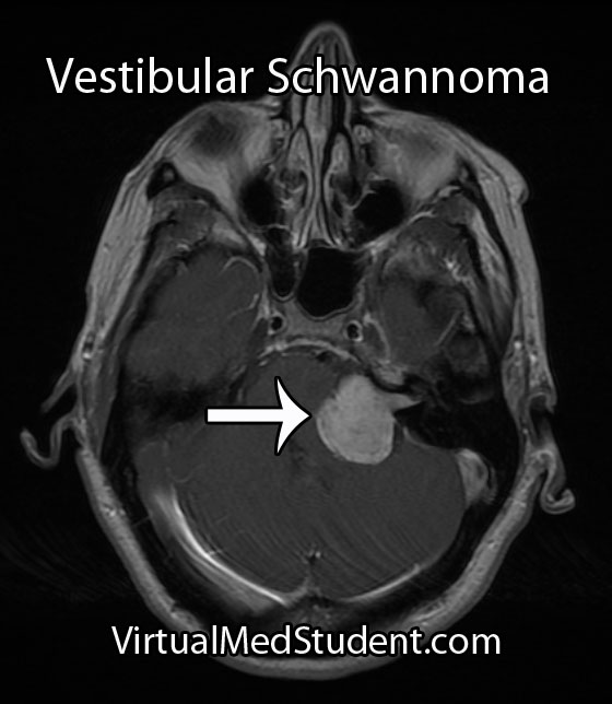
Vestibular Schwannoma
MRI is the imaging study of choice. It will show a well encapsulated tumor that sits in the cerebellopontine angle and/or involves the internal acoustic meatus.

Audiometric analysis is important in order to document hearing loss and for monitoring treatment outcomes. The most useful test is a pure tone audiogram. Differences in hearing ability between the two ears is suspicious for an acoustic neuroma, but not specific.

Although these tumors are commonly diagnosed from characteristic MRI findings, the definitive diagnosis is made when a pathologist looks at the tumor under a microscope.

A common classification system known as the Koos grading scale is frequently used. Grade 1 tumors involve only the internal auditory canal. Grade 2 tumors extend into the cerebellopontine angle, but do not encroach on the brainstem. A grade 3 tumor fills the entire cerebellopontine angle and a grade 4 tumor displaces the brainstem and adjacent cranial nerves.

Treatment

Treatment of these tumors depends on several factors, such as how large the tumor is, and whether or not the patient has symptoms from it (ie: hearing loss, face weakness, etc). If the tumor is small it can be followed with repeat MRI to monitor for enlargement. If the tumor grows, or begins to cause symptoms, then definitive treatment should be provided.

The two most commonly used treatment modalities are surgical resection and radiation. Surgery is most useful for very large tumors or when the patient is clinically deaf. Radiation comes in two flavors: single session stereotactic radiosurgery and fractionated radiotherapy.

Stereotactic radiosurgery is a single dose of radiation delivered directly to the tumor, typically with a dose of 12 to 13 Gy. The ability to preserve useful hearing with radiosurgery ranges from 32 to 71%. For tumors less than 3 cm in diameter, the ability of radiosurgery to halt the growth of the tumor has been shown to be between 92 and 100%.

Radiation can be harmful, especially when large doses are used in one session. Inadvertent injury to the facial nerve, acoustic nerve, trigeminal nerve, and brainstem are all possible adverse events. The use of fractionated radiotherapy has been tried to decrease these risks while still delivering large doses of radiation to the tumor.

Fractionated radiotherapy spreads the total radiation dose over multiple distinct sessions. For example, a total of 40 to 58 Gy can be delivered to the tumor in 2 Gy sessions over the course of several weeks. This is more radiation delivered to the tumor compared to single session radiosurgery (13 Gy), but it is delivered over a longer time frame, which helps mitigate the risk of damaging the adjacent cranial nerves and brainstem. Hearing preservation with fractionated radiotherapy has been shown to be superior to single-session radiosurgery.

Overview

A vestibular schwannoma is a benign tumor that arises from the vestibular portion of the 8th cranial nerve. It cause hearing loss and may cause compression of adjacent cranial nerves. It is diagnosed by clinical history, audiometric studies, and MRI. Treatment consists of surgical excision, radiation therapy, or both depending on the clinical situation.

More Brain Tumors…

References and Resources

  • Ferrer M, Schulze A, Gonzalez S, et al. Neurofibromatosis type 2: molecular and clinical analyses in Argentine sporadic and familial cases. Neurosci Lett. 2010 Aug 9;480(1):49-54. Epub 2010 Jun 8.
  • Cayé-Thomasen P, Borup R, Stangerup SE, et al. Deregulated genes in sporadic vestibular schwannomas. Otol Neurotol. 2010 Feb;31(2):256-66.
  • Harner SG, Laws ER Jr. Clinical findings in patients with acoustic neurinoma. Mayo Clin Proc. 1983 Nov;58(11):721-8.
  • Bederson JB, von Ammon K, Wichmann WW, et al. Conservative treatment of patients with acoustic tumors. Neurosurgery. 1991 May;28(5):646-50; discussion 650-1.
  • Kumar V, Abbas AK, Fausto N. Robbins and Cotran Pathologic Basis of Disease. Seventh Edition. Philadelphia: Elsevier Saunders, 2004.
  • Koos WT, Day JD, Matula C, et al. Neurotopographic considerations in the microsurgical treatment of small acoustic neurinomas. J Neurosurg. 1998 Mar;88(3):506-12.
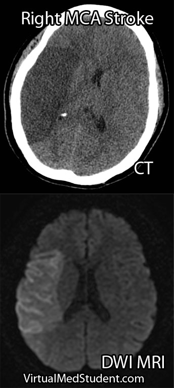
Brain Boo-Boos: Cerebrovascular Accidents (Stroke)

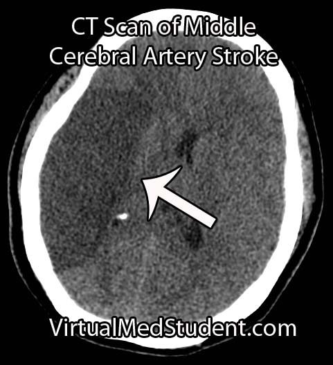
MCA Stroke CT Scan

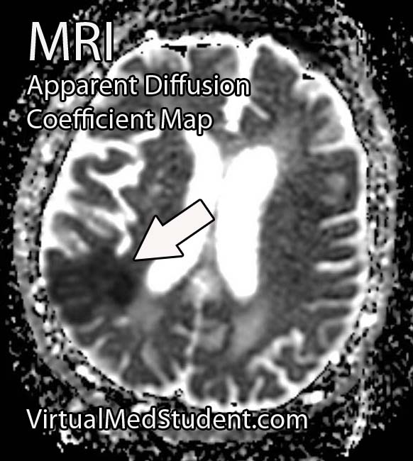
Stroke - ADC Map

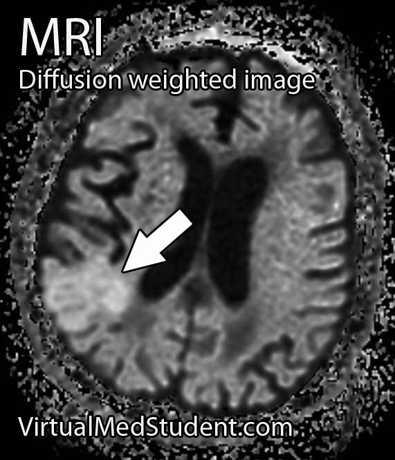
Stroke - Diffusion Weighted
A cerebrovascular accident, commonly known as a stroke, occurs when blood flow stops reaching brain tissue. If the entire brain is involved it is referred to as a "global" stroke; if a specific region of brain is involved it is referred to as a "focal" or "territorial" stroke. There are three broad causes of territorial strokes: thrombotic, embolic, and hemorrhagic.

A thrombotic stroke occurs when a blood clot forms in a blood vessel that supplies brain tissue. This is similar to what happens in cardiac infarction (ie: heart attacks). Thrombi are most commonly caused by atherosclerotic disease of the cerebral blood vessels. Thrombi usually form at areas of turbulent blood flow and at locations where vessels form branch points.

Embolic strokes are similar because they are technically blood clots. However, an embolus is a fragment of a clot (thrombus) that formed in another part of the body. Those fragments break free from the original clot and travel to blood vessels in the brain. They get lodged at some point and prevent blood from flowing resulting in a stroke if treatment is not obtained quickly.

Strokes can also be caused by bleeding into brain tissue. These type of strokes are called “hemorrhagic stroke”. Bleeding can occur in people with long standing untreated high blood pressure, or in those that have underlying structural disorders of the blood vessels in the brain (ie: aneurysms or arteriovenous malformations).

Diagnosis

Speedy diagnoses of stroke is extremely important because brain tissue dies quickly if it doesn’t receive adequate oxygen.

The first test that is done in cases of suspected stroke is a CT scan of the head. The purpose of the CT scan is not necessarily to "see" the stroke, but rather to rule out some other cause (ie: tumor, subdural hematoma, etc) for the symptoms. If bleeding is present on the CT scan the treatment algorithm becomes much different. If no bleeding is seen on CT then the second scan is usually an MRI.

An MRI takes longer than a CT scan, but it gives a much more detailed picture of the brain. In addition, it can pick up ischemia (ie: cell death related to decreased blood flow) much earlier than CT.

The best sequences to detect a stroke on an MRI are the diffusion weighted images and apparent diffusion coefficient maps. Stroked brain tissue will appear “bright” on diffusion weighted imaging and “dark” on the apparent diffusion coefficient map (see images to the left).

In addition, the carotid arteries are scanned using ultrasound in order to detect potential narrowing from atherosclerotic disease. Atherosclerotic carotid arteries are a potential source of emboli.

Sometimes a procedure known as transcranial doppler, which also uses ultrasound technology, is used to detect blood flow in the individual blood vessels of the brain. This can sometimes help determine the specific location of the thrombus/embolus.

Cerebral angiograms are much more invasive tests, but give a detailed view of which vessels are blocked. Cerebral angiograms can also be used to treat some strokes by directly removing clot from the affected blood vessel.

Most patients should undergo a thorough work up for atherosclerotic disease including a fasting lipid panel and hemoglobin A1C levels (a marker of diabetes).

If the heart is a suspected source of emboli than transthoracic echocardiography (ie: an ultrasound of the heart) is often done as well.

Signs and Symptoms

Cerebrovascular accidents present with a wide variety of signs and symptoms. It is entirely dependent on the blood vessel, and therefore, region of the brain involved. For example, strokes in the left middle cerebral artery will often cause significant language impairments if left untreated. Middle cerebral artery strokes usually cause contralateral paresis as well (usually the face and arm are more affected than the leg). Strokes in the frontal lobes caused by blockage of the anterior cerebral arteries can cause personality changes, as well as paresis/paralysis of the contralateral lower extremity.

Suffice it to say that there are a variety of possible clinical presentations in patients suffering from stroke. These presentations generally correlate with our understanding of brain anatomy and function.

Treatment

Prompt treatment of stroke is critical for preserving viable brain tissue. If a stroke is due to a blood clot (ie: thrombus or embolus) the treatment is with a drug known as tissue plasminogen activator (tPA). tPA is a medication that helps break up the clot.

It can be a dangerous medication because it can cause serious bleeding, but if given early enough, and in the right patient, it can completely prevent brain tissue death. There are numerous contraindications to giving tPA so caution must be used. The traditional teaching is that is should be given within three hours of symptom onset (this is the FDA approved indication); however, up to 4.5 hours from symptom onset has become common in clinical practice (but this is not FDA approved).

Endovascular therapies that mechanically remove the clot are becoming more common, especially for large vessel disease. However, this type of treatment requires specialized interventional neuro-radiologists and is not available in all medical centers. Endovascular therapy with a clot retrieving device is usually indicated up to 6 hours post symptom onset for large vessel occlusions. More distal (ie: further out) occlusions are not candidates for this type of procedure yet.

If a patient survives their first stroke, they are often started on medications to decrease their risk of having a second stroke. One of the most common medications used to prevent a second stroke is aspirin.

However, other medications like ticlopidine and clopidogrel (Plavix®) are also frequently used. All three of these medications prevent platelets (ie: one of the bodies natural ways of forming blood clots) from clumping together. In addition, aspirin is often mixed with another medication called dipyridamole (dipyridamole + aspirin = Aggrenox® in the United States). Patients who have suffered a minor stroke or have high risk transient ischemic attacks should be started on aspirin and clopidogrel and then transitioned to aspirin alone at 21 days.

If atherosclerosis is believed to be the cause of the stroke patients are often started on a statin. This helps slow the process of atherosclerosis and can help prevent another stroke from occurring.

If an embolus was the cause of the stroke patients are often started on an anticoagulant. The most common one used is warfarin (although there are many others). Warfarin is also used to treat a common cause of embolic stroke, an abnormal heart rhythm known as atrial fibrillation.

Overview

Strokes can be caused by thrombi or emboli which are blood clot that block blood flor, or from hemorrhage into brain tissue. Diagnosis is made by CT and MRI scans. Additional studies including carotid ultrasound, cerebral angiography, echocardiography, fasting lipid profiles, and tests for diabetes are also frequently performed.

Treatment depends on the etiology. Tissue plasminogen activator (tPA) is given if thrombi or emboli are the cause, and symptoms began less than 3 hours prior to presentation (4.5 hours is becoming the standard of care). Mechanical endovascular removal of the clot is also possible in some medical centers with specialized equipment.

Prevention of secondary strokes involve the use of anti-platelet (ie: aspirin, clopidogrel), anti-coagulant (ie: warfarin), and anti-atherosclerotic medications depending on the etiology of the previous stroke.

Related Articles

References and Resources

Book Review: The White Coat Investor

The White Coat Investor: A Doctor’s Guide To Personal Finance And Investing written by Dr. James M. Dahle is an excellent overview of many of the financial issues that face physicians. At 157 pages, it is not meant to be an exhaustive review of each topic; that being said, I found the length of the book to be just right, especially for a busy practicing physician.

The book starts with medical school and progresses through the attending years. Each chapter addresses an important topic that is salient to physicians. I found the chapters on life insurance, taxes, and estate planning very interesting because I had little understanding of these topics.

If you are ignorant like me about many financial concepts, this book is a must have! You may need to read deeper on certain topics, but again, this book is not meant to be exhaustive; it provides a much needed solid foundation on which doctors can build their financial futures. A 5 star book that I will read and re-read as the years go on.

Related Articles

References and Resources

Ependymoma: Myxopapillary, Anaplastic, and Perivascular Pseudorosettes

Ependymomas are tumors that develop from cells known as ependymal cells (duh!). Ependymal cells are a type of glial cell that line the ventricles (ie: fluid filled cavities) of the brain and central canal of the spinal cord.

Normal ependyma have cilia and microvilli on the side of the cell that faces cerebrospinal fluid (ie: the "apical" side). Cilia are hair like extensions that are believed to "beat" cerebrospinal fluid around the ventricles. Microvilli are folds in the cellular membrane that are thought to aid in the reabsorption of cerebrospinal fluid.

Unlike other epithelial cells in the body, of which ependyma are considered a subgroup, they do not rest on a basement membrane. Instead their basal surfaces (the surface not in contact with cerebrospinal fluid) intertwine with the overlying brain tissue.

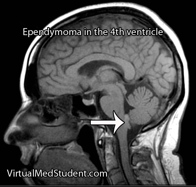
Like any other cell in the body, ependymal cells can decide to turn naughty and form a tumor. Ependymomas can occur anywhere there are ependymal cells, and therefore develop in both the brain and spinal cord. Intracranial ependymomas are more common in younger age groups, whereas spinal forms are more common in older individuals. Of those that form within the confines of the skull, the most common location is in the fourth ventricle near the brainstem.

There are three "grades" of ependymoma. There are two subsets of grade one: myxopapillary and subependymomas. The second grade of ependymoma has four distinct variants. They are cellular, papillary, clear cell, and tanycytic. The third grade is also referred to as "anaplastic" ependymoma. Regardless of the grade, each type has its own distinct characteristics when viewed under the pathology microscope.

Surgical specimens of ependymomas are often "stained" by pathologists to help aid in diagnosis, and more importantly, distinguish them from other tumor types. Ependymomas stain positive for the glial fibrillary acidic protein (GFAP), as well as phosphotungstic acid hematoxylin (PTAH).

Ependymomas may have perivascular pseudorosettes, which helps support the diagnosis. Pseudorosettes may not be apparent in tumors with dense cellularity such as anaplastic ependymomas.

In addition, ependymomas can spread throughout the cerebrospinal fluid space. For example, a tumor that arises in the fourth ventricle may "drop" tumor cells down into the spinal cord forming a secondary tumor. These secondary tumors are referred to as "drop mets".

Signs and Symptoms

The signs and symptoms depend on the location of the ependymoma.

The most common symptom of intracranial ependymoma is headache associated with nausea and/or vomiting. These symptoms occur when the ependymoma blocks the flow of cerebrospinal fluid, which causes a condition known as non-communicative hydrocephalus.

You can think of non-communicative hydrocephalus as a clog in a pipe. Everything upstream of the clog starts to back up, which eventually leads to increasing pressures. When this increased pressure occurs in the ventricular system of the brain it causes worsening headaches, nausea, and vomiting. This is especially true if the ependymoma is in the fourth ventricle of the brain, which even without tumor, is an anatomically narrow "pipe" to begin with.

Additionally, if the tumor pushes on brainstem structures a patient may present with dysfunction of the nerves that go to the various muscles of the head and face. The most commonly involved nerves are the facial nerve, which can cause weakness of the face, as well as the abducens nerve, which can cause weakness of the eye.

Tumors located in the spinal cord cause weakness and sensory disturbances.

Diagnosis

Ependymoma

MRI scans can be very useful and can support (but not prove) the diagnosis of ependymoma, especially when the tumor is in a common anatomical location.

If there is a high index of suspicion for ependymoma then the entire neuro-axis, meaning the brain and entire spinal column, should be imaged using MRI. This will detect “drop” mets, which, if present, further support the diagnosis.

Diagnosis can only be officially made when a sample of tumor (either surgical or at autopsy) is seen under the pathology microscope.

Treatment

Treatment of ependymoma is with surgical resection followed by radiation therapy. Patient outcome is most effective if the entire tumor can be removed during surgery. This is known as "gross total resection". However, the extent of surgical resection should always be weighed against the risk of harming the patient, especially if the tumor has invaded vital structures like the brainstem.

Fortunately, ependymomas are very radio-sensitive, which means that they respond well to getting zapped with radiation. Chemotherapy is not typically helpful except in very young children where the effects of radiation can be devastating.

Overview

Ependymomas arise from the cells that line the ventricular system of the brain and spinal cord. There are different subtypes depending on what it looks like under the pathology microscope. Diagnosis is based on pathological analysis and characteristic MRI findings. Treatment is with surgery and radiation.

Other Diseases You Should Know About…

References and Resources

Cerebral Ateriovenous Malformations: A Disease of Eloquence

A cerebral arteriovenous malformation is an abnormal tangle of blood vessels within the brain.

In order to understand these tangles we have to first understand normal blood flow. Blood flows from arteries to smaller arteries and then into capillary beds. In the capillary beds, gas, nutrients, and "wastes" are exchanged between the blood and adjacent body tissue. Once past the capillaries, the blood drains into successively larger veins where it eventually returns to the heart to be re-oxygenated.

In arteriovenous malformations there are no capillaries. Because of this, blood is shunted from the high pressure arterial system directly into the low pressure venous system. The "shunted" blood is unable to deliver its nutrients or oxygen to the nearby brain.

The risk of an arteriovenous malformation rupturing is relatively high because the pressure of arterial blood is "banging" into the walls of low pressure veins. The body tries to compensate for this by "arterializing" the blood vessels associated with the AVM.

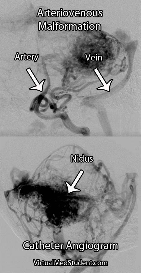
The term "nidus" is often used to describe the center of the malformation. This is the point where the arterial feeding vessels meet the draining venous structures.

In addition, any brain tissue around, or within the AVM is usually gliotic (a term used to describe scarring within the brain). Macrophages are sometimes present and are usually there to "gobble up" hemosiderin (a breakdown product of blood).

Signs and Symptoms

The signs and symptoms of cerebral arteriovenous malformations are dependent on the location of the malformation.

Most patients discover they have an AVM after it bleeds into the surrounding brain tissue. Patients can present with everything from a mild headache to a severe neurological deficit depending on the location and size of the malformation.

In addition, AVMs may cause transient neurological symptoms. These transient symptoms are caused by blood being shunted away from the surrounding normal brain tissue. Again, the location of the AVM dictates what symptoms may develop (ie: weakness if near the motor strip, difficulty with speech if located near Wernicke’s or Broca’s area, balance problems if in the cerebellum, disturbances in sensation if in the parietal lobe, etc., etc.).

Patients may also present with seizures as a result of irritation of the surrounding cortex by hemosiderin (a breakdown product of blood). In fact, seizures are the second most common presenting symptom.

Interestingly, headache is an uncommon symptom of arteriovenous malformations.

Diagnosis and Classification

Cerebral Arteriovenous Malformation
Diagnosis is made with special imaging studies like CT angiography, MR angiography, and formal catheter angiography (formal angiography is the gold standard).

AVMs are characterized by an abnormal tangle of blood vessels. The tell tale sign of an AVM on an angiogram is that both arterial and venous structures are seen at the same time (normally the venous phase follows the arterial phase).

The Spetzler-Martin grading system helps guide treatment decisions. This system takes into account the size, location, and type of venous drainage (see the first reference below).

Treatment

Treatment is highly individualized. There are currently three accepted treatment strategies: surgery, radiation, and embolization.

Surgery is still the treatment of choice, especially for AVMs near the surface of the brain or in non-eloquent cortex. Surgery is also considered "definitive" therapy (ie: the AVM is removed all at once), which is ideal for lesions considered high risk for rupture. Patient’s with deep seated lesions (ie: basal ganglia, thalamus, etc.), or those located in very "eloquent" cortical areas may be better treated with radiation or embolization.

Radiation works by causing changes in the vessels of the AVM. Over the course of several months to years the vessels are "cooked" by the radiation. This effectively eliminates blood flow into the AVM. Since the effects of radiation take months to years to shut down the AVM, the patient remains at risk for rupture. In addition, side effects from radiation may be permanent in a small percentage of patients.

Embolization is usually used as an adjunct to surgical resection. During embolization, various substances are injected into the AVM. These substances deprive the AVM of its arterial blood flow. This can be very useful prior to surgery to help with intra-operative blood loss (especially for very large AVMs!). Embolization is less commonly used as a stand alone treatment.

Overview

Arteriovenous malformations are abnormal tangles of blood vessels within the brain tissue. They have no intervening capillary bed so arterial blood flows directly into dilated veins. The main risk of an arteriovenous malformation is when it ruptures and bleeds into the surrounding brain. They can cause numerous signs and symptoms depending on their location. They are diagnosed with CT angiograms, MR angiograms, or formal catheter angiograms. Treatment is with surgery, radiation, and/or embolization depending on the risk of rupture and the location of the lesion.

Other Interesting Neurovascular Diseases…

References and Resources

  • Spetzler RF, Martin NA. A proposed grading system for arteriovenous malformations. J Neurosurg. 1986 Oct;65(4):476-83.
  • Ding D, Yen CP, Xu Z, et al. Radiosurgery for patients with unruptured intracranial arteriovenous malformations. J Neurosurg. 2013 May;118(5):958-66
  • Fokas E, Henzel M, Wittig A, et al. Stereotactic radiosurgery of cerebral arteriovenous malformations: long-term follow-up in 164 patients of a single institution. J Neurol. 2013 May 28.
  • Albuquerque FC, Ducruet AF, Crowley RW, et al. Transvenous to arterial Onyx embolization. J Neurointerv Surg. 2013 Mar 6.
  • Nataraj A, Mohamed MB, Gholkar A, et al. Multimodality Treatment of Cerebral Arteriovenous Malformations. World Neurosurg. 2013 Feb 20.

The Flaws of RVUs: Why They Fall Short in Measuring Physician Worth

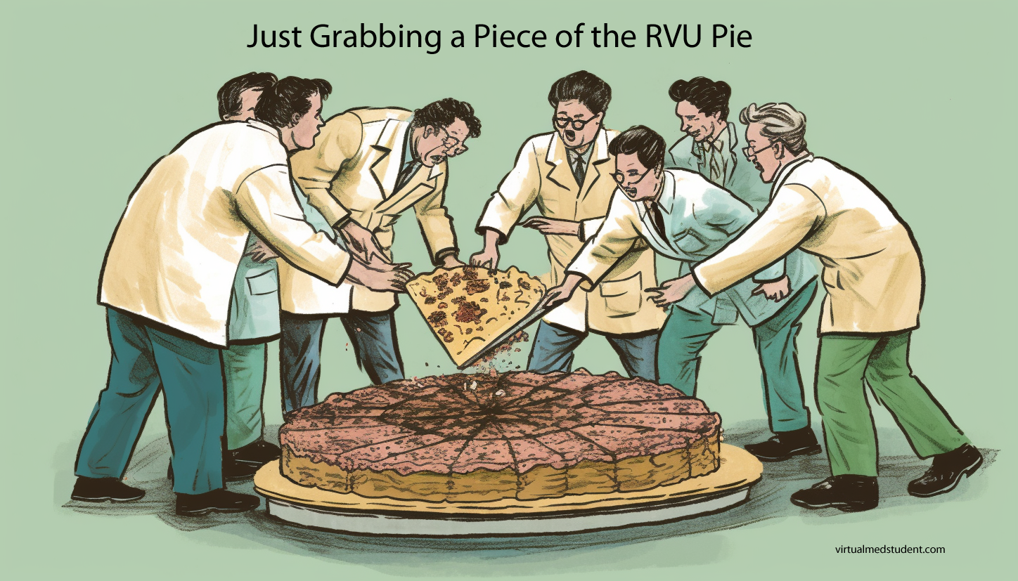
Relative Value Units (RVUs) have been utilized in the United States healthcare system for several decades as a standardized method for quantifying the “value” of medical services. They have become an important component of determining physician reimbursement. While the RVU system was initially designed to bring uniformity and transparency to physician payments, it has been increasingly criticized for its shortcomings in accurately reflecting physician worth. This article will discuss the key reasons why RVUs are a flawed measure of physician worth and explore alternative methods of evaluation.

Focus on Quantity over Quality

One of the most significant criticisms of the RVU system is its emphasis on the volume of services provided rather than the quality of care. By assigning higher RVU values to more complex and time-consuming procedures, the system inadvertently incentivizes physicians to prioritize high-revenue-generating procedures over less profitable, but potentially necessary, patient care activities. This focus on quantity can compromise the quality of care and hinder a physician’s ability to provide patient-centered care.

Lack of Personalized Care

RVUs are based on standardized averages, which do not account for individual variations in patient needs, physician skill, and the complexity of cases. Consequently, the RVU system fails to recognize the nuances of personalized care that physicians provide. High-quality care often involves tailoring treatment plans to each patient’s unique circumstances, which may not align with the fixed values assigned by RVUs. For example, if a primary care physician spends an entire hour discussing a patient’s mental health issues in a caring and compassionate way they are reimbursed substantially less than an orthopedic surgeon who takes an hour to pin a fracture or a neurosurgeon who spends an hour doing a cervical discectomy and fusion.

Limited Scope of Measurement

The RVU system only captures a narrow scope of a physician’s worth by focusing solely on clinical procedures and services. It does not account for the many other essential aspects of healthcare, such as patient education, interdisciplinary collaboration, care coordination, and research contributions. By overlooking these non-clinical activities, the RVU system fails to provide a comprehensive evaluation of a physician’s value.

Inadequate Incentive for Preventative Care

Preventative care plays a critical role in promoting public health and reducing healthcare costs. However, the RVU system does not adequately incentivize physicians to engage in these activities, as they are generally assigned lower RVUs. This discrepancy may lead to an underemphasis on preventative care and a potential increase in long-term healthcare costs.

Perpetuation of Disparities

RVUs can contribute to healthcare disparities by allocating resources based on service volume rather than patient need. Physicians practicing in underserved areas may find it challenging to generate high RVU values due to lower patient volume or a greater focus on primary care. This imbalance in resource distribution may inadvertently widen the gap in healthcare access and quality.

Lack of Collegiality

RVUs also tend to foster a lack of collegiality amongst physicians. In my experience, especially doctors on an “eat what you kill” compensation model, meaning they are paid for the number of RVUs they produce, are much less likely to refer to colleagues who may have more expertise or skill in a particular area. In other words, the doctor may “hold on” to patients to generate more RVUs rather than getting them to a colleague who may be able to provide a higher level of care for their particular ailment.

While RVUs were initially intended to standardize physician reimbursement and provide a transparent measure of physician worth, their shortcomings have become increasingly apparent over time. By focusing on quantity over quality and failing to capture the full spectrum of a physician’s value, the RVU system has inadvertently compromised patient care and perpetuated healthcare disparities. It is essential for the healthcare industry to consider alternative methods of evaluation and reimbursement that better align with the goals of patient-centered care, quality improvement, and equitable resource distribution.

It can be a fine line between incentivizing physicians to “work harder” and “earn more” while maintaining a high level of care and maintaining a highly ethical medical practice. I don’t think RVUs are a great answer to this dilemma, but for the time being it is the best we have in the United States. Let’s here your thoughts below…