Acoustic Neuroma, and Really We Mean Vestibular Schwannoma

Both the term “acoustic” and “neuroma” are incorrect ways of describing a tumor that arises from the 8th cranial nerve (vestibulocochlear nerve). An "acoustic neuroma" is a tumor that arises from Schwann cells that myelinate the peripheral portion of the nerve; this technically makes them “schwannomas”.

In addition, the tumor does not arise from the acoustic division of the 8th cranial nerve (ie: the portion of the nerve responsible for hearing), but instead arises most commonly from the vestibular division (ie: the balance portion of the nerve). Therefore, the appropriate medical term given to these tumors is a “vestibular schwannoma”.

These tumors are frequently caused by mutations in genes responsible for controlling cell cycle, cell morphogenesis, cell development, cell death, and cell adhesion. A well known cause of vestibular schwannomas occurs in patients with neurofibromatosis (NF) type II.

In this condition, which is responsible for about 5% of acoustic neuromas, a mutation in the NF gene on chromosome 22 causes an absent or dysfunctional protein product. This protein normally serves as a tumor suppressor; once mutated, it is no longer able to suppress tumor growth. The growth of various cells, including Schwann cells, becomes unchecked. The end result? A vestibular schwannoma.

When viewed under a pathology microscope, vestibular schwannomas are composed of different patterns of tissue. The first pattern is referred to as Antoni A; it consists of densely packed, elongated cells with nuclear free areas of cytoplasmic extensions referred to as "Verocay bodies". The second pattern is, you guessed it – Antoni B. This pattern has fewer cells and appears "looser" than the type A pattern.

These tumors are considered "benign", which means that they do not spread (metastasize) to other areas of the body. Overall, acoustic neuromas increase in size at the rate of roughly 1mm per year, but about 50% of tumors show no growth at all! Although they are not malignant tumors they can still cause symptoms.

Signs and Symptoms

Vestibular schwannomas cause local signs and symptoms. Since they arise from the 8th cranial nerve (vestibulocochlear nerve), which is responsible for hearing and balance, almost all patients present with some degree of hearing loss. In type II neurofibromatosis acoustic neuromas arising from both vestibulocochlear nerves may cause deafness. Some patients have tinnitus (ie: ear ringing), as well as a sense of vertigo.

Symptoms that are less common are a result of the tumor pressing on adjacent cranial nerves. Dysfunction of the 7th cranial nerve (facial nerve) can cause weakness of the facial muscles. If the tumor presses on the 5th cranial nerve (trigeminal nerve) it can cause face numbness; if it touches the 6th nerve (abducens nerve) diploplia (ie: double vision) may occur.

Finally, if the tumor continues to grow, it can cause compression of the brainstem. This can block the flow of cerebrospinal fluid (CSF) leading to a condition called hydrocephalus. These patients often have headaches, nausea, and vomiting secondary to increased pressure within the skull.

Diagnosis and Classification

Vestibular Schwannoma
MRI is the imaging study of choice. It will show a well encapsulated tumor that sits in the cerebellopontine angle and/or involves the internal acoustic meatus.

Audiometric analysis is important in order to document hearing loss and for monitoring treatment outcomes. The most useful test is a pure tone audiogram. Differences in hearing ability between the two ears is suspicious for an acoustic neuroma, but not specific.

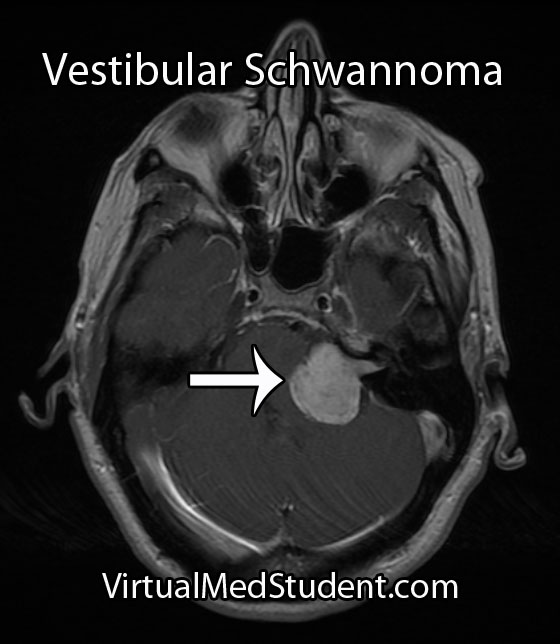
Although these tumors are commonly diagnosed from characteristic MRI findings, the definitive diagnosis is made when a pathologist looks at the tumor under a microscope.

A common classification system known as the Koos grading scale is frequently used. Grade 1 tumors involve only the internal auditory canal. Grade 2 tumors extend into the cerebellopontine angle, but do not encroach on the brainstem. A grade 3 tumor fills the entire cerebellopontine angle and a grade 4 tumor displaces the brainstem and adjacent cranial nerves.

Treatment

Treatment of these tumors depends on several factors, such as how large the tumor is, and whether or not the patient has symptoms from it (ie: hearing loss, face weakness, etc). If the tumor is small it can be followed with repeat MRI to monitor for enlargement. If the tumor grows, or begins to cause symptoms, then definitive treatment should be provided.

The two most commonly used treatment modalities are surgical resection and radiation. Surgery is most useful for very large tumors or when the patient is clinically deaf. Radiation comes in two flavors: single session stereotactic radiosurgery and fractionated radiotherapy.

Stereotactic radiosurgery is a single dose of radiation delivered directly to the tumor, typically with a dose of 12 to 13 Gy. The ability to preserve useful hearing with radiosurgery ranges from 32 to 71%. For tumors less than 3 cm in diameter, the ability of radiosurgery to halt the growth of the tumor has been shown to be between 92 and 100%.

Radiation can be harmful, especially when large doses are used in one session. Inadvertent injury to the facial nerve, acoustic nerve, trigeminal nerve, and brainstem are all possible adverse events. The use of fractionated radiotherapy has been tried to decrease these risks while still delivering large doses of radiation to the tumor.

Fractionated radiotherapy spreads the total radiation dose over multiple distinct sessions. For example, a total of 40 to 58 Gy can be delivered to the tumor in 2 Gy sessions over the course of several weeks. This is more radiation delivered to the tumor compared to single session radiosurgery (13 Gy), but it is delivered over a longer time frame, which helps mitigate the risk of damaging the adjacent cranial nerves and brainstem. Hearing preservation with fractionated radiotherapy has been shown to be superior to single-session radiosurgery.

Overview

A vestibular schwannoma is a benign tumor that arises from the vestibular portion of the 8th cranial nerve. It cause hearing loss and may cause compression of adjacent cranial nerves. It is diagnosed by clinical history, audiometric studies, and MRI. Treatment consists of surgical excision, radiation therapy, or both depending on the clinical situation.

More Brain Tumors…

References and Resources

  • Ferrer M, Schulze A, Gonzalez S, et al. Neurofibromatosis type 2: molecular and clinical analyses in Argentine sporadic and familial cases. Neurosci Lett. 2010 Aug 9;480(1):49-54. Epub 2010 Jun 8.
  • Cayé-Thomasen P, Borup R, Stangerup SE, et al. Deregulated genes in sporadic vestibular schwannomas. Otol Neurotol. 2010 Feb;31(2):256-66.
  • Harner SG, Laws ER Jr. Clinical findings in patients with acoustic neurinoma. Mayo Clin Proc. 1983 Nov;58(11):721-8.
  • Bederson JB, von Ammon K, Wichmann WW, et al. Conservative treatment of patients with acoustic tumors. Neurosurgery. 1991 May;28(5):646-50; discussion 650-1.
  • Kumar V, Abbas AK, Fausto N. Robbins and Cotran Pathologic Basis of Disease. Seventh Edition. Philadelphia: Elsevier Saunders, 2004.
  • Koos WT, Day JD, Matula C, et al. Neurotopographic considerations in the microsurgical treatment of small acoustic neurinomas. J Neurosurg. 1998 Mar;88(3):506-12.

Brain Boo-Boos: Cerebrovascular Accidents (Stroke)

MCA Stroke CT Scan

Stroke - ADC Map

Stroke - Diffusion Weighted
A cerebrovascular accident, commonly known as a stroke, occurs when blood flow stops reaching brain tissue. If the entire brain is involved it is referred to as a "global" stroke; if a specific region of brain is involved it is referred to as a "focal" or "territorial" stroke. There are three broad causes of territorial strokes: thrombotic, embolic, and hemorrhagic.

A thrombotic stroke occurs when a blood clot forms in a blood vessel that supplies brain tissue. This is similar to what happens in cardiac infarction (ie: heart attacks). Thrombi are most commonly caused by atherosclerotic disease of the cerebral blood vessels. Thrombi usually form at areas of turbulent blood flow and at locations where vessels form branch points.

Embolic strokes are similar because they are technically blood clots. However, an embolus is a fragment of a clot (thrombus) that formed in another part of the body. Those fragments break free from the original clot and travel to blood vessels in the brain. They get lodged at some point and prevent blood from flowing resulting in a stroke if treatment is not obtained quickly.

Strokes can also be caused by bleeding into brain tissue. These type of strokes are called “hemorrhagic stroke”. Bleeding can occur in people with long standing untreated high blood pressure, or in those that have underlying structural disorders of the blood vessels in the brain (ie: aneurysms or arteriovenous malformations).

Diagnosis

Speedy diagnoses of stroke is extremely important because brain tissue dies quickly if it doesn’t receive adequate oxygen.

The first test that is done in cases of suspected stroke is a CT scan of the head. The purpose of the CT scan is not necessarily to "see" the stroke, but rather to rule out some other cause (ie: tumor, subdural hematoma, etc) for the symptoms. If bleeding is present on the CT scan the treatment algorithm becomes much different. If no bleeding is seen on CT then the second scan is usually an MRI.

An MRI takes longer than a CT scan, but it gives a much more detailed picture of the brain. In addition, it can pick up ischemia (ie: cell death related to decreased blood flow) much earlier than CT.

The best sequences to detect a stroke on an MRI are the diffusion weighted images and apparent diffusion coefficient maps. Stroked brain tissue will appear “bright” on diffusion weighted imaging and “dark” on the apparent diffusion coefficient map (see images to the left).

In addition, the carotid arteries are scanned using ultrasound in order to detect potential narrowing from atherosclerotic disease. Atherosclerotic carotid arteries are a potential source of emboli.

Sometimes a procedure known as transcranial doppler, which also uses ultrasound technology, is used to detect blood flow in the individual blood vessels of the brain. This can sometimes help determine the specific location of the thrombus/embolus.

Cerebral angiograms are much more invasive tests, but give a detailed view of which vessels are blocked. Cerebral angiograms can also be used to treat some strokes by directly removing clot from the affected blood vessel.

Most patients should undergo a thorough work up for atherosclerotic disease including a fasting lipid panel and hemoglobin A1C levels (a marker of diabetes).

If the heart is a suspected source of emboli than transthoracic echocardiography (ie: an ultrasound of the heart) is often done as well.

Signs and Symptoms

Cerebrovascular accidents present with a wide variety of signs and symptoms. It is entirely dependent on the blood vessel, and therefore, region of the brain involved. For example, strokes in the left middle cerebral artery will often cause significant language impairments if left untreated. Middle cerebral artery strokes usually cause contralateral paresis as well (usually the face and arm are more affected than the leg). Strokes in the frontal lobes caused by blockage of the anterior cerebral arteries can cause personality changes, as well as paresis/paralysis of the contralateral lower extremity.

Suffice it to say that there are a variety of possible clinical presentations in patients suffering from stroke. These presentations generally correlate with our understanding of brain anatomy and function.

Treatment

Prompt treatment of stroke is critical for preserving viable brain tissue. If a stroke is due to a blood clot (ie: thrombus or embolus) the treatment is with a drug known as tissue plasminogen activator (tPA). tPA is a medication that helps break up the clot.

It can be a dangerous medication because it can cause serious bleeding, but if given early enough, and in the right patient, it can completely prevent brain tissue death. There are numerous contraindications to giving tPA so caution must be used. The traditional teaching is that is should be given within three hours of symptom onset (this is the FDA approved indication); however, up to 4.5 hours from symptom onset has become common in clinical practice (but this is not FDA approved).

Endovascular therapies that mechanically remove the clot are becoming more common, especially for large vessel disease. However, this type of treatment requires specialized interventional neuro-radiologists and is not available in all medical centers. Endovascular therapy with a clot retrieving device is usually indicated up to 6 hours post symptom onset for large vessel occlusions. More distal (ie: further out) occlusions are not candidates for this type of procedure yet.

If a patient survives their first stroke, they are often started on medications to decrease their risk of having a second stroke. One of the most common medications used to prevent a second stroke is aspirin.

However, other medications like ticlopidine and clopidogrel (Plavix®) are also frequently used. All three of these medications prevent platelets (ie: one of the bodies natural ways of forming blood clots) from clumping together. In addition, aspirin is often mixed with another medication called dipyridamole (dipyridamole + aspirin = Aggrenox® in the United States). Patients who have suffered a minor stroke or have high risk transient ischemic attacks should be started on aspirin and clopidogrel and then transitioned to aspirin alone at 21 days.

If atherosclerosis is believed to be the cause of the stroke patients are often started on a statin. This helps slow the process of atherosclerosis and can help prevent another stroke from occurring.

If an embolus was the cause of the stroke patients are often started on an anticoagulant. The most common one used is warfarin (although there are many others). Warfarin is also used to treat a common cause of embolic stroke, an abnormal heart rhythm known as atrial fibrillation.

Overview

Strokes can be caused by thrombi or emboli which are blood clot that block blood flor, or from hemorrhage into brain tissue. Diagnosis is made by CT and MRI scans. Additional studies including carotid ultrasound, cerebral angiography, echocardiography, fasting lipid profiles, and tests for diabetes are also frequently performed.

Treatment depends on the etiology. Tissue plasminogen activator (tPA) is given if thrombi or emboli are the cause, and symptoms began less than 3 hours prior to presentation (4.5 hours is becoming the standard of care). Mechanical endovascular removal of the clot is also possible in some medical centers with specialized equipment.

Prevention of secondary strokes involve the use of anti-platelet (ie: aspirin, clopidogrel), anti-coagulant (ie: warfarin), and anti-atherosclerotic medications depending on the etiology of the previous stroke.

Related Articles

References and Resources

Ependymoma: Myxopapillary, Anaplastic, and Perivascular Pseudorosettes

Ependymomas are tumors that develop from cells known as ependymal cells (duh!). Ependymal cells are a type of glial cell that line the ventricles (ie: fluid filled cavities) of the brain and central canal of the spinal cord.

Normal ependyma have cilia and microvilli on the side of the cell that faces cerebrospinal fluid (ie: the "apical" side). Cilia are hair like extensions that are believed to "beat" cerebrospinal fluid around the ventricles. Microvilli are folds in the cellular membrane that are thought to aid in the reabsorption of cerebrospinal fluid.

Unlike other epithelial cells in the body, of which ependyma are considered a subgroup, they do not rest on a basement membrane. Instead their basal surfaces (the surface not in contact with cerebrospinal fluid) intertwine with the overlying brain tissue.

Like any other cell in the body, ependymal cells can decide to turn naughty and form a tumor. Ependymomas can occur anywhere there are ependymal cells, and therefore develop in both the brain and spinal cord. Intracranial ependymomas are more common in younger age groups, whereas spinal forms are more common in older individuals. Of those that form within the confines of the skull, the most common location is in the fourth ventricle near the brainstem.

There are three "grades" of ependymoma. There are two subsets of grade one: myxopapillary and subependymomas. The second grade of ependymoma has four distinct variants. They are cellular, papillary, clear cell, and tanycytic. The third grade is also referred to as "anaplastic" ependymoma. Regardless of the grade, each type has its own distinct characteristics when viewed under the pathology microscope.

Surgical specimens of ependymomas are often "stained" by pathologists to help aid in diagnosis, and more importantly, distinguish them from other tumor types. Ependymomas stain positive for the glial fibrillary acidic protein (GFAP), as well as phosphotungstic acid hematoxylin (PTAH).

Ependymomas may have perivascular pseudorosettes, which helps support the diagnosis. Pseudorosettes may not be apparent in tumors with dense cellularity such as anaplastic ependymomas.

In addition, ependymomas can spread throughout the cerebrospinal fluid space. For example, a tumor that arises in the fourth ventricle may "drop" tumor cells down into the spinal cord forming a secondary tumor. These secondary tumors are referred to as "drop mets".

Signs and Symptoms

The signs and symptoms depend on the location of the ependymoma.

The most common symptom of intracranial ependymoma is headache associated with nausea and/or vomiting. These symptoms occur when the ependymoma blocks the flow of cerebrospinal fluid, which causes a condition known as non-communicative hydrocephalus.

You can think of non-communicative hydrocephalus as a clog in a pipe. Everything upstream of the clog starts to back up, which eventually leads to increasing pressures. When this increased pressure occurs in the ventricular system of the brain it causes worsening headaches, nausea, and vomiting. This is especially true if the ependymoma is in the fourth ventricle of the brain, which even without tumor, is an anatomically narrow "pipe" to begin with.

Additionally, if the tumor pushes on brainstem structures a patient may present with dysfunction of the nerves that go to the various muscles of the head and face. The most commonly involved nerves are the facial nerve, which can cause weakness of the face, as well as the abducens nerve, which can cause weakness of the eye.

Tumors located in the spinal cord cause weakness and sensory disturbances.

Diagnosis

Ependymoma

MRI scans can be very useful and can support (but not prove) the diagnosis of ependymoma, especially when the tumor is in a common anatomical location.

If there is a high index of suspicion for ependymoma then the entire neuro-axis, meaning the brain and entire spinal column, should be imaged using MRI. This will detect “drop” mets, which, if present, further support the diagnosis.

Diagnosis can only be officially made when a sample of tumor (either surgical or at autopsy) is seen under the pathology microscope.

Treatment

Treatment of ependymoma is with surgical resection followed by radiation therapy. Patient outcome is most effective if the entire tumor can be removed during surgery. This is known as "gross total resection". However, the extent of surgical resection should always be weighed against the risk of harming the patient, especially if the tumor has invaded vital structures like the brainstem.

Fortunately, ependymomas are very radio-sensitive, which means that they respond well to getting zapped with radiation. Chemotherapy is not typically helpful except in very young children where the effects of radiation can be devastating.

Overview

Ependymomas arise from the cells that line the ventricular system of the brain and spinal cord. There are different subtypes depending on what it looks like under the pathology microscope. Diagnosis is based on pathological analysis and characteristic MRI findings. Treatment is with surgery and radiation.

Other Diseases You Should Know About…

References and Resources

Cerebral Ateriovenous Malformations: A Disease of Eloquence

A cerebral arteriovenous malformation is an abnormal tangle of blood vessels within the brain.

In order to understand these tangles we have to first understand normal blood flow. Blood flows from arteries to smaller arteries and then into capillary beds. In the capillary beds, gas, nutrients, and "wastes" are exchanged between the blood and adjacent body tissue. Once past the capillaries, the blood drains into successively larger veins where it eventually returns to the heart to be re-oxygenated.

In arteriovenous malformations there are no capillaries. Because of this, blood is shunted from the high pressure arterial system directly into the low pressure venous system. The "shunted" blood is unable to deliver its nutrients or oxygen to the nearby brain.

The risk of an arteriovenous malformation rupturing is relatively high because the pressure of arterial blood is "banging" into the walls of low pressure veins. The body tries to compensate for this by "arterializing" the blood vessels associated with the AVM.

The term "nidus" is often used to describe the center of the malformation. This is the point where the arterial feeding vessels meet the draining venous structures.

In addition, any brain tissue around, or within the AVM is usually gliotic (a term used to describe scarring within the brain). Macrophages are sometimes present and are usually there to "gobble up" hemosiderin (a breakdown product of blood).

Signs and Symptoms

The signs and symptoms of cerebral arteriovenous malformations are dependent on the location of the malformation.

Most patients discover they have an AVM after it bleeds into the surrounding brain tissue. Patients can present with everything from a mild headache to a severe neurological deficit depending on the location and size of the malformation.

In addition, AVMs may cause transient neurological symptoms. These transient symptoms are caused by blood being shunted away from the surrounding normal brain tissue. Again, the location of the AVM dictates what symptoms may develop (ie: weakness if near the motor strip, difficulty with speech if located near Wernicke’s or Broca’s area, balance problems if in the cerebellum, disturbances in sensation if in the parietal lobe, etc., etc.).

Patients may also present with seizures as a result of irritation of the surrounding cortex by hemosiderin (a breakdown product of blood). In fact, seizures are the second most common presenting symptom.

Interestingly, headache is an uncommon symptom of arteriovenous malformations.

Diagnosis and Classification

Cerebral Arteriovenous Malformation
Diagnosis is made with special imaging studies like CT angiography, MR angiography, and formal catheter angiography (formal angiography is the gold standard).

AVMs are characterized by an abnormal tangle of blood vessels. The tell tale sign of an AVM on an angiogram is that both arterial and venous structures are seen at the same time (normally the venous phase follows the arterial phase).

The Spetzler-Martin grading system helps guide treatment decisions. This system takes into account the size, location, and type of venous drainage (see the first reference below).

Treatment

Treatment is highly individualized. There are currently three accepted treatment strategies: surgery, radiation, and embolization.

Surgery is still the treatment of choice, especially for AVMs near the surface of the brain or in non-eloquent cortex. Surgery is also considered "definitive" therapy (ie: the AVM is removed all at once), which is ideal for lesions considered high risk for rupture. Patient’s with deep seated lesions (ie: basal ganglia, thalamus, etc.), or those located in very "eloquent" cortical areas may be better treated with radiation or embolization.

Radiation works by causing changes in the vessels of the AVM. Over the course of several months to years the vessels are "cooked" by the radiation. This effectively eliminates blood flow into the AVM. Since the effects of radiation take months to years to shut down the AVM, the patient remains at risk for rupture. In addition, side effects from radiation may be permanent in a small percentage of patients.

Embolization is usually used as an adjunct to surgical resection. During embolization, various substances are injected into the AVM. These substances deprive the AVM of its arterial blood flow. This can be very useful prior to surgery to help with intra-operative blood loss (especially for very large AVMs!). Embolization is less commonly used as a stand alone treatment.

Overview

Arteriovenous malformations are abnormal tangles of blood vessels within the brain tissue. They have no intervening capillary bed so arterial blood flows directly into dilated veins. The main risk of an arteriovenous malformation is when it ruptures and bleeds into the surrounding brain. They can cause numerous signs and symptoms depending on their location. They are diagnosed with CT angiograms, MR angiograms, or formal catheter angiograms. Treatment is with surgery, radiation, and/or embolization depending on the risk of rupture and the location of the lesion.

Other Interesting Neurovascular Diseases…

References and Resources

  • Spetzler RF, Martin NA. A proposed grading system for arteriovenous malformations. J Neurosurg. 1986 Oct;65(4):476-83.
  • Ding D, Yen CP, Xu Z, et al. Radiosurgery for patients with unruptured intracranial arteriovenous malformations. J Neurosurg. 2013 May;118(5):958-66
  • Fokas E, Henzel M, Wittig A, et al. Stereotactic radiosurgery of cerebral arteriovenous malformations: long-term follow-up in 164 patients of a single institution. J Neurol. 2013 May 28.
  • Albuquerque FC, Ducruet AF, Crowley RW, et al. Transvenous to arterial Onyx embolization. J Neurointerv Surg. 2013 Mar 6.
  • Nataraj A, Mohamed MB, Gholkar A, et al. Multimodality Treatment of Cerebral Arteriovenous Malformations. World Neurosurg. 2013 Feb 20.

Atlanto-Occipital Dislocation (Internal Decapitation)

Atlanto-occipital dislocations (AODs) occur when there is severe damage to the ligaments that connect the skull to the cervical spine. The most common cause of an AOD is trauma.

There are several classifications of atlanto-occipital dislocations. In type 1 dislocations the base of the skull moves anteriorly (ie: forward) relative to the dens of the second cervical vertebrae. In type 2 dislocations the skull moves superiorly (ie: upwards) relative to the cervical spine. In type 3 dislocations the skull moves posteriorly (ie: backwards) relative to the cervical spine.

Often times more than one classification may exist in a single patient. For example, a patient may have both a type 1 and type 2 AOD if the patient’s skull has moved forward and upwards relative to the cervical spine.

Signs and Symptoms

Most cases of complete atlanto-occipital dislocation are fatal. Severe damage to the spinal cord around the cranio-cervical junction leads to respiratory paralysis and death if mechanical ventilation is not started rapidly. Patients are often quadriplegic (ie: unable to move their upper and lower extremities) because of damage to the descending motor axons of the corticospinal tract. Incomplete injuries can also occur with varying degrees of cervical spine injury.

The lower cranial nerves are sometimes involved in the injured segment. The most commonly injured cranial nerves are the abducens (VI), vagus (X), and hypoglossal (XII) nerves. Abducens nerve injury causes an inability to deviate the eye outwards. Damage to the hypoglossal nerve causes deviation of the tongue towards the side of the injured nerve (“lick your wound”).

Surprisingly, some patients with atlanto-occipital dislocation may have no neurologic symptoms! It is therefore important to rule out ligamentous injury, especially in patients who have had severe head and neck trauma.

Diagnosis and Classification

BDI for AOD
BAI AOD
Power's Ratio
Diagnosis is made after appropriate imaging studies are obtained. Plain films and CT scans of the cervical spine are the most commonly obtained initial imaging studies.

There are several methods for determining if atlanto-occipital dislocation has occurred. The first is the “BAI-BDI method”. Two distances are measured. The BDI, or basion-dental interval, measures the distance between the most anterior and inferior portion of the skull (ie: “basion”) and the tip of the dens, which is a superior extension of the second cervical vertebrae. The BDI should normally be less than 12mm. In the image below the BDI is 14mm and is indicative of atlanto-occipital dislocation.

The second distance that is measured is the BAI, or basion-axial interval. The BAI is the distance between the basion and a line drawn straight up from the posterior (ie: back) portion of the dens. The normal interval is less than 12mm. In the image below the BAI is 14.2mm indicating likely atlanto-occipital dislocation.

A second method of assessing atlanto-occipital dislocation is known as Power’s ratio. Power’s ratio is most useful if the dislocation is anterior (ie: the head moves forward relative to the spine). Two distances are measured. The first is the distance between the opisthion and the anterior ring of C1; the second is the distance between the basion and the posterior ring of C1. The second distance is then divided by the first to obtain the ratio. A “normal” ratio is less than 1.0; if the ratio is greater than 1.0 then suspicion for dislocation should be high.

X-rays and CT show bony anatomy very well, but do not demonstrate ligamentous injury. Many patients with suspected AOD may also get an MRI to assess ligamentous damage. MRI will usually show severe injury to the ligaments that connect the first and second cervical vertebrae to the skull base.

There are two commonly used classification systems for describing atlanto-occipital dislocations. The Traynelis system is purely descriptive and is based on which way the head moves relative to the spine.

Traynelis Classification
Type 1 Head moves forward relative to the spine
Type 2 Head moves upwards relative to the spine
Type 3 Head moves backwards relative to the spine

The Bellabarba system is based on CT findings and is used to determine stability; it is also useful because it helps guide treatment decisions. A stable injury is one in which the dislocation is within 2mm of a normal BDI/BAI value and does not get worse with a traction test. Unstable injuries are present when the dislocation gets worse with a traction test; these injuries may be reduced at baseline (ie: within 2mm of a normal BDI/BAI) or grossly abnormal. That being said, it is important to note that very few doctors perform traction tests in patients with suspected AOD as this can worsen the condition!

Bellabarba Classification
Type Description Stability
Type 1 Alignment within 2mm of normal BAI/BDI interval; distracts less than 2mm with traction Stable
Type 2 Reduced at baseline (ie: within 2mm of normal BDI/BAI), but significant distraction with traction test Unstable
Type 3 Severely misaligned relative to normal BDI/BAI values Unstable

Treatment

Treatment of atlanto-occipital dislocations involves spinal immobilization. This is usually done via a surgical procedure known as an occipital-cervical fusion in which the back of the skull is fused to the cervical spine. Some patients are also put in a halo immobilization device.

Overview

Atlanto-occipital dislocation occurs after severe injury to the ligaments that connect the skull to the cervical spine. It most commonly occurs after severe head or neck trauma. Death secondary to ventilatory failure, quadriplegia, and cranial nerve deficits are common symptoms. Diagnosis is based on characteristic findings found on CT, x-rays, and MRI scans. Treatment is with spinal immobilization, either with a halo apparatus, or a surgical procedure known as occipital-cervical fusion.

Some More Useful Learning Material…

References and Resources

  • Garrett M, Consiglieri G, Kakarla UK, et al. Occipitoatlantal dislocation. Neurosurgery. 2010 Mar;66(3 Suppl):48-55.
  • Greenberg MS. Handbook of Neurosurgery. Sixth Edition. New York: Thieme, 2006. Chapter 25.
  • Deliganis AV, Mann FA, Grady MS. Rapid diagnosis and treatment of a traumatic atlantooccipital dissociation. AJR Am J Roentgenol. 1998 Oct;171(4):986.
  • Traynelis VC, Marano GD, Dunker RO, et al. Traumatic atlanto-occipital dislocation. Case report. J Neurosurg. 1986 Dec;65(6):863-70.
  • Bellabarba C, Mirza SK, West GA, et al. Diagnosis and treatment of craniocervical dislocation in a series of 17 consecutive survivors during an 8-year period. J Neurosurg Spine. 2006 Jun;4(6):429-40.
  • Horn EM, Feiz-Erfan I, Lekovic GP, et al. Survivors of occipitoatlantal dislocation injuries: imaging and clinical correlates. J Neurosurg Spine. 2007 Feb;6(2):113-20.

The Anterior Choroidal Artery: Small but Mighty

The anterior choroidal arteries are small, but vital blood vessels in the brain. They are branches of the internal carotid arteries. They arise proximal to the splitting of the internal carotid into the anterior and middle cerebral arteries.

The anterior choroidal arteries deliver blood to vital brain structures. These structures include the posterior limbs of the internal capsules, portions of the thalami, optic tracts, middle third of the cerebral peduncles, portions of the temporal lobes (ie: parts of the pyriform cortex, uncus, and amygdala), substantia nigra, portions of the globus pallidus, as well as the choroid plexus in the lateral ventricles.

The anterior choroidal artery forms connections (anastamoses) with the posterior lateral choroidal arteries. Cerebral angiograms are the best way to visualize the anterior choroidal arteries.

Importance in Disease

Blockage of the anterior choroidal artery can cause a stroke. The most common symptoms of an anterior choroidal stroke are hemiparesis (weakness on the opposite side of the body), hemianesthesia (decreased sensation on the opposite side of the body), and a homonymous hemianopsia (loss of a portion of the visual field of both eyes). High blood pressure is the most common underlying factor in people with anterior choroidal artery strokes.

The hemiparesis is a result of damage to the posterior limb and genu of the internal capsule. The posterior limb contains the corticospinal tracts, which send information about movement from the brain to the spinal cord.

The hemianesthesia is a result of damage to the ventral posterolateral nucleus of the thalamus. This nucleus contains neurons that receive information from the spinal cord about sensation from the body. This symptom is less common than weakness, and occurs in roughly half of patients with an anterior choroidal stroke.

Cerebral angiogram showing anterior choroidal artery.

The final symptom, homonymous hemianopsia, is caused by damage to the optic tracts and lateral geniculate nucleus of the thalamus. Patients lose the ability to see objects on the left or right side (depending on which anterior choroidal artery is involved) in both eyes. This is an even more uncommon symptom, which occurs in less than 10% of patients with an anterior choroidal artery stroke.

Strokes of the anterior choroidal artery rarely cause all three symptoms. This is because the brain tissue served by the anterior choroidals also receives blood flow from other arteries.

Aneurysms of the anterior choroidal arteries are rare and will not be discussed in this article.

Overview

The anterior choroidal arteries are paired structures that arise from the internal carotid arteries. They supply blood to many important structures within the brain. Stroke is the most common pathological disease related to this blood vessel and frequently causes weakness of the opposite side of the body. High blood pressure is the most common underlying disease seen in people who have a stroke in this vascular distribution.

Other Stuff Worth Looking At…

References and Resources

  • Pezzella FR, Vadalà R. Anterior choroidal artery territory infarction. Front Neurol Neurosci. 2012;30:123-7. Epub 2012 Feb 14.
  • Bruno A, Graff-Radford NR, Biller J, et al. Anterior choroidal artery territory infarction: a small vessel disease. Stroke. 1989 May;20(5):616-9.
  • Baehr M, Frotscher M. Duus’ Topical Diagnosis in Neurology: Anatomy, Physiology, Signs, Symptoms. Fourth Edition. Stuttgart: Thieme, 2005.
  • Nolte J. The Human Brain: An Introduction to its Functional Anatomy. Sixth Edition. Philadelphia: Mosby, 2008.
  • Baskaya MK, Coscarella E, Gomez F, et al. Surgical and angiographic anatomy of the posterior communicating and anterior choroidal arteries. Neuroanatomy (2004) v3:38-42.

The Internal Capsule: Some Pricey Brain Real Estate

The internal capsule is one pricey piece of brain real estate! It contains all of the pathways that allow information to be transferred between the cerebral cortex and the spinal cord, brainstem, and subcortical structures (ie: thalamus, basal ganglia). It is divided into an anterior limb, posterior limb, and genu (ie: the area where the anterior and posterior limbs meet).

The anterior limb contains axons that send information between the thalamus and the cingulate gyrus and pre-frontal cortex. It also contains axons in the frontopontine pathway (ie: axons going from the frontal cortex to a portion of the brainstem known as the pons).

The genu contains the corticobulbar tract, which originate in the motor areas of the frontal lobes and extend to the cranial nerve nuclei in the brainstem. It also contains axons that connect the motor section of the thalamus (ie: VA and VL nuclei) with the motor areas of the frontal cortex.

The posterior limb contains the corticospinal tract, which are axons that come from the motor area of the frontal cortex and travel all the way to the anterior horns of the spinal cord where α-motor neurons are located. The posterior limb also contains sensory information coming from the body via the medial lemniscus and the anterolateral (aka: spinothalamic tract) systems.

Internal Capsule MRI

The blood supply to most of the internal capsule comes from the lenticulostriate arteries. These small arteries originate from the first portion of the middle cerebral artery. Two other important arteries also supply portions of the internal capsule: the anterior choroidal artery and the recurrent artery of Heubner. The anterior choroidal artery is a branch of the internal carotid. It supplies the inferior portion of the posterior limb. The recurrent artery of Heubner is a branch of the anterior cerebral artery. It supplies the inferior portions of the anterior limb and the genu.

Anatomy of the Internal Capsule
Division Major Communication Tracts Blood Supply
Anterior limb

– Tracts between the frontal lobe and pons (brainstem)

– Tracts between the thalamus and prefrontal cortex

– Tracts between the thalamus and cingulate gyrus

– Lenticulostriate arteries (branches of the middle cerebral artery)

– Recurrent artery of Heubner (branch of the anterior cerebral artery)

Genu – Tracts between the motor cortex in the frontal lobe and the cranial nerve nuclei in the brainstem (aka: corticobulbar tract)

– Lenticulostriate arteries (branches of the middle cerebral artery)

– Recurrent artery of Heubner (branch of the anterior cerebral artery)
Posterior limb

– Tracts between the motor cortex of frontal lobe and anterior horn of spinal cord (aka: corticospinal tract)

– Medial lemniscus tract (a continuation of the dorsal columns), which carries information about light touch, vibration, and pressure sensation from the body and spinal cord.

– Anterolateral (aka: spinothalamic) tract, which carries pain and temperature information

– Lenticulostriate arteries (branches of the middle cerebral artery)

– Anterior choroidal artery (branch of the internal carotid)

Importance in Disease

Thalamic Hemorrhage
Thalamic intracerebral hematoma
compressing the posterior limb
of the internal capsule

Damage to the internal capsule can be devastating neurologically because it contains so many vital tracts.

For example, a stroke of the anterior choroidal artery can lead to posterior limb damage. This can cause paralysis of the contralateral (ie: opposite) arm and leg secondary to interruption of the corticospinal tract.

Posterior limb disruption can also cause co-existent sensory deficits including an inability to feel light touch, pain, and temperature due to damage of the spinothalamic and medial lemniscal pathways.

Hypertensive hemorrhages in the thalamus or basal ganglia can compress the adjacent fibers of the internal capsule leading to similar clinical findings. The head CT to the right shows a thalamic hemorrhage secondary to severely elevated blood pressure. The patient had compression of the posterior limb of the internal capsule. As a result she was unable to move her left arm and leg, and could not feel pain or light touch on the left side of her body.

Overview

The internal capsule is a vital structure. It contains many communication pathways between the brain’s cortex, brainstem, spinal cord, and subcortical nuclei (ie: thalamus, basal ganglia). Its blood supply comes from branches of the middle cerebral artery (ie: lenticulostriates), anterior cerebral artery (ie: recurrent artery of Heubner), and the internal carotid (ie: anterior choroidal artery). Lesions in this area caused by strokes or hypertensive hemorrhages can have devastating clinical consequences.

Other Pertinent Articles…

References and Resources

  • Greenberg MS. Handbook of Neurosurgery. 9th Edition. New York: Thieme, 2006. Chapter 25.
  • Chowdhury F, Haque M, Sarkar M, et al. White fiber dissection of brain; the internal capsule: a cadaveric study. Turk Neurosurg. 2010 Jul;20(3):314-22. doi: 10.5137/1019-5149.JTN.3052-10.2.
  • Simon RP, Aminoff MJ, Greenberg DA. Clinical Neurology, Seventh Edition (LANGE Clinical Medicine). Seventh Edition. New York: McGraw Hill, 2009.
  • Nolte J. The Human Brain: An Introduction to its Functional Anatomy. Sixth Edition. Philadelphia: Mosby, 2008.
  • Bickley LS, Szilagyi PG. Bates’ Guide to Physical Examination and History Taking. Ninth Edition. New York: Lippincott Williams and Wilkins, 2007.

Peroneal (Fibular) Nerve and the Dropped Foot

The peroneal nerve is one of the branches of the sciatic nerve. It receives most of its innervation from the L4, L5, and S1 nerves. The common peroneal nerve (aka: common fibular nerve) wraps around the outside of the knee over the head of the fibula. It then branches into two separate nerves: the superficial peroneal nerve and the deep peroneal nerve.

The superficial peroneal nerve innervates two muscles: peroneus longus and brevis (aka: fibularis longus and brevis). These muscles allow you to evert (ie: move your foot outwards) and plantarflex (ie: help you step on the gas pedal) the foot. This nerve also provides the sensation to the outside half (ie: lateral half) of the lower leg, as well as the top of most of the foot (ie: the dorsum of the foot).

The deep peroneal nerve innervates three muscles in the leg: tibialis anterior, extensor digitorum longus, and extensor hallucis longus. Tibialis anterior allows you to dorsiflex (ie: lift your foot off the ground) and invert (ie: bringing your big to closer to the middle of the body) your foot. Extensor digitorum longus helps you extend your toes (ie: the opposite of curling them), as well as evert, and dorsiflex your foot. The third muscle, extensor hallucis longus, allows you to extend your big toe.

The deep peroneal nerve also innervates two muscles in the foot: extensor digitorum brevis (also helps to extend the toes) and extensor hallucis brevis (also helps to extend the big toe).

Importance in Disease

The peroneal nerve is most frequently compressed over the fibular head. Compression typically affects the deep peroneal nerve rather than the common or superficial nerve; however, all of the nerves may be involved.

When the deep peroneal nerve is compressed, the foot is unable to dorsiflex secondary to dysfunction of the tibialis anterior muscle. Additionally, the patient is unable to extend the toes. Sensation may be decreased on a small patch of skin between the big toe and second toe; pain in the area of the lateral lower leg may also be present.

When the superficial peroneal nerve is compressed, the peroneus longus and brevis muscles are affected. Dysfunction of these muscles prevents the patient from everting their foot. Sensation over the lateral half of the lower leg and top of the foot may also be decreased.

Placing the nerve under passive, or active, stretch by placing the patient’s foot in inversion will often reproduce the symptoms. Percussing the nerve over the fibular head (Tinel’s test) can reproduce the symptoms.

It is important to distinguish a peroneal nerve palsy from a herniated L4-L5 disc. A patient with a herniated L4-L5 disc – causing an L5 radiculopathy – will not only have difficulty dorsiflexing the foot and toes (secondary to dysfunction of the L5 component of the peroneal nerve), but will also have difficulty inverting the foot (secondary to dysfunction of the L5 component of the tibial nerve).

Wait a second! This still shouldn’t make sense if you are actually thinking about it, and this is where things get tricky… Both the anterior tibialis (deep peroneal nerve innervated) and the posterior tibialis (tibial nerve innervated) help invert the foot. So how do we know that the weakness in inversion is related to an L5 disc herniation, a peroneal nerve palsy, or a tibial nerve palsy? You need to have the patient attempt to invert the foot while plantarflexing! The posterior tibialis is an inverter and plantarflexor of the foot so if dorsiflexion is weak (L5 –> deep peroneal nerve –> anterior tibialis) and the patient cannot invert (L5 –> tibial nerve –> posterior tibialis OR L5 –> deep peroneal nerve –> anterior tibialis) well while the foot is plantarflexed you probably have an L5 radiculopathy and not a peroneal nerve palsy.

To make it a bit easier clinically… A foot drop WITH inversion weakness is most likely an L5 radiculopathy (most commonly from a herniated disc) because you are getting the invertors for both the deep peroneal nerve (tibialis anterior muscle) AND the tibial nerve (tibialis posterior muscle), which both have get fibers from the L5 nerve root. However, injury to the sciatic nerve proximally in the leg could also give you this, but it would be associated with significant plantarflexion weakness too (due to gastroc weakness from S1)! So therefore a foot drop with eversion weakness and toe extension weakness is also suspicious for a peroneal nerve injury.

Overview

The peroneal nerve splits at the level of the knee from the sciatic nerve. It then further divides into the superficial and deep peroneal nerves. The superficial branch controls the evertors of the foot (peroneus longus and brevis) and provides sensation over the lateral aspect of the lower leg and top of the foot. The deep branch controls the dorsiflexors of the foot, and the extensor muscles of the toes. The common peroneal, and either of its branches, is most commonly compressed near the fibular head.

More Important Anatomy to Master…

References and Resources

  • Greenberg MS. Handbook of Neurosurgery. Sixth Edition. New York: Thieme, 2006. Chapter 25.
  • Prakash, Bhardwaj AK, Devi MN. Sciatic nerve division: a cadaver study in the Indian population and review of the literature. Singapore Med J. 2010 Sep;51(9):721-3.
  • Yuen EC, So YT. Sciatic neuropathy. Neurol Clin. 1999 Aug;17(3):617-31, viii.
  • Simon RP, Aminoff MJ, Greenberg DA. Clinical Neurology, Seventh Edition (LANGE Clinical Medicine). Seventh Edition. New York: McGraw Hill, 2009.
  • Netter FH. Atlas of Human Anatomy: with Student Consult Access (Netter Basic Science). Fifth Edition. Philadelphia: Saunders Elsevier, 2010.
  • Bickley LS, Szilagyi PG. Bates’ Guide to Physical Examination and History Taking. Ninth Edition. New York: Lippincott Williams and Wilkins, 2007.

An Overview of the Sciatic Nerve

The sciatic nerve is a collection of nerve fibers that exit the spinal column between the 4th lumbar and 3rd sacral levels (L4-S3).

In the upper leg, the sciatic nerve gives off branches to the hamstring muscles. These muscles, which include the semimembranous, semitendinous, biceps femoris and part of the adductor magnus, are powerful flexors of the knee joint.

After sending off branches to the hamstrings, the sciatic nerve hits the back of the knee. At the popliteal fossa the sciatic splits into two distinct nerves: the common peroneal nerve and the tibial nerve.

The first nerve, the common peroneal (aka: common fibular), wraps around the outside of the knee and over the head of the fibula. It then branches into two separate nerves: the superficial peroneal and deep peroneal nerves.

The superficial peroneal nerve innervates two muscles: peroneus longus and brevis (aka: fibularis longus and brevis). These muscles allow you to evert (ie: allow you to lift your "pinky" toe higher than your "big" toe) and plantarflex (ie: help you step on the gas pedal) the foot. This nerve also provides the sensation to the outside half (ie: lateral half) of the lower leg, as well as the top of the foot.

The deep peroneal nerve innervates three muscles in the leg: tibialis anterior, extensor digitorum longus, and extensor hallucis longus. Tibialis anterior allows you to dorsiflex (ie: lift your foot off the ground) and invert (ie: bring your "big" toe higher than your "pinky" toe) your foot. Extensor digitorum longus helps you extend your toes (ie: the opposite of curling them), as well as evert, and dorsiflex your foot. The third muscle, extensor hallucis longus allows you to extend your big toe.

The deep peroneal nerve also innervates two muscles in the foot: extensor digitorum brevis (also helps to extend the toes) and extensor hallucis brevis (also helps to extend the big toe).

Peroneal Nerve and its Branches
Branch Muscles
Superficial peroneal – Peroneus longus
– Peroneus brevis
Deep peroneal
(leg branches)
– Tibialis anterior
– Extensor digitorum longus
– Extensor hallucis longus
Deep peroneal
(foot branches)
– Extensor digitorum brevis
– Extensor hallucis brevis

The other branch of the sciatic nerve, the tibial nerve, dives deep into the lower part of the leg where it acts on muscles in the calf and foot. In the calf it innervates the gastrocnemius (commonly called the "calf" muscle), soleus, popliteus, plantaris, flexor digitorum longus, flexor hallucis longus, and tibialis posterior. The gastrocnemius, soleus, and tibialis posterior are important plantarflexors of the foot (ie: allow you to "step on the gas pedal"). Flexor digitorum longus and flexor hallucis help flex (ie: curl) the toes.

The tibial nerve also sends branches into the foot. It further branches into the medial plantar and lateral plantar nerves, which innervate numerous muscles (see table below) in the foot itself.

The tibial nerve branches in the foot, namely the medial plantar and lateral plantar nerves also provide sensation to the sole of the foot.

Tibial Nerve and its Branches
Branch Muscles
Tibial nerve (leg branches) – Gastrocnemius
– Soleus
– Popliteus
– Plantaris
– Flexor digitorum longus
– Flexor hallucis longus
– Tibialis posterior

Tibial nerve (foot branches) -> medial plantar nerve

– Abductor hallucis
– Flexor digitorum brevis
– First lumbrical
Tibial nerve (foot branches) ->
lateral plantar nerve
– Flexor digiti minimi
– Adductor hallucis
– Interossei
– 2nd, 3rd, 4th lumbricals
– Abductor digiti minimi

Importance in Disease

The most well known problem of the sciatic nerve is a condition termed "sciatica". Sciatica is not a disease or disorder in itself, but rather a symptom of some underlying spine or nerve pathology. It is most often due to compression of one or more of the nerve roots that contribute axons to the sciatic nerve (L4-S3).

Compression of the nerve roots can be caused by many different pathologies, of which the most common is a herniated disc. Other causes include spinal stenosis and spondylolisthesis. Rarely, the nerve itself is compressed at some point as it travels down the leg.

A common symptom of sciatica is a severe and sharp pain that starts in the lower back and shoots down the buttock and back of the leg. If the involved nerve roots are severely compressed, weakness may also occur. This can cause a "foot drop" (ie: an inability to lift your foot off the ground).

The sciatic nerve may also get compressed as it passes through the piriformis muscle in the pelvis. When this occurs weakness of the hamstrings, lower leg, and foot muscles occurs; in addition, sensation of the outside of the lower leg, the calf, and the sole of the foot may also be affected.

Overview

The sciatic nerve is really two nerves that split at the level of the knee. The two main branches are the common peroneal (aka: common fibular) and tibial nerves, each of which branch again to innervate different muscles in the lower leg and foot. The most common pathologic problem with the sciatic nerve is a constellation of symptoms termed "sciatica". Sciatica is caused, most commonly, by compression of the nerve roots (L4-S3) in the spine that give rise to the sciatic nerve.

More Anatomy You Should Know…

References and Resources

The Median Nerve: Anatomy, Function, and Clinical Relevance

Radial Nerve Course
In order to appreciate the median nerve, we have to first understand the brachial plexus. The brachial plexus can be thought of as a massive system of highway intersections, in which numerous highways come together and then split apart.

The "highways" merging into the brachial plexus are the 5th, 6th, 7th, and 8th cervical nerve roots, as well as the 1st thoracic nerve root. These nerve roots mix together to form trunks, divisions, cords, and finally branches. The median nerve is one of the final branches of the brachial plexus. It is composed of fibers from the 6th, 7th and 8th cervical nerve roots, as well as the 1st thoracic nerve root.

After branching from the brachial plexus, the median nerve courses along the front aspect of the humerus in the upper arm. At the elbow it branches to supply the pronator teres, flexor carpi radialis, palmaris longus, and flexor digitorum superficialis muscles.

In the forearm the nerve divides into a branch called the anterior interosseous nerve, which supplies the flexor pollicis longus, flexor digitorum profundus (more specifically the lateral head, the medial being supplied by the ulnar nerve), and the pronator quadratus muscles.

The other forearm branch travels into the hand where it supplies the abductor pollicis brevis (abductor pollicis longus is supplied by the radial nerve), flexor pollicis brevis, opponens pollicis, and the first and second lumbrical muscles. This branch also supplies the skin on the palmar side of the hand including the thumb, index, middle, and lateral half of the ring finger (see picture below).

For the most part the median nerve and its branches are involved in muscles that allow joints to flex, especially at the wrist and fingers.

Muscles Innervated by the Median Nerve and its Branches
Muscle Action of Muscle
Pronator teres Pronates the hand (ie: facing the palm towards the floor as if you were patting a dog)
Flexor carpi radialis Flexion of the wrist
Flexor digitorum superficialis Flexion of the proximal interphalangeal joints primarily (ie: flexion of the second knuckles of the fingers)
Palmaris longus Variable, frequently not even present in some people
Flexor digitorum profundus (lateral half of the muscle) Helps flex all the fingers joints (ie: as if you were squeezing something or making a fist)
Flexor pollicis longus Helps flex the thumb
Pronator quadratus Helps pronate the hand (as if you were "patting" a dog)
Opponens pollicis Helps oppose the thumb (ie: bringing the thumb towards the little finger)
Lumbricals (1st and 2nd) Help flex the metacarpophalangeal joints and extend the interphalangeal joints of the index and middle fingers
Abductor pollicis brevis Helps abduct the thumb (ie: moving your thumb further from the palm)
Flexor pollicis brevis Helps flex the thumb

Importance in Disease

Median Nerve Sensory Distribution in Hand
Damage to the median nerve occurs at one of three places along its course. It may be compressed by the two heads of the pronator teres muscle near the elbow, or at the wrist in the carpal tunnel. Additionally, the anterior interosseous nerve may become compressed in the forearm.

Patients with compression of the nerve by the pronator teres muscle have an inability to flex the index or middle fingers. This is due to dysfunction in the flexor digitorum profundus and first and second lumbricals. The ability to flex or oppose the thumb is also affected because of flexor pollicis longus and brevis dysfunction, as well as opponens pollicis muscle dysfunction. Abduction of the thumb is moderately affected because abductor pollicis brevis is affected; however, abductor pollicis longus is innervated by the radial nerve so some thumb abduction may be possible. The muscles affected above can cause the "hand of Benediction" sign when the patient is asked to make a fist. Finally, there is decreased sensation in the distribution of the median nerve in the hand (see image to right).

Compression of the anterior interosseous nerve can occur in the forearm. The flexor digitorum profundus and flexor pollicis longus muscles are affected, which causes a decreased ability to flex the thumb, index, and middle fingers. This causes an abnormal "pinch attitude" when the patient is asked to make an "OK" sign with their index finger and thumb. Sensation is normal.

The nerve can also get trapped in the carpal tunnel near the wrist. Patients typically have weakness in the abductor pollicis brevis, flexor pollicis brevis, opponens pollicis, and the first two lumbricals. This causes decreased thumb, index, and middle finger function. In addition, sensation is affected. Patients with carpal tunnel syndrome typically have pain, numbness, and tingling in their affected hand, which is worst at night. This is in contrast to pronator teres compression in which the sensation changes are not exacerbated at night.

Overview

The median nerve is one of the terminal branches of the brachial plexus. It sends branches to the main wrist and finger flexors, as well as many of the muscles that control thumb function. It provides sensation to most of the palmar side of the hand. It gives rise to three syndromes: pronator teres syndrome, anterior interosseous syndrome, and carpal tunnel syndrome depending on where along its course the nerve is affected.

Related Anatomy You Should Know About…

References and Resources