Barrett’s Esophagus: Epithelial Metaplasia

The mucosal layer of the human esophagus is lined with cells known as stratified squamous epithelium. This epithelium is ideal for areas with lots of abrasion and use (ie: swallowing food and drink) because it is easily shed and replaced.

However, in Barrett’s esophagus the squamous epithelium is replaced by another cell type known as columnar epithelium interspersed with goblet cells (ie: cells that secrete mucous). This type of epithelium normally lines the intestine. This abnormal change in esophageal cell type is called "metaplasia".

Long standing acid reflux is responsible for the metaplasia that occurs in the esophagus. Poorly controlled reflux allows stomach acid to regurgitate into the lower esophagus. After many years of this the esophagus responds by changing its lining to be more like an intestinal cell type; this is referred to as “intestinal metaplasia”. The intestinal cell types, including goblet cells, secrete mucous and bicarbonate ions to neutralize the refluxed acid. This change is believed to help prevent damage to the esophagus.

Why is Barrett’s esophagus a bad thing? Occasionally, the metaplastic cells undergo a process known as dysplasia in which they assume pre-cancerous characteristics. Many patients do not progress past low grade dysplasia, and therefore their risk of developing esophageal cancer is minimal. However, a small percentage of patients will go on to develop high grade dysplasia, which puts them at significant risk for developing esophageal cancer.

Signs and Symptoms

Barrett’s esophagus does not cause any symptoms per se. Rather the symptoms are usually related to gastroesophageal reflux disease (GERD). Most patients with GERD have “heart burn” symptoms, especially after eating spicy foods. Heart burn refers to a burning pain behind the sternum. In addition, patients with GERD frequently have nausea and mild amounts of regurgitation.

Interestingly, patients who develop Barrett’s esophagus often have improvement in GERD symptoms. This is because the intestinal type cells are able to neutralize the acid more effectively.

Diagnosis

Diagnosis of Barrett’s esophagus usually occurs after years of uncontrolled GERD symptoms. Patient’s with severe reflux can undergo upper endoscopic visualization of the esophagus (aka: swallowing a "roto-rooter" with a camera on the end of it). At this time biopsies can be taken to look for metaplasia and dysplasia. A pathologist reviews the biopsy samples and is able to tell if Barrett’s esophagus is present.

Treatment

Treatment is highly variable and depends on the severity of dysplasia seen. For simple metaplasia and low grade dysplasia treating the acid reflux with medications such as H2-blockers (ie: ranitidine, famotidine, etc.) and proton pump inhibitors (omeprazole, lansoprazole, etc.) can often reverse the changes.

In patients with high grade dysplasia numerous options exist including surgery, radiofrequency ablation of the esophageal lining, and close endoscopic monitoring with frequent repeat biopsies. Treatment is highly tailored to the individual patient.

Overview

Barrett’s esophagus is intestinal metaplasia of the cells lining the esophagus. It normally occurs after years of uncontrolled acid reflux. The metaplastic cells can undergo a pre-cancerous change, which can ultimately lead to esophageal cancer in a certain subset of individuals. Treatment is highly individualized and is dictated by the degree of dysplasia and risk of developing esophageal cancer.

References and Resources

  • Shaheen NJ, Sharma P, Overholt BF, et al. Radiofrequency Ablation in Barrett’s Esophagus with Dysplasia. N Engl J Med 360:22, p2277-88. May 28, 2009.
  • Flynn JA. Oxford American Handbook of Clinical Medicine (Oxford American Handbooks of Medicine). First Edition. Oxford University Press, 2007.
  • Kumar V, Abbas AK, Fausto N. Robbins and Cotran Pathologic Basis of Disease. Seventh Edition. Philadelphia: Elsevier Saunders, 2004.
  • Blackbourne LH. Surgical Recall, Fifth North American Edition (Recall Series). Fifth Edition. Philadelphia: Lippincott Williams and Wilkins, 2009.

Ossified Posterior Longitudinal Ligament: Its Clinical Significance

The posterior longitudinal ligament is a long ligament that runs from the base of the skull to the sacrum. It provides a significant amount of mechanical integrity to the spinal column. The ligament runs down the back of the vertebral bodies just in front of the spinal cord itself.

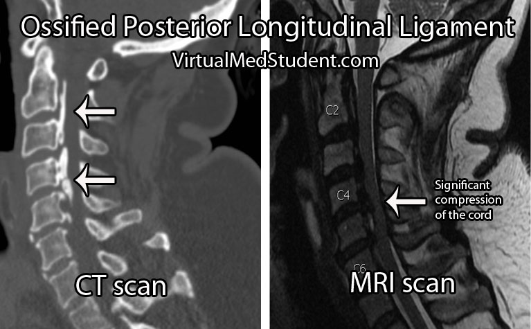
In some individuals this ligament becomes ossified, which means that it takes on a bone like quality. As a result, its overall size and hardness increase. Given the ligament’s location adjacent to the spinal cord, any increase in the girth or flexibility of the ligament can cause injury to the cord.

Ossification of the posterior longitudinal ligament can occur at multiple spots along the spinal column. The most common spot is in the cervical spine (usually C2 to C6), but ossification can also occur in the thoracic (usually T4 to T7) and lumbar spine as well.

Exactly why the longitudinal ligament ossifies in some people is unknown. Current thinking is that a combination of genetic and lifestyle factors play a role in its pathology. For example, family studies have shown increased rates of OPLL in first degree relatives of people known to have the disorder. OPLL is also more common in people of Japanese and Korean descent.

Both familial and racial linkage usually indicate a genetic component to the disease. In fact, patients with OPLL have higher rates of dysfunctional collagen gene regulation (specifically, type XI and VI collagens). Linkage to specific human leukocyte antigen haplotypes on chromosome 6 have also been implemented.

Lifestyle factors such as diet have also been shown to increase risk. Patient’s who are diabetic or pre-diabetic have higher rates of OPLL compared to the rest of the population. High protein diets seems to decrease the risk, whereas high salt diets seem to increase risk.

Overall, the reasons why the posterior longitudinal ligament ossifies in some people, but not others remains an area of ongoing debate and research.

Signs and Symptoms

When an ossified posterior longitudinal ligament pushes on the spinal cord it causes numerous signs and symptoms. The constellation of clinical findings seen in patient’s with symptomatic ossified posterior longitudinal ligaments is known as myelopathy.

Myelopathic patients present with a combination of weakness, clumsiness (ie: decreased ability to hold objects), bowel or bladder dysfunction, spasticity – which is manifested as increased reflexes, as well as changes in sensation (ie: numbness, tingling, etc.).

Ossified Posterior Longitudinal Ligament
Myelopathy can be subtle at first, but can become debilitating depending on how much compression of the cord is present.

Diagnosis and Classification

Diagnosis of an ossified posterior longitudinal ligament is usually made when a patient presents with myelopathic features, or after neurological injury from a traumatic event.

Imaging studies such as xrays and CT scans illustrate the bony quality of the ligament. MRIs are frequently ordered to assess how "squashed" the spinal cord is. CT myelograms also provide excellent detail of cord compression when MRI is not feasible.

The ossification is classified according to its anatomic location and continuity. There are four distinct patterns. They include a continuous pattern, in which there is ossification behind both the vertebral bodies and the disc spaces. The second pattern is known as segmental; in this type the ossification is only present behind the vertebral bodies and does not span the disc spaces. The third pattern is localized, which means that the ossified ligament is present and localized behind only one vertebral body. Finally, there is a mixed type, which is a combination of continuous and segmental.

Treatment

The problem with ossification of the posterior longitudinal ligament is that it progresses over time. Slowly, the ligament will compress the spinal cord. Therefore, surgical treatment is typically offered at the first sign of spinal cord compression (ie: myelopathy).

The best surgical treatment is controversial. Approaching the spine from the front (ie: anterior approach) allows direct removal of the ossified ligament. However, it is important to note that the ligament is often stuck to the dura mater overlying the spinal cord which can make dissection extremely difficult. A cerebrospinal fluid leak is not uncommon when an anterior approach is taken, and there is an increased risk of inadvertent injury to the spinal cord. That being said, when successful, surgery from the front offers several distinct advantages. The first is that myelopathic symptoms seem to respond better to this type of approach; in addition, removal of the ossified ligament can retard further progression of the disease.

The second surgical option is to approach the spine from the back (ie: posterior approach). Removing the bone behind the spinal cord allows the cord to "drift" backwards away from the ossified ligament. This approach is considered more safe because there is no direct removal of the ossified ligament, and therefore there is a decreased risk of inadvertent cord injury. That being said, progression of the ossification can still occur and myelopathic symptoms do not respond as well to this type of surgery.

Ultimately, the type of surgery offered – anterior versus posterior – is dictated by the severity of the ossification and symptoms present, as well as the preferred approach of the surgeon.

Overview

Ossification of the posterior longitudinal ligament is seen more commonly in patients of Japanese descent. There are genetic factors that appear to increase risk. Symptoms are related to compression of the spinal cord by the ossified ligament. Ossification is progressive in nature. Treatment consists of surgery from either an anterior, posterior, or combined approach.

Related Articles

References and Resources

What Do New Orleans and Canada Have in Common? Head CT Rules

Background

CT scans have become the sine-qua-non of assessing traumatic brain injury. CT scans of the head are fast and relatively inexpensive. In addition, they are able to pick up injuries that require emergent intervention.

CT scanners are so ubiquitous, at least in the United States, that it is easy to order a scan regardless of whether or not it is clinically indicated. The “scan everybody with a head bonk” mentality is dangerous for several reason. First, it exposes the patient to unnecessary radiation. Second, the term “relatively” inexpensive is exactly that, “relative”. CT scans are still costly by comparison, and every unnecessary scan only adds to the economic health care crisis.

So who should we scan? The literature on who to scan is based heavily on the Glasgow Coma Scale (GCS). This scale divides patients with head trauma into three categories: mildly injured, moderately injured, or severely injured.

The Glasgow Coma Score is based on three behavioral components: eye opening, verbal performance, and motor responsiveness. Scores range from 3 to 15, with 15 being a "normal" score and 3 being completely comatose (or even dead!). Patients with a score of 8 or less are considered severely injured. Those with a score of 9 to 12 are considered moderately injured, and those with a score of 13 to 15 are considered mildly injured.

Patients with moderate to severe GCS scores should always be scanned. These people are at high risk for clinically important brain injury.

That brings us to the next question… What should we do with all the mild head bonks? The mildly injured patient usually looks good clinically (ie: a normal neurological examination), but may still harbor intracranial nastiness! So how do we determine which mild injuries to scan and which ones to send home?

The New Orleans Criteria

New Orleans Criteria:
Scan if GCS 15 and
any of the following
are present…

– Headache
– Vomiting
– Age 60 or older
– Short term memory
problems
– Seizure
– Intoxicated
– Visible injury above
clavicles
The answer lies in two commonly used guidelines. The first set of guidelines is known as the "New Orleans Criteria for Minor Head Injury". The New Orleans Criteria state that anyone with a normal GCS should be scanned if any of the following criteria are present: headache, vomiting, 60 years of age or older, short term memory problems, seizure, intoxication (ie: alcohol or drugs), or visible injuries above the clavicles.

The Canadian Head CT Rules

The second set of guidelines is known as the "Canadian Head CT Rules in Minor Head Injury". This set of guidelines states that any patient with a mild GCS score (ie: a 13, 14 or 15) are at high risk for neurosurgical intervention if the following factors are present: GCS score of less than 15 for longer than 2 hours after the injury, open or depressed skull fracture, signs of basilar skull fracture on physical examination, greater than two episodes of vomiting, and age greater than 65. In addition, patients at risk for brain injury (although not necessarily requiring surgical intervention) include those with amnesia longer than 30 minutes from the injury, or those involved in a dangerous mechanism of injury.

Comparing the Two Criteria

Canadian Head CT Rules:
Scan if GCS 13, 14, or 15 and any
of the following are present…

– GCS < 15 at 2 hours
– Open/depressed skull fracture
– Vomiting > 2 times
– Signs of basilar skull fracture
– Age 65 or older
– Dangerous mechanism
– Antegrade amnesia > 30 minutes
The two sets of criteria for scanning are surprisingly different. I would argue that the New Orleans Criteria are more "loose" compared to the Canadian Rules. For example, most people have a "headache" after traumatic injury, and based on the New Orleans criteria this alone would be enough to warrant a scan. In a head-to-head comparison both criteria were very sensitive at picking up clinically important head injuries, but the Canadian Rules were more specific.

Overview

Deciding who to scan after mild head injuries has been studied extensively. Currently, two common sets of criteria are used to decide who gets a CT scan. Both sets of criteria are sensitive in picking up clinically significant head injury, but the Canadian Head CT Rules are more specific than the New Orleans Criteria and may help further reduce unnecessary scanning.

References and Resources

  • Washington CW, Grubb RL Jr. Are routine repeat imaging and intensive care unit admission necessary in mild traumatic brain injury? J Neurosurg. 2012 Mar;116(3):549-57.
  • Stiell IG, Clement CM, Rowe BH, et al. Comparison of the Canadian CT Head Rule and the New Orleans Criteria in patients with minor head injury. JAMA. 2005 Sep 28;294(12):1511-8.
  • Teasdale G, Jennett B. Assessment of coma and impaired consciousness. A practical scale. Lancet. 1974 Jul 13;2(7872):81-4. This is the original GCS paper.
  • Bouida W, Marghli S, Souissi S, et al. Prediction value of the Canadian CT head rule and the New Orleans criteria for positive head CT scan and acute neurosurgical procedures in minor head trauma: a multicenter external validation study. Ann Emerg Med. 2013 May;61(5):521-7.

Tuberculous Meningitis: Basal Cisterns, Strokes, Hydrocephalus

Tuberculosis is one of the most common infectious diseases in the world. It is caused by a bacteria of the genus mycobacterium. Tuberculosis usually infects the lungs, but may also infect the lymph nodes, vertebral bodies, kidneys, gastrointestinal system, or central nervous system.

Central nervous system disease comes in two flavors: a focal abscess-like lesion known as a tuberculoma or tuberculosis meningitis. The remainder of this article will focus on tuberculosis meningitis, which is an uncommon (although not rare!) form of extra-pulmonary tuberculosis.

Let’s start by discussing how the bacteria get into the central nervous system. After being inhaled the bacteria infect cells known as macrophages. The infected macrophages move towards lymph nodes, and eventually end up in the blood stream. Once in the blood stream, the mycobacterium-infected macrophages can travel anywhere in the body!

One spot the bacterium hitch a ride to is the lining of the brain (aka: the meninges). Collections of mycobacterium-laden macrophages (“Rich foci”) can rupture into the subarachnoid space causing an inflammatory reaction (ie: a "meningitis", or inflammation of the meninges).

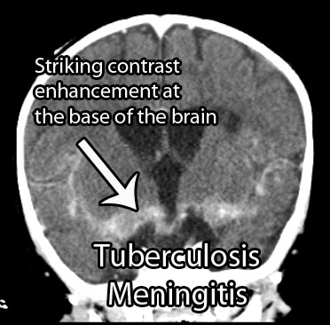
For unclear reasons, the inflammation seen in tuberculosis meningitis preferentially affects the basal cisterns and base of the brain. Autopsy specimens show a gelatinous material coating the undersurface of the brain. The inflammatory reaction is what causes the signs and symptoms of tuberculosis meningitis.

Signs and Symptoms

Tuberculosis meningitis can present in a number of different ways. Many patients present with days to months of non-specific symptoms such as headache, lethargy, nausea, and vomiting.

Cranial neuropathies are commonly seen, especially since tuberculosis meningitis affects the basal cisterns and base of the brain, which is where many of the cranial nerves run.

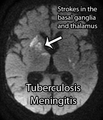
Additionally, up to 40% of patients may present with stroke. Strokes occur because the inflammation can "eat up" the linings of small blood vessels. The basal ganglia, internal capsule, and thalamus are the most common locations where strokes occur in tuberculosis meningitis.

Diagnosis

The clinical history is extremely important. Tuberculosis meningitis may affect both immunocompetent and immunocompromised (ie: think HIV/AIDs, diabetics, people on immunosuppressives, etc.) people. If the clinical history and physical exam findings are concerning for meningitis, than confirmatory studies should be performed.

TB Meningitis MRI
TB Meningitis CT
Cerebrospinal fluid (CSF) analysis shows elevated opening pressures, increased cellularity with inflammatory cells like neutrophils (earlier) and lymphocytes (later), an increased amount of protein, and a decreased amount of glucose. An acid-fast stain of the CSF to look for bacteria is sometimes positive. Culturing the CSF for mycobacterium is routinely done, but results can take weeks to months, and therefore is not useful in deciding whether or not to start treatment. Polymerase chain reactions (PCR) to look for mycobacterium DNA are also commonly used, and are much quicker than culturing.

Appropriate imaging studies include CT or MRI scans with contrast. The basal cisterns and base of the brain will "light up" with contrast because of inflammation. Imaging may also show strokes and hydrocephalus. Under the right clinical scenario imaging studies can help support the diagnosis, but are not specific.

Treatment

Treatment should consist of antibiotics that target mycobacterium tuberculosis. Commonly used antibiotics include rifampin, isoniazid, streptomycin, and pyrazinamide. Other antibiotics may be necessary if the strain of bacteria is resistant to these drugs.

Steroids are also frequently given to help reduce inflammation. Dexamethasone, a commonly used steroid, has been shown to improve survival rates (although it may not affect outcome in those that survive).

The intense inflammatory reaction seen in tuberculosis meningitis may gunk up the re-absorption of cerebrospinal fluid and cause a communicating hydrocephalus. Hydrocephalus occurs in 70% of cases and may require surgically inserted shunts to fix.

Overview

Tuberculosis meningitis is a devastating manifestation of a common infectious disease. It can cause cranial neuropathies, strokes, and hydrocephalus. Prompt diagnosis is mandatory and is made from the clinical history, CSF analysis, and imaging studies. Treatment is with anti-TB medications and steroids. Many survivors have long term disabilities despite appropriate treatment.

Related Articles

References and Resources

Appendicitis: A Vestigial Remnant to Belly Pain

The appendix is a small out-pouching off a part of the large intestine known as the cecum. It functions similar to normal large intestine by secreting mucous and absorbing water. Its overall importance, however, is not well understood, and it is likely a vestigial remnant from a distant ancestor. Unfortunately for some unlucky folk it can become inflamed; when this occurs it is called “appendicitis”.

Appendicitis occurs when something blocks the opening of the appendix into the cecum. There are numerous causes. The most common in younger individuals is a mass of inflammatory cells known as lymphoid hyperplasia, which can occur after a viral or bacterial infection of the gut. In older individuals, the most common cause is a small, hard piece of poop known as a “fecalith”.

When lymphoid hyperplasia or a fecalith (or any other obstructing thing) blocks the opening of the appendix, any mucous secreted by it gets trapped. When the appendix becomes distended enough, it literally chokes off its own blood supply and starts to die.

The dying appendix sets off a cascade of inflammation. Bacteria within the intestine are able to move in and wreak further havoc! The end result is a nasty inflammatory process, that if left unchecked, can lead to a very serious surgical illness.

Signs and Symptoms

Appendicitis initially presents with periumbilical pain (ie: pain around the belly button) that quickly migrates to involve the right lower quadrant of the abdomen.

The reason pain occurs in this sequence is because the initial discomfort of appendicitis is due to inflammation of the visceral peritoneum and appendix itself. The visceral peritoneum is a layer of tissue that envelopes the gut tube. This type of pain is carried back to the spinal cord by autonomic nerves, and due to their embryological origin, that pain gets referred to the midline of the abdomen (the belly button).

Over the course of the disease, the parietal peritoneum eventually becomes inflamed. This pain is carried by somatosensory nerves with a very specific dermatomal distribution, thus the pain gets localized directly to the anatomical location of the appendix – the right lower quadrant. This pain is often very well localized at an area known as McBurney’s point.

Patients often have nausea, vomiting, and a decreased appetite. In fact, if patients are hungry and want to eat, appendicitis becomes a highly unlikely diagnosis for abdominal pain. This is referred to informally as the "cheeseburger sign".

There are also several physical exam findings. Pain with palpation of the right lower quadrant associated with rebound tenderness (ie: pain that occurs when you release pressure during palpation) is frequently seen. Sometimes palpating the left lower quadrant (ie: the area on the other side of the abdomen from the appendix) will cause discomfort in the right lower quadrant. This is known as "Rovsing’s sign".

Any maneuvers that irritate the peritoneum will also cause discomfort. The first of these signs occurs when you flex the hip. This causes the iliopsoas muscle to rub up against an inflamed peritoneum. This is known as the "psoas sign". Another way to irritate the peritoneum is to internally rotate the leg while the patient’s knee and hip are flexed. This is known as the "obturator sign".

Diagnosis

Appendicitis CT Scan
Appendicitis is first and foremost a clinical diagnosis. Therefore, in patients with a history of periumbilical pain that migrates to the right lower quadrant appendicitis is the most likely diagnosis.

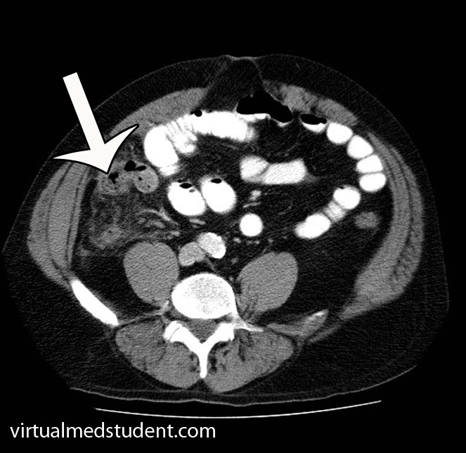
However, in a world with advanced imaging technologies we can quickly get pictures that support the diagnosis. Both ultrasound (frequently used in children, pregnant patients, and younger adults) or CT scans can be obtained quickly and inexpensively. The CT scan will show fat stranding and fluid around an enlarged appendix (see image to the left).

Blood tests such as a complete blood count (CBC) will show an elevated white blood cell count (ie: the cells that fight off infection and are responsible for inflammation).

Treatment

Treatment is surgical (ie: appendectomy)! Get that nasty inflamed appendix out of there STAT! In addition, patients are started on intravenous fluids and antibiotics that cover anaerobic organisms.

Commonly used antibiotics for a non-ruptured appendix are metronidazole, ampicillin/sulbactam, and ciprofloxacin. If the appendix has ruptured, broad spectrum coverage with piperacillin/tazobactam or a combination of ampicillin, ciprofloxacin, and clindamycin is started and continued for at least 5 days.

Occasionally a ruptured appendix will wall itself off into an abscess. If this is the case, the abscess must be drained with a needle. Antibiotics are started, and the patient is taken to surgery roughly six weeks later to remove the appendix after the inflammation has "calmed down".

Overview

Appendicitis occurs when something blocks the opening of the appendix into the cecum. Progressive enlargement of the appendix occurs eventually chocking off its own blood supply. Pain in the right lower quadrant of the abdomen is a common symptom of appendicitis. Diagnosis is clinical, but ultrasound and CT scanning can be helpful in elucidating unclear cases. Treatment is appendectomy (removal of the appendix), IV fluids, and antibiotics.

References and Resources

  • Merlin MA, Shah CN, Shiroff AM. Evidence-based appendicitis: the initial work-up. Postgrad Med. 2010 May;122(3):189-95.
  • Kim JK, Ryoo S, Oh HK, et al. Management of appendicitis presenting with abscess or mass. J Korean Soc Coloproctology. 2010 Dec;26(6):413-9. Epub 2010 Dec 31.
  • Lee SL, Islam S, Cassidy LD. Antibiotics and appendicitis in the pediatric population: an American Pediatric Surgical Association Outcomes and Clinical Trials Committee systematic review. J Pediatr Surg. 2010 Nov;45(11):2181-5.
  • Markides G, Subar D, Riyad K. Laparoscopic versus open appendectomy in adults with complicated appendicitis: systematic review and meta-analysis. World J Surg. 2010 Sep;34(9):2026-40.
  • Grundmann RT, Petersen M, Lippert H, et al. The acute (surgical) abdomen – epidemiology, diagnosis and general principles of management. Z Gastroenterol. 2010 Jun;48(6):696-706. Epub 2010 Jun 1.

Aortic Dissection: Intima, Media, DeBakey

Pathology

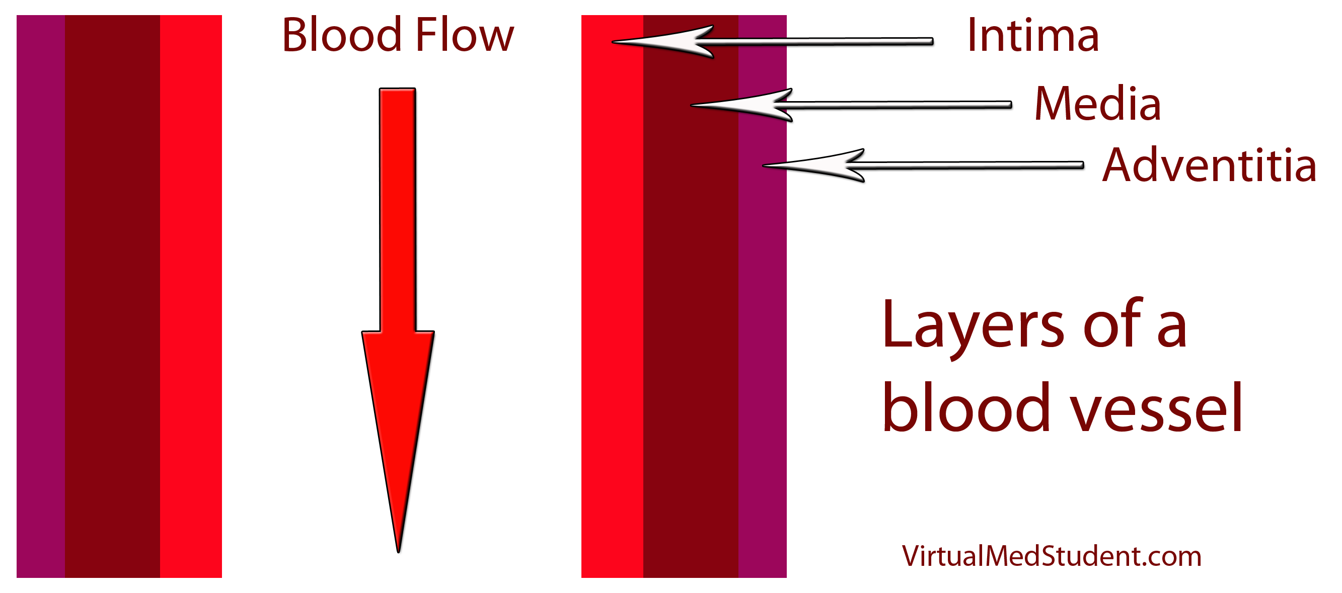
Aortic dissection occurs when blood flows into a “false” pathway created by damage to a layer of the aorta. In order to understand dissections, we have to first appreciate the layers of the aorta. Most blood vessels have three layers: the intima, media, and adventitia (see image below). You can think of these layers as insulation around a pipe. The adventitia is the outermost portion of the “pipe”; it helps connect the blood vessel to adjacent structures in the body. The media is the middle most layer, and is composed of smooth muscle; it helps control the diameter, and therefore pressure within the vessel. The intima is the layer that is immediately adjacent to blood flow.

Blood Vessel Layers
It is this intimal layer where all the “action” in aortic dissection occurs. Damage to the intima is the initiating event in a dissection, and can be caused by many different things. The most common cause is years and years of untreated high blood pressure (ie: hypertension). The hypertension “beats” down the intimal layer making it more likely to tear. In addition, other less common causes such as Marfans Disease or Ehlers-Danlos Syndrome can also cause aortic dissection. These genetic disorders result in weak connective tissue that can predispose the intimal layer to tearing.

And this is precisely what happens in aortic dissection. The intimal layer of the vessel tears. As a result blood can now take one of two possible pathways:

(1) Down into the aorta (ie: its normal path).

(2) Into the “false” space between the intimal and medial layer (aka: a "false lumen").

If blood takes the later pathway it ultimately “dissects” the intimal and medial layer away from one another, hence the name aortic “dissection”. Ultimately, the classification and treatment of aortic dissections depends on where along the aorta the dissection occurs.

Classification Systems

Aorta Schematic
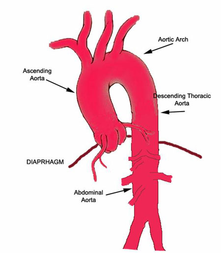
There are two different systems for classifying aortic dissections based on their location along the aorta. The first system is the DeBakey system (named after a world renowned heart surgeon, Michael DeBakey). It is divided into 3 subtypes:

– DeBakey type 1 -> involves the ascending and descending aorta.
– DeBakey type 2 -> involves the ascending aorta only.
– DeBakey type 3 -> involves the descending aorta only.

The second classification system is more simple. It is called the Stanford classification, and is divided into 2 subtypes:

– Stanford type A -> Involves the ascending aorta (may or may not involve the descending aorta)

– Stanford type B -> involves the descending aorta (does not involve the ascending aorta)

The reason these classification systems exist is because the location of the dissection dictates treatment. Ascending dissections are treated much differently than descending dissections.

Signs and Symptoms

The symptoms of aortic dissection depend on its location. If the dissection affects the ascending aorta the classical presentation is a tearing or ripping chest pain. If the descending aorta is involved the pain is often referred to the upper back between the scapulae. In addition, patients usually come in with elevated blood pressures (which is usually the precipitating cause in most cases). However, the blood pressure readings can be different between arms. Pulses from blood vessels that originate before the dissection are often stronger. This can lead to pulse asymmetry on physical exam.

New heart murmurs can also occur. The most common one is the murmur of aortic regurgitation. New murmurs occur when the aortic root is involved in the dissection. This can cause mechanical damage to the aortic valve leading to abnormal function. Blood is then able to regurgitate (ie: flow backwards) into the heart due to the deficient valve.

Since the aortic arch (the portion between the ascending and descending sections) contains vessels that eventually go to the brain, patients sometimes have neurological symptoms as well, although this is a relatively uncommon finding in clinical practice.

Diagnosis

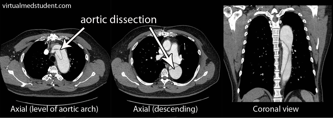
The diagnosis of aortic dissection is based on clinical suspicion combined with imaging studies. Chest x-ray will sometimes show a “widened mediastinum”. This occurs because the enlarged aorta casts a larger shadow on the x-ray detector. If this is seen, and there is a high clinical suspicion of a dissection, a CT scan of the chest with intravenous contrast is ordered (see image).

Aortic Dissection

The CT scan will show the true and false lumens associated with dissection. Transesophageal echocardiography (TEE) is another way of visualizing the aorta and is highly sensitive and specific for detecting dissections.

Treatment

Treatment is dependent on the location of the dissection along the aorta. Dissections of the ascending aorta (DeBakey type 1 and 2, and Stanford class A) are surgical emergencies and require operative repair.

Descending aortic dissections (DeBakey type 3, and Stanford class B) are often managed medically through blood pressure control.

With the advent of endovascular techniques, many dissections can be treated endovascularly (ie: "through the groin"), which may be superior to open operative techniques for certain types of dissections.

Overview

Aortic dissections occur after damage to the intimal layer of the blood vessel. There are different classification systems depending on what part of the aorta is involved. Symptoms are usually a severe ripping chest pain that can radiate to the back. Pulses and blood pressures may vary between the right and left arms. In addition, neurological symptoms may occur if the dissections includes the vessels leading to the head. If the ascending aorta is involved surgery is indicated; descending aortic dissections are usually managed with aggressive blood pressure control.

References and Resources

  • Kumar V, Abbas AK, Fausto N. Robbins and Cotran Pathologic Basis of Disease. Seventh Edition. Philadelphia: Elsevier Saunders, 2004.
  • Lilly LS, et al. Pathophysiology of Heart Disease: A Collaborative Project of Medical Students and Faculty. Fourth Edition. Lippincott Williams and Wilkins, 2006.
  • Flynn JA. Oxford American Handbook of Clinical Medicine (Oxford American Handbooks of Medicine). First Edition. Oxford University Press, 2007.
  • Blackbourne LH. Surgical Recall, Fifth North American Edition (Recall Series). Fifth Edition. Philadelphia: Lippincott Williams and Wilkins, 2009.
  • Hata M, Sezai A, Yoshitake I, et al. Clinical trends in optimal treatment strategy for type a acute aortic dissection. Ann Thorac Cardiovasc Surg. 2010 Aug;16(4):228-35.
  • Nordon IM, Hinchliffe RJ, Loftus IM, et al. Management of Acute Aortic Syndrome and Chronic Aortic Dissection. Cardiovasc Intervent Radiol. 2010 Nov 12. [Epub ahead of print].

Atherosclerosis: Gruel and Hardening

Pathology

What does the term atherosclerosis mean? If we break the term down into its components, "athero" is Greek for a gruel or paste, and sclerosis means hardening. This is precisely what is happening in the blood vessels of people with this disease; a paste-like material hardens to form a plaque. The specifics of how that paste-like material forms are much more complicated.

The paste-like material is composed of several different elements. The first, and perhaps most important element, is low density lipoprotein (ie: LDL). LDL, or the “bad cholesterol” as it is commonly referred, is a mixture of lipid (ie: fat and cholesterol) and protein. These molecules are highly atherogenic, which means they accelerate the plaque forming process.

How does LDL lead to a plaque? The first step involves disruption of part of the blood vessel known as the intima. Blood vessels have three layers: intima, media, and adventitia. The intimal layer is the portion of the vessel directly in contact with blood flow. It is composed of endothelial cells, which have numerous important functions. The medial layer is smooth muscle that controls the diameter of the blood vessel; the adventitial layer is a connective tissue coating (ie: similar to the plastic coating surrounding an electrical wire) that anchors the vessel to adjacent structures in the body.
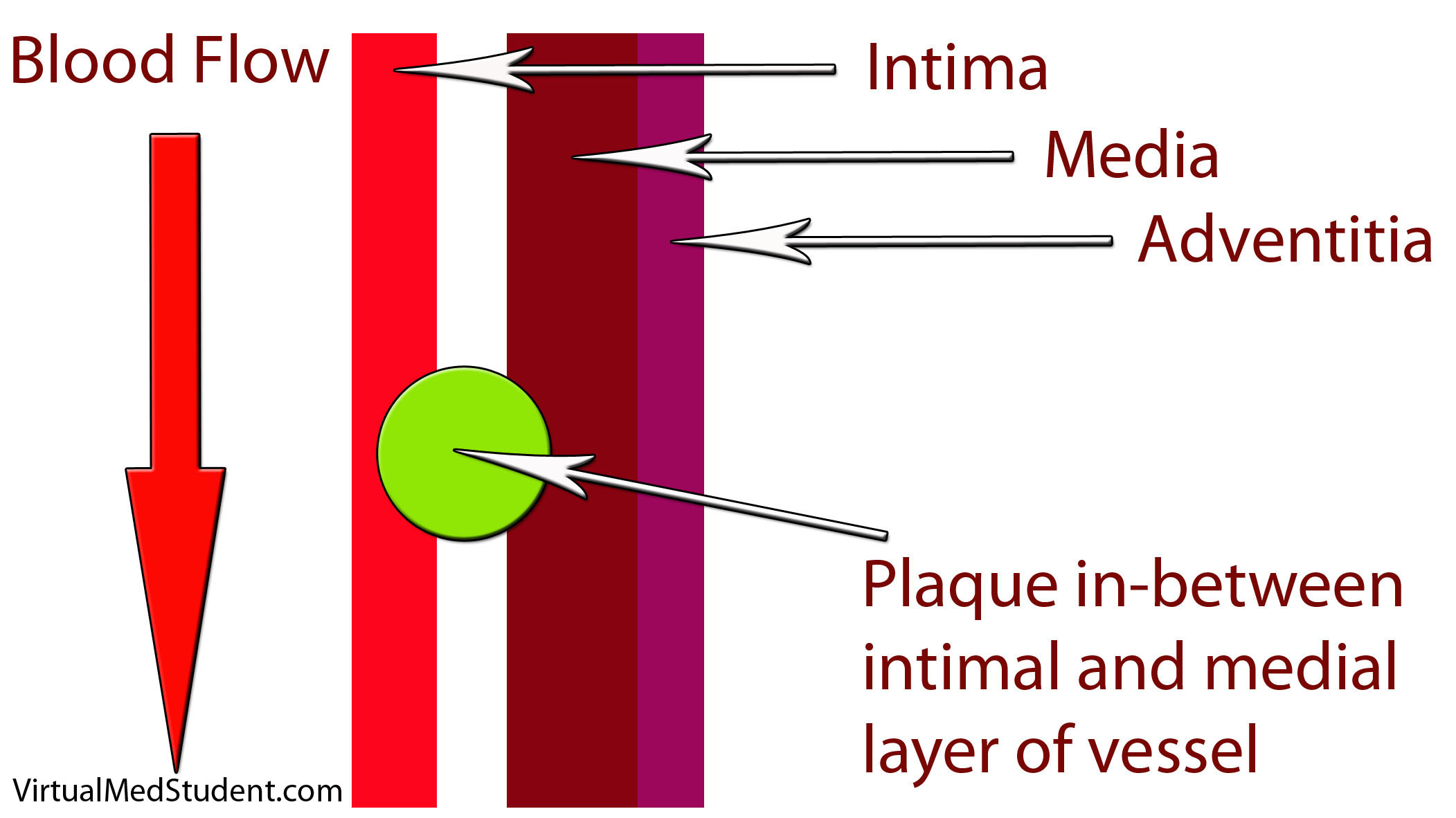
Blood Vessel with Plaque

When the intimal layer is disrupted, LDL particles floating in the blood get trapped in the vessel wall. Once trapped, they start an inflammatory reaction. White blood cells (a key component of inflammation) known as macrophages are recruited to the vessel’s walland gobble up the intruding LDL particles. At this point the macrophages are known as “foam cells”. Foam cells get enmeshed in a web of scar and smooth muscle. All of this together becomes a "plaque".

Symptoms

The symptoms of atherosclerosis depend on how large the plaques are, and where they are located. The three common symptoms associated with atherosclerosis:

(1) Angina (chest pain) – read about acute coronary syndromes
(2) Transient ischemic attacks (TIA)
(3) Vascular claudication

Angina refers to chest pain that occurs with exercise (by exercise we refer to any activity above normal daily activity). The pain is a result of atherosclerotic plaque blocking the increased blood flow needed to supply the heart during strenuous activities. In essence, what is happening is that the heart is "starving" for oxygen, which results in chest pain. When the patient stops exercising the heart no longer needs as much oxygen; the amount of blood flow is again adequate, and the chest pain ceases. Angina is a harbinger of a potential heart attack.

Transient ischemic attacks are due to atherosclerosis in the blood vessels leading to the brain. The temporary decrease in blood flow to the brain caused by the blockages can result in stroke-like symptoms that resolve within twenty-four hours. TIAs are harbingers of potential strokes in the future.

If atherosclerosis develops in the vessels going to the lower extremities, vascular claudication occurs. Claudication refers to pain in the legs that worsens while walking (or running). Similar to angina, the pain is due to blocked blood flow to the legs secondary to the plaques. A condition known as "Leriche’s syndrome" occurs when there is decreased blood flow to not only the legs, but also the vessels leading to the penis. This causes not only lower extremity claudication, but also impotence.

The complications of atherosclerosis stem from the symptoms. They include myocardial infarction (eg: heart attack), of which angina is a warning sign; as well as cerebrovascular accidents (eg: strokes), of which transient ischemic attacks serve as a warning. Worsening disease in the lower extremities can lead to gangrenous limbs and the need for potential amputation.

(1) Myocardial infarction (eg: heart attack)
(2) Cerebrovascular accidents (eg: stroke)
(3) Worsening lower extremity disease -> amputations

Diagnosis

Diagnosis of atherosclerotic disease depends on the location of symptoms. If anginal chest pain is the main symptom several different studies can be done. One such study is the “stress test”. There are several different permutations of the stress test.

All of them include two components. The first is a method of "visualizing" the heart. This can be done by ECG, echocardiography (ie: ultrasound of the heart), or nuclear/radioactive scans. The second component involves "stressing" the heart. This is most commonly done by exercising on a treadmill, but medications like dobutamine can also be used if the patient cannot exercise for whatever reason.

Stress Testing
Methods for visualizing the heart’s function – Electrocardiogram (ECG)
– Echocardiogram (echo)
– Nuclear studies
   
Methods for stressing the heart – Exercise (ie: treadmill)
– Medications (ie: dobutamine, persantine, etc.)

If the stress test is positive, or there is a high index of suspicion for atherosclerosis involving the coronary arteries, the next test performed is an angiogram. This involves injecting radio-opaque contrast material into the coronary arteries through a tiny catheter. The coronary arteries can then be visualized using x-rays. Areas where there is less contrast in the vessel indicate coronary atherosclerotic disease. The image below is an example of narrowing in the circumflex artery (indicated by the arrow).
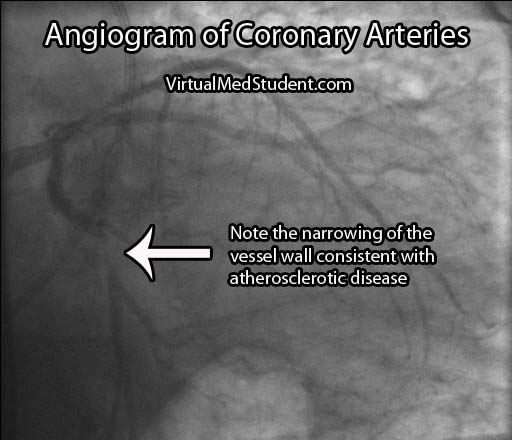
Angiogram Circumflex Atherosclerosis

If the involved areas are the carotid arteries, a test known as "carotid artery duplex scanning" can be done. This involves using ultrasound to detect the amount of blood flow present in the vessel(s). Decreased rates are often attributable to atherosclerotic disease.

Finally, if disease is thought to be present in the lower extremities, a test known as the arterial brachial-ankle index (ABI) is obtained. This test involves taking the blood pressure in the arm and the leg and comparing the results. Blood pressure should be about equal in the upper and lower extremities resulting in a brachial to ankle ratio of 1.0. However, if there is atherosclerotic diseases in the lower extremities the ABI decreases to less than 1.0.

Location of suspected atherosclerotic disease: Initial test performed:
– Coronary arteries – Stress test
– Carotid arteries – Duplex scanning
– Arteries of lower extremities – Ankle-Brachial index (ABI)

Occasionally cardiologists or vascular surgeons will recommend a more invasive procedure known as a "catheterization". In this procedure a small catheter is threaded from a blood vessel in the groin to the area of interest (ie: the heart, carotids, vessels of the lower extremities). From there radio-opaque dye is injected. A fancy X-ray machine can then pick up locations of significant plaque formation.

Treatment

Treatment is designed to prevent further plaque formation. Since LDL plays a crucial role in this process, it is a target of medical therapy, but in order to understand treatment we have to first understand how cholesterol gets into the body. Cholesterol is either (a) absorbed by the gut from your diet, or (b) produced naturally by your body through a series of biosynthetic reactions.

The first interventions for decreasing cholesterol in the body are, you guessed it, dietary modifications. By decreasing the amount of cholesterol in the diet it is possible to decrease the amount of cholesterol, either free or in the form of LDL, that is in the blood stream. There is also some evidence that replacing saturated fats with polyunsaturated fats (ie: α-linolenic acid, an ω-3 fatty acid) can decrease atherosclerotic plaque formation.

Unfortunately, diet and exercise are not always enough to decrease blood cholesterol levels to acceptable limits. Several medications have been developed, and as one could imagine, they target either cholesterol in the gut, or the biosynthetic machinery that produces cholesterol naturally.

There are several types of drugs that bind cholesterol in the gut to keep it from being absorbed. They are referred to as "bile acid binding medications" or "bile acid sequestrants". It is important to realize that bile acids, which are secreted by the liver and gallbladder, are rich in cholesterol.

Under normal circumstances they are reabsorbed by the gut and recycled back into the bile pathway. By inhibiting their re-absorption, cholesterol from the blood stream is recruited by the liver to replace the bile acids that were previously being recycled. The ultimate effect is that these drugs decrease the amount of cholesterol in the blood stream. There are three common medications in this category: colesevelam, cholestyramine, and colestipol.

Another medication, ezetimibe, directly binds to free cholesterol in the gut. As a result, less cholesterol is delivered to the liver. To compensate, the liver increases its ability to absorb LDL and other forms of cholesterol from the blood resulting in decreased plasma levels.

Atherosclerosis
treatments:

(1) diet and
      exercise
(2) bile acid
      sequestrants
(3) statins
(4) niacin
(5) fibrates
Perhaps the most widely used class of medications used to treat atherosclerosis are known as “statins.” Statins such as atorvastatin and pravastatin inhibit an enzyme (ie: HMG-CoA reductase) that normally produces cholesterol in the body. These medications dramatically reduce the risk of stroke and heart attack in patients with atherosclerotic disease.

A vitamin, niacin (ie: vitamin B3), can also lead to favorable effects on the lipid profile of the blood. It increases "good" cholesterol (ie: HDL). It is believed to perform these feats by increasing the activity of an enzyme, lipoprotein lipase, which normally breaks down VLDL particles (a precursor of LDL).

A final category of medications known as the "fibrates" decrease fat content in the blood. They do not necessarily affect LDL levels significantly, but they do decrease another atherosclerotic forming fat type known as triglycerides. The two common fibrates in use today are gemfibrozil and fenofibrate.

Overview

Atherosclerosis is due to LDL trapping in blood vessel walls. This trapping results in inflammation and the formation of a fibromuscular plaque. Symptoms include angina (ie: chest pain), lower extremity claudication, and transient ischemic attacks. Diagnosis is made by stress testing, carotid ultrasound, and ankle-brachial index; more invasive testing with groin catheters can be performed as well. Treatment is based on decreasing cholesterol absorption from the gut, or decreasing the bodies natural mechanism for creating cholesterol.

References and Resources

  • Negi S, Nambi V. Coronary heart disease risk stratification: pitfalls and possibilities. Methodist Debakey Cardiovasc J. 2010 Dec;6(4):26-32.
  • Amarenco P, Lavallée PC, Labreuche J, et al. Prevalence of Coronary Atherosclerosis in Patients With Cerebral Infarction. Stroke. 2010 Nov 18. [Epub ahead of print].
  • Kumar V, Abbas AK, Fausto N. Robbins and Cotran Pathologic Basis of Disease. Seventh Edition. Philadelphia: Elsevier Saunders, 2004.
  • Lilly LS, et al. Pathophysiology of Heart Disease: A Collaborative Project of Medical Students and Faculty. Fourth Edition. Lippincott Williams and Wilkins, 2006.
  • Flynn JA. Oxford American Handbook of Clinical Medicine (Oxford American Handbooks of Medicine). First Edition. Oxford University Press, 2007.
Atrioventricular Heart Block: PR, QRS, Mobitz

Heart block, also known as atrioventricular (AV) conduction system block, refers to decreased conduction of electrical impulses through the heart. This decreased conduction occurs at the atrioventricular node of the heart’s conduction system. Heart block occurs in three flavors: type 1, type 2, and type 3. Each type has different causes and different treatments.

Pathology and Types

In type 1 AV block the PR interval on an ECG is longer than normal (> 200 msec), but every QRS complex has a preceding P-wave indicating that the electrical impulses from the atria (ie: the top chambers in the heart) are "making" it through the AV node to the ventricles. In type 1 block, the AV nodal conduction is slowed compared to normal "healthy" individuals, but the impulses are still able to get all the way through the conduction system of the heart.

Type 1 AV node block = PR interval > 200 msec, no missed beats.

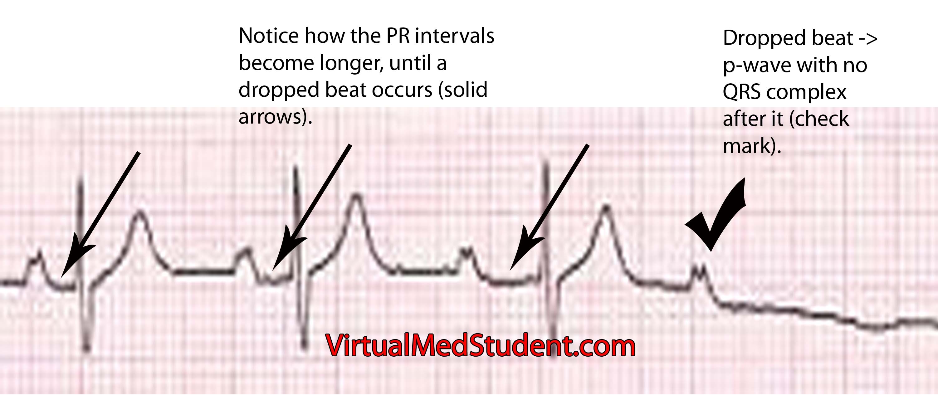
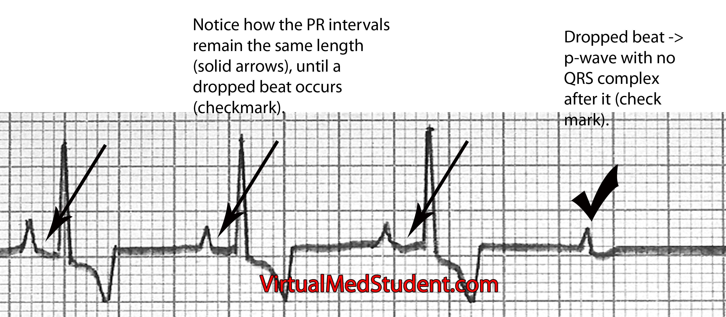
Type 2 heart block occurs in 2 flavors: Mobitz type 1 and type 2. In Mobitz type 1 the amount of delay between the P-wave and QRS complex (ie: the PR interval) gradually increases until a beat is dropped. It is caused by impaired conduction in the AV node. Mobitz type 2 occurs when the PR interval remains stable, and then out of the blue a missed beat occurs; this is caused most commonly by slowed conduction through the bundle of His or bundle branches of the heart’s conduction system.

Mobitz type 1 = increasing PR interval until dropped beat occurs (image below). Mobitz type 1 is also referred to as "Wenckeback" block.

Mobitz Type 1

Mobitz type 2 = PR interval remains the same until a dropped beat occurs (image below). Mobitz type 2 is also referred to as "Hay" block.

Mobitz Type 2

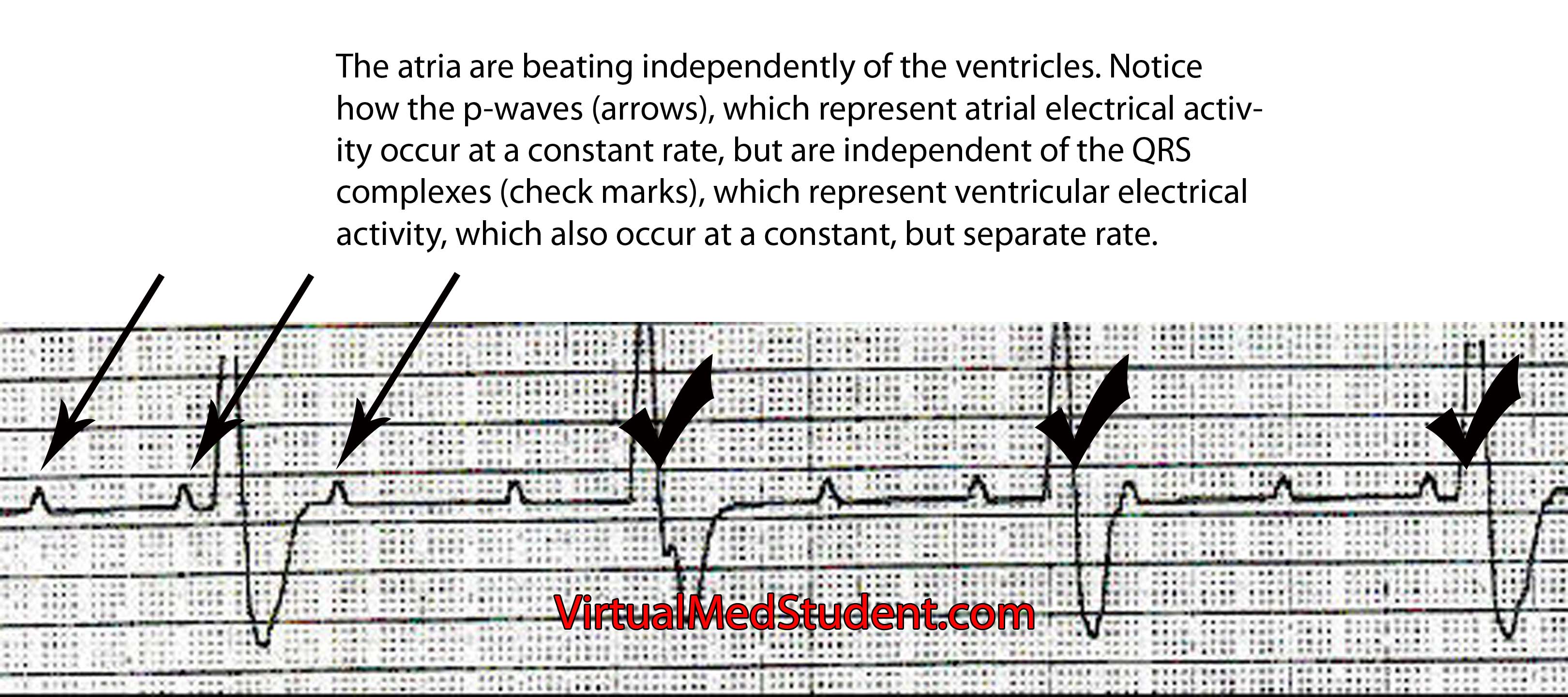
Finally, type 3 AV nodal block occurs when the atria and ventricles of the heart beat independently of one other. The most common causes of 3rd degree block are severe diseases of the AV nodal system due to age, previous heart attack, drug and medication toxicity (ie: digitalis), and untreated Lyme disease. The atria generally beat at a faster rate than the ventricles. In other words, the ECG will show more P-waves than QRS complexes. The QRS complexes may be narrow or wide depending on where in the conduction system the escape rhythm originates.

Type 3 heart block = atria (p-waves) and ventricles (QRS) beat independently of each other; no impulses from the atria make it to the ventricles (image below).

AV nodal block type 3

Symptoms

The symptoms of heart block depend on how slow the rate becomes. Patients with type 1 block rarely become symptomatic because the atria are still setting the pace of the entire hearts rhythm. However, in Mobitz type 2 and type 3 block the heart rate can become very slow. When this occurs symptoms such as syncope (ie: fainting), extreme fatigue, shortness of breath, and dizziness can occur. These symptoms occur because the heart is not pumping blood fast enough to keep up with the body’s demand; the result is a decrease in cardiac output.

Diagnosis

Diagnosis of heart block can be made by looking at the characteristic findings on ECG.

Treatment

Treatment of AV nodal dysfunction is dependent on the type. Most patients with type 1 heart block do not need treatment. Type 1 heart block can be considered a "healthy" variant of AV nodal conduction, which is slower than most people in the population. The only instance where type 1 block may need further work up is in elderly patients who have other signs or symptoms of coronary artery disease.

Type Treatment
Type 1

Usually none, look for causes such as coronary artery disease in elderly patients

Type 2 – Mobitz type 1

Usually none, but pacemaker if symptomatic. Also discontinue any drugs that slow AV node.
Type 2 – Mobitz type 2 Pacemaker
Type 3 Pacemaker

Mobitz type 1 (ie: one of the 2 subgroups of type 2 heart block) also generally requires no treatment. If patients are symptomatic pacemaker insertion may be considered. Mobitz type 2 is more worrisome because it often progresses to 3rd degree heart block. In these patients, pacemakers are often inserted to ensure that the heart beats at a pre-defined and safe rate.

Type 3 heart block is almost always controlled with pacemaker insertion. The reason is that the ventricles often beat at a rate that is too slow for normal daily activity. A pacemaker will keep the heart beating at a pre-defined rate to ensure that symptoms do not develop.

Overview

Heart block (aka: atrioventricular block) occurs when impulses travel too slowly, or not at all through the AV node. There are different types depending on how severe the block is. In type 1 AV block impulses are slowed, but cause no conduction block. In type 2 missed beats can occur, and in type 3 the atria beat independently of the ventricles indicating complete conduction block. Symptoms depend on the severity of block but can include fainting, shortness of breath, dizziness, and fatigue. Diagnosis is made by ECG. Treatment is dependent on the type of block, but can include placement of a pacemaker.

References and Resources

Hypertension: Understanding and Managing High Blood Pressure

The pathology of essential hypertension is not well understood. However, there are numerous theories, each with supporting evidence.

Genetic causes are supported by the fact that children of hypertensive parents have an increased risk of developing the disease. However, specific known genetic mutations are not common. When mutations are responsible for essential hypertension they often involve the sodium and chloride channels in kidney cells, or mutations in the genes responsible for producing the proteins of the renin-angiotensin-aldosterone system. Sodium, renin, angiotensin, and other molecules play a vital role in blood pressure. In some hypertensive patients these systems are abnormal and can lead to elevated blood pressure.

Some patients with essential hypertension appear to be “salt sensitive”. Salt, or more specifically sodium, appears to play a role in the development of hypertension. Some patients likely have a genetic predisposition to retain sodium. Excess sodium enters the blood stream where it exerts an osmotic pull on water in adjacent tissues. Fluid from body tissues is "sucked" into the blood stream resulting in increased blood volumes. The expanded blood volume increases the pressure within the blood vessel causing hypertension. Although salt plays a role in hypertension, new research has shown its importance may not be as clear as once thought.

There are other known causes of hypertension, but they constitute a relatively small proportion of cases. They are discussed in separate articles.

Signs and Symptoms

Essential hypertension in its earliest stages does not cause any signs or symptoms. Many people do not know they have high blood pressure. However, if left untreated hypertension can cause serious long term consequences.

One such consequence is an increase in the risk of cardiovascular disease. The risk of cardiovascular disease doubles with each 20/10 mmHg increase in blood pressure beyond a "baseline" of 115/75 mmHg! After years of pumping at elevated pressures the heart undergoes physical changes. Like any good muscle, it becomes larger because it is having to pump harder than normal. The result is a process known as concentric hypertrophy. The added muscle mass of the heart results in increased oxygen demand and the potential for heart attacks and heart failure.

Overall, nearly a third of heart attacks are attributable to high blood pressure. In addition, the risk of stroke is also dramatically higher in untreated hypertensive patients. The small blood vessels in the kidney and retina can also be damaged by years of high blood pressure resulting in kidney failure (hypertensive nephropathy) and blindness (hypertensive retinopathy).

Diagnosis

The diagnostic parameters of hypertension are constantly shifting. Currently the diagnosis can be made if a patient presents with a severely elevated blood pressure (systolic blood pressure > 200 and/or a diastolic blood pressure > 120) and/or signs or symptoms referable to the elevated blood pressure.

Severity Classification:
(1) Pre-hypertensive:
       130-140/80-90
(2) Grade 1:
       140-160/90-100
(3) Grade 2:
        >160/>100
In patients with less severe elevations, it is generally recommended that the blood pressure be measured several times over a period of multiple weeks. If the average of these readings is a systolic blood pressure of 140 or greater, or a diastolic blood pressure of 90 or greater than the diagnosis can be made. If the blood pressue stays between 120 and 130 systolic, and 80 to 90 diastolic the diagnosis of "pre-hypertension" is made meaning the patient is at risk of becoming hypertensive.

Some patients may have "white coat" hypertension simply by being in a doctor’s office (and the anxiety that this can provoke! Gosh, I hate going to the doctor’s office!). If this is the case the patient should be instructed to take their blood pressure at home and keep a log of the results.

Treatment

Treatment of hypertension consists of lifestyle modifications and/or using medications from several different pharmacologic categories. These medications may be used alone, or in combination, depending on each individual patient’s needs.

Patients who are pre-hypertensive or have stage 1 hypertension should be started on “lifestyle” modification therapy for at least 6 months prior to starting medicines (assuming there are no other health issues like diabetes or coronary artery disease). Lifestyle therapy consists of increasing aerobic exercise, as well as adhering to the “DASH” diet. DASH stands for "dietary approaches to stop hypertension" and consists of eating fruits, vegetables, low-fat dairy, whole grains, poultry and fish while reducing (or eliminating) red meat and sugars. This intervention alone can decrease systolic pressures over 10 mmHg and diastolic pressures over 5 mmHg.

Sometimes diet and exercise aren’t enough so we may have to resort to medications to control blood pressure. The first category of medications are known as diuretics, most commonly of the thiazide class. Thiazide diuretics work by inhibiting the re-absorption of sodium at the distal convoluted tubule of the kidney. Less sodium means less circulating blood volume, and therefore decreased blood pressure. Hydrochlorothiazide is a commonly used thiazide diuretic.

The second category of medications is known as β-blockers. β-blockers lower blood pressure by slowing the heart rate and indirectly lowering the amount of angiotensin II (angiotensin II is a potent natural blood vessel constrictor produced by the body). β-blockers reduce renin synthesis by specialized cells in the kidney.

A third category of medications known as angiotensin converting enzyme inhibitors (ACEIs) interfere with the formation of angiotensin II. ACEIs inhibit an enzyme present in the lung, which converts angiotensin I into the more potent vasoconstrictor angiotensin II. Interestingly, ACEIs also increase the level of bradykinin; this molecule causes blood vessels to dilate, which helps to further lower pressure.

There are also medications called direct angiotensin receptor blockers (ARBs), which inhibit the actions of angiotensin II. They do this by blocking angiotensin IIs ability to bind to its normal receptor sites.

Some medications, the calcium channel blockers (CCBs), lower blood pressure by inhibiting the contraction of smooth muscle cells that line the blood vessel walls. A specific subcategory of calcium channel blockers known as dihydropyridines block calcium from flowing into vascular smooth muscle cells. This results in decreased contraction of the muscle surrounding the vessel. As a result the vessel remains dilated, which lowers pressure.

Overview

The pathology of essential hypertension is not well understood. Several theories have been postulated, but it is likely a combination of genetic predispositions that result in this type of high blood pressure. Years of elevated blood pressure lead to cardiovascular disease including heart failure, heart attacks, and stroke, as well as damage to the kidneys and retinas. Treatment is with lifestyle modifications, diuretics, β-blockers, calcium channel blockers, angiotensin converting enzyme inhibitors, and angiotensin receptor blockers.

References and Resources

Monro-Kellie and Their Doctrine: Blood, Brain, Spinal Fluid

The Monro-Kellie doctrine states that three things exist within the fixed dimensions of the skull: blood, cerebrospinal fluid, and brain. An increase in any one component must necessarily lead to a decrease in one (or both) of the other components, otherwise intracranial pressure will increase.

Increases in one of the three components can take many different shapes and sizes. For example, abnormal bleeding within the cranium such as in epidural and subdural hematomas are common examples, which typically occur after traumatic events. Bleeding within the brain tissue itself – known as an intraparenchymal or intracerebral hematoma – can also occur, especially in patients with untreated high blood pressure. Brain tumors of any type effectively increase the amount of brain tissue. And last, but not least, the cerebrospinal fluid can back up in a condition known as hydrocephalus.

Regardless of the cause, the end result is an abnormal increase in either blood, brain, or cerebrospinal fluid within the confines of the skull.

So what’s the big deal? If the abnormality becomes large enough, the pressure within the skull can increase rapidly. Eventually the pressure can become so great that the brain gets squished, and will pop over rigid boundaries and out the small holes within the skull.

This is known as “herniating” the brain tissue. It can occur in numerous places depending on where the pressure is greatest. However, the most important herniation clinically occurs at the base of the skull where a hole known as the foramen magnum exists.

When the brain herniates here it really pisses off a vital structure known as the brainstem. The brainstem is responsible for all the stuff we don’t consciously think about (heart rate, breathing, etc.), which ultimately keeps us alive. When herniation of the brainstem through the foramen magnum occurs it stretches all the “wires” that allow our brainstem to function properly. If severe enough, all those autopilot functions (ie: breathing, beating of the heart, etc.) stop working and brain death occurs.

Overview

The skull contains three components within it: blood, brain, and cerebrospinal fluid. An abnormal increase in any one of these components causes an increase in pressure, which if severe enough, can cause herniation of brain tissue out of the skull. This can lead to coma and brain death.

References and Resources